
Abstract
To determine the role of oxidative DNA damage and repair in brain injury after focal ischemia and reperfusion, the authors investigated DNA base damage and DNA base excision repair (BER) capacity, the predominant repair mechanism for oxidative DNA lesions, in the rat model of temporary middle cerebral artery occlusion. Contents of 8-hydroxyl-2′-deoxyguanosine (8-oxodG) and apurinic/apyrimidinic abasic site (AP site), hallmarks of oxidative DNA damage, were quantitatively measured in nuclear DNA extracts from brains 0.25 to 72 hours after 1 hour of middle cerebral artery occlusion. In parallel to the detection of DNA lesions, the capacity for 8-oxodG- or AP site-dependent DNA repair synthesis was measured in nuclear protein extracts using specific in vitro DNA repair assays. After postischemic reperfusion, the levels of 8-oxodG and AP sites were markedly increased in ischemic tissues. In frontal/parietal cortex, regions that survived ischemia, 8-oxodG and AP sites were efficiently repaired during reperfusion. However, in the caudate, a region that was destined to infarct, the DNA lesions were poorly repaired. In consistent with the patterns of endogenous lesion repair, a markedly induced and long-lasting (at least 72 hours) BER activity was detected in the cortex but not in the caudate after ischemia. The induced BER activity in ischemic cortex was attributed to the upregulation of gene expression and activation of selective BER enzymes, particularly DNA polymerase-β and OGG1. These results strongly suggest that inducible DNA BER constitutes an important endogenous mechanism that protects brain against ischemia-induced oxidative neuronal injury.
Endogenous oxidative damage to nuclear DNA is a severe consequence of oxidative stress that, when unrepaired, may result in cell death by triggering various intracellular signaling pathways (Payne et al., 1995). Oxidative DNA damage is a prominent feature of ischemic brain injury and has been reproducibly detected in brain cells after transient or permanent cerebral ischemia (Chen et al., 1997; Cui et al., 1999; Huang et al., 2000; Liu et al., 1996; Nagayama et al., 2000a). Ischemia-induced oxidative DNA damage consists of highly specific chemical lesions such as hydroxyl radical-modified bases, apurinic/apyrimidinic abasic sites (AP sites), and strand damage (Chen et al., 1997; Huang et al., 2000; Liu et al., 1996; Nagayama et al., 2000a). This damage occurs in neurons as early as minutes after transient ischemia and is accumulated in brain regions that eventually develop DNA fragmentation and neuronal cell death (Chen et al., 1997; Cui et al., 2000). Thus, accumulation of endogenous oxidative DNA lesions could be an important contributory factor in ischemic cell death in the brain.
Several types of oxidative DNA damage may have specific detrimental effects on the fate of ischemic neurons. Formation of 8-hydroxyl-2′-deoxyguanosine (8-oxodG), the most common oxidative base modification resulting from direct attacks by hydroxyl radicals, has been associated with gene mutagenesis (Dizdaroglu, 1991; Gajewski et al., 1990; Grollman and Moriya, 1993), and the accumulation of 8-oxodG may result in partial or complete loss of the functional properties of the damaged genes (Cui et al., 1999; Mazzarello et al., 1992; Schneider et al., 1990). AP sites are generated by spontaneous base loss, by hydroxyl radical-induced base loss, or as intermediates of base excision repair (BER) of damaged DNA bases by specific N-glycosylases (Demple and Harrison, 1994; Nakamura and Swenberg, 1999; Nakamura et al., 2000). AP sites, upon accumulation, prevent the process of DNA synthesis or gene transcription through the lesion site, and thus are directly lethal to cells (Janssen et al., 1993; Moran and Wallace, 1985; Schaaper and Loeb, 1981). DNA strand damage, such as single-strand breaks, is also a potent blocker of DNA synthesis and gene transcription. The accumulation of DNA strand breaks may directly trigger cell death by activating both p53-dependent and p53-independent pathways (Eliasson et al., 1997; Endres et al., 1997; Miyashita and Reed, 1995; Nelson and Kastan, 1994; Payne et al., 1995).
Most, if not all, DNA lesions induced by reactive oxygen species (ROS) are repaired via the BER pathway in mammalian cells (Demple and Harrison, 1994; Frosina et al., 1996; Lindahl et al., 1997). BER proceeds by the sequential actions of a group of essential DNA repair enzymes, including specific DNA glycosylases, AP endonuclease, DNA polymerase-β (β-pol), DNA ligase I, or DNA ligase III and XRCC1 (x-ray cross-complementing group 1) (Frosina et al., 1996; Klungland and Lindahl, 1997; Srivastava et al., 1998). In the brain, BER activity is essential for neurons to survive and maintain the integrity of normal neuronal functions because neuronal genomic DNA constantly encounters numerous attacks by DNA-damaging ROS, even under normal physiologic conditions (Ames et al., 1993; Nakamura and Swenberg, 1999). Therefore, there is a great demand for increased BER capacity in the brain after an ischemic insult, given that oxidative DNA lesions are markedly increased in the brain within minutes after ischemia (Chen et al., 1997; Cui et al., 1999; Liu et al., 1996). It can be speculated that neurons failing to match the demand for increased BER capacity after ischemia would suffer from the accumulation of oxidative DNA lesions, impairment of genomic integrity, and eventually cell death. However, despite our increasing understanding of the role of oxidative DNA damage in mediating cell death and the importance of BER activity in protecting neurons against oxidative injury, little is known about how the neuronal BER mechanism in the brain responds to oxidative DNA damage after ischemic injury.
In this study, we investigated the postischemic regulation of cellular BER activity in a rat model of transient focal cerebral ischemia and reperfusion. The temporal and regional alterations in cellular BER activity after ischemia were correlated to the induction of several types of oxidative DNA damage measured in the same brains under the same injury paradigms.
MATERIALS AND METHODS
Animal model of focal ischemia and reperfusion
All experimental procedures were performed under protocols approved by the Animal Care Committee of the University of Pittsburgh and in accordance with the principles outlined in the National Institute of Health's Guide for the Care and Use of Laboratory Animals. All experiments were performed on adult male Sprague-Dawley rats each weighing 275 to 300 g (Charles River Sprague-Dawley). Anesthesia was induced with 5% isoflurane, and rats were then intubated and ventilated with 1.5% isoflurane in an air/oxygen mixture (90%: 8.5%). Blood pressure, blood gases, and blood glucose concentration were maintained in the normal range throughout the experiments. Temporalis muscle and rectal temperatures were maintained in the range of 36.5°C to 37.5°C using a heating pad and a temperature-regulated heating lamp. Focal cerebral ischemia was induced by intraluminal middle cerebral artery occlusion (MCAO) as previously described (Chen et al., 1996; Chen et al., 1997). In this study, the middle cerebral artery was occluded for 1 hour, resulting in infarction in the caudate-putamen, but sparing the ischemic frontal-parietal cortex (Chen et al., 1997). Sham operation was performed in control animals using the same anesthesia and surgical exposure procedures except that the middle cerebral artery was not occluded.
To ensure the induction of ischemia by MCAO and restoration of blood flow after reperfusion, regional cerebral blood flow was monitored in eight rats using laser Doppler flowmetry (PeriFlux System 5000, PERINMED) equipped with PROBE 403 (Fiber separation = 0.25 mm) as previously described (Nagayama et al., 2000a). Two parietal cortical regions ipsilateral and contralateral to the ischemic hemisphere were measured at the following coordinates: 0.5 mm anterior and 5.5 mm lateral to the bregma. Cortical blood flow was measured (2-Hz sampling rate) at 5-minute intervals 15 minutes before and up to 100 minutes after middle cerebral artery occlusion (MCAO). Blood flow values were averaged during a 30-second period at each time point, and the data were expressed as the percentage changes from pre-ischemic values.
To verify the reproducibility of the ischemic lesions in the brain produced by MCAO, 6 rats were sacrificed by an overdose IP injection of 8% chloral hydrate 72 hours after ischemia. The brains were quickly removed, frozen at −30°C in 2-methylbutane and then in powdered dry ice, and processed for brain cutting. Twenty-micrometer-thick serial coronal sections were obtained every 0.4 mm between the levels of +5.0 and −5.0 mm (anterior-posterior) from the bregma. The sections were stained with cresyl violet for computerized image analysis of infarct volume. Infarct sizes were measured and calculated as previously described (Chen et al., 1996).
Measurement of the 8-OhdG:2-dG ratio
The content of 8-hydroxyl-2′-deoxyguanosine (8-OHdG), a hallmark of oxidative DNA base damage, was measured in nuclear DNA extracts from brain tissues using high-performance liquid chromatography with electrochemical detection as previously described (Lan et al., 2000; Nagayama et al., 2000a). Rats were killed 0, 15, and 30 minutes or 2, 4, 8, 24, and 72 hours after 1 hour of focal ischemia, or 24 hours after sham operation (n = 6–8 per time point). The brains were rapidly removed, and the cortex and caudate-putamen were separately dissected and stored at −80°C. The methods for nuclear DNA extraction and digestion were as previously described (Lan et al., 2000). This protocol avoided the use of phenol or any known oxidants that may induce background oxidative lesions 8-OHdG or AP sites.
The isocratic analysis was carried out on a CoulArray system (Model 5600) equipped with a dual piston (Model 580) and a PEEK pulse damper (ESA, Inc., Chelmsford, MA, U.S.A.). The analysis was performed using two coulometer array cell columns. The data were acquired and analyzed using the CoulArray software, and expressed as the number of 8-OHdG in 105 2-dG determined in the same sample.
Quantitative measurement of AP sites in nuclear DNA
Nuclear DNA isolated from ischemic and sham brain tissues (n = 6–8 per time point) were subjected to quantitative measurement of AP sites using the calorimetric assay previously described (Nagayama et al., 2000a). A biotin-labeled reagent specific for the aldehyde group in the ring-open form of AP site, designated as the aldehyde reactive probe (ARP), was used for the detection of AP sites (Dojindo Molecular Technologies, Gaithersburg, MD, U.S.A.). ARP specifically binds to AP sites in isolated genomic DNA, and the biotin molecular in ARP can then be detected calorimetrically using a streptavidin/biotin complex conjugated to horseradish peroxidase as the indicator enzyme (Kubo et al., 1992). In brief, the purified DNA isolated from brain tissue (ratio of OD260nm/280nm>1.8) was dissolved at a concentration of 100 μg/mL in TE, and 10 μL of DNA solution was incubated with the same volume of ARP solution (5 mmol/L) at 37°C for 1 hour. The ARP-labeled DNA was then ethanol precipitated, and the DNA pellet was suspended in TE and subjected to DNA concentration determination. ARP in the labeled DNA was measured using an ELISA-like assay in a microtiter plate, which was irradiated overnight with a 40-W unfiltered lamp (Nagayama et al., 2000a). The ARP-labeled DNA (30 ng in 60 μL) and 90 μL of DNA binding solution (Dojindo Molecular Technologies) were added to each well, and the plate was covered and incubated in the dark at 37°C overnight. This was followed by incubation in 100 μL of ABC solution (Vector, Burlingame, CA, U.S.A.) at 37°C for 1 hour, and then in the K-Blue substrate solution (Neogen Corporation, Lexington, KY, U.S.A.) at room temperature for 1 hour. The wells were then subjected to optical density measurement at 650 nm. All ARP assays were performed in triplicate, and the means were calculated. The data, expressed as the amount of AP sites per 105 nucleotides, were calculated based on the linear calibration curve generated for each experiment using ARP-DNA standard solutions.
In selective time points after ischemia, the amounts of AP sites of different forms (5′-incised or intact or 3′-incised AP sites) were determined separately using the AP site cleavage assay as previously described (Nakamura and Swenberg, 1999). Briefly, for the 5′-cleavage assay (for the detection of intact or 3′-incised AP sites), 2 μg of DNA from each brain sample and 30 U of Escherichia coli exonuclease III (Exo III) were incubated in 40 μL of 10-mmol/L Tris-HCl/KOH (pH 7.5) containing 50-mmol/L NaCl and 5-mmol/L MgCl2 at 37°C for 1 minutes and immediately subjected to AP site measurement as described previously. For the 3′-cleavage assay (for the detection of 5′-incised AP sites), 2 μg of DNA, 10-mmol/L EDTA, and 100-mmol/L putrescine were incubated in 40 μL of 10-mmol/L Tris-HCl/KOH (pH 7.5) at 37°C for 30 minutes and then analyzed for the amounts of AP sites. The data are expressed as the number of 5′-incised and intact AP sites plus 3′-incised AP sites, respectively, under each experimental condition studied (Nakamura et al., 2000).
Measurement of DNA repair activities in nuclear protein extracts
In vitro DNA incorporation repair assay: detection of overall BER activity. This assay examined the ability of nuclear protein extracts to incorporate [32P]dGTP into oxidatively damaged plasmids. The DNA repair substrate used in the assay was pcDNA plasmids containing the oxidative adduct 8-oxodG, a DNA lesion known to be repaired via the BER pathway. These experiments provided an estimation of overall BER activity in cells (Chen et al., 2002; Chen et al., 2000; Nagayama et al., 2000b).
The DNA repair substrate was prepared using photoactivated methylene blue to induce 8-oxodG in purified plasmids (Chen et al., 2000; Schneider et al., 1990). The contents of 8-oxodG in the plasmids were verified using high-performance liquid chromatography with electrochemical detection. Typically, under the described conditions (Chen et al., 2000), incubation of methylene blue for 30 minutes induced approximately 150 to 200 8-oxodG/105dG in the plasmids, representing approximately 50 to 75-fold increases compared with normal undamaged plasmids.
To perform the repair assay, nuclear protein extracts (quantity of protein as indicated) from cortical or caudate tissues obtained at 0.5, 2, 8, 24, or 72 hours after 1 hour of ischemia or 24 hours after sham operation (n = 4 per group) were incubated for 60 minutes at 32°C in 50 μL of reaction mixture. The reaction mixture consisted of 0.3 μg 8-oxodG-rich plasmids, 45-mmol/L HEPES-KOH, 70-mmol/L KCl, 5-mmol/L MgCl2, 1-mmol/L dithiothreitol, 0.4-mmol/L EDTA, 2-mmol/L ATP, 20 μM each dATP, dTTP, and dCTP, 8-μmol/L dGTP, 2 μCi of [α-32P]GTP (NEN), 40-mmol/L phosphocreatine, 2.5 μg of creatine phosphokinase, 3% glycerol, 20-μg/mL bovine serum albumin, 2-mmol/L NAD+, and 1-mmol/L β-mercaptoethanol. The reaction was terminated by the addition of proteinase K (240 μg/mL), 1% SDS, and 20-mmol/L EDTA, and the DNA was phenol-extracted from the mixture and dissolved in TE buffer. The samples were treated with 10 U of BamHI (Gibco BRL) overnight at 37°C to linearize the DNA, and then separated by electrophoresis on a 1% agarose gel. DNA bands on the gel were visualized using ultraviolet light and photographed. Radioactive nucleotide incorporation into the DNA was detected using autoradiography and analyzed by gel densitometry using the Microcomputer Imaging Device (MCID) image analysis system (St. Catharine's, Ontario, Canada). All densitometric values for DNA radiolabels were normalized to values for ultraviolet photographs of DNA bands on the same lane.
In vitro oligonucleotide incision assays: detection of repair activities for 8-oxodG or AP site. The following experiments, including the oligonucleotide incision assay, DNA-polymerase-mediated repair synthesis assay, and DNA ligation assay, were designed to detect the repair activities of nuclear extracts at each step along the BER pathway (Chen et al., 2000). The oligonucleotide incision assay was performed to estimate the ability of nuclear protein extracts to recognize and remove two types of DNA lesions, 8-oxodG and AP site. The repair substrate used in this assay was a 50-mer oligonucleotide with a precisely positioned 8-oxodG or synthetic AP site at position 26 (5′-TCG GTA CCC GGG GAT CCT CTA GAG TOG ACC TGC AGG CAT GCA AGC TTG GC-3′; O = 8-oxodG or AP site). The oligonucleotide was 5′-end-labeled using T4 polynucleotide kinase and [γ-32P]ATP, and the reaction mixture was passed through a G-25 spin column to remove the free unlabeled [γ-32P]ATP. The labeled oligonucleotide was annealed to the complementary oligonucleotides in 100-mmol/L KCl, 10-mmol/L Tris (pH 7.8), and 1-mmol/L EDTA by heating the samples to 80°C (for 8-oxodG-containing oligonucleotides) or 55°C (for AP site-containing oligonucleotides), and then allowing them to cool down slowly to room temperature. The reaction mixture for the incision assay contained 40-mmol/L HEPES-KOH (pH 7.6), 75-mmol/L KCl, 2-mmol/L DTT, 1-mmol/L EDTA, 0.1-mg/mL BSA, 2-mmol/L CaCl, 20μmol/L zinc acetate, 10% glycerol, 300 fmol of 32P-labeled DNA duplex, and nuclear protein extracts in the indicated amounts. The reaction was incubated at 32°C for 60 minutes and then terminated. The DNA was ethanol-precipitated and then resuspended in formamide dye containing 90% formamide, 0.002% bromphenol blue, and 0.002% xylene cyanol. The samples were heated to 80°C for 2 minutes and subjected to electrophoresis on a denaturing 20% polyacrylamide gel containing 7-mol/L urea. The incision products were analyzed by autoradiography and densitometry analysis.
Detection of DNA polymerase-β activity
The principle of the repair assay (Fig. 6) uses a synthesized DNA duplex as the repair substrate. Autoradiography of this assay shows specific repair products, either the 26-mer (representing the DNA polymerase activity) or the 50-mer (representing combined DNA polymerase and DNA ligase activities), with high resolution. To perform the assay, the 50-mer oligonucleotide (300 fmol) containing a uracil at position 26 (sequence: 5′-TCG GTA CCC GGG GAT CCT CTA GAG TUG ACC TGC AGG CAT GCA AGC TTG GC-3′) was annealed to the complementary oligonucleotide. The DNA duplex was subjected to lesion-digestion using purified UDG (5 U) and endonuclease IV (10 U) at 37°C for 15 minutes in a buffer containing 50-mmol/L HEPES-KOH (pH 7.6), 5-mmol/L MgCl2, 50-mmol/L KCl, 0.05% Triton X-100, and 0.1-mg/mL BSA, and then the mixture was heated to 55°C for 10 minutes to inactivate UDG and endonuclease IV. This reaction produced a single-nucleotide nick at position 26 in the DNA duplex, which subsequently served as the repair substrate for DNA polymerase and DNA ligase. Nuclear extracts at the indicated protein concentrations were then incubated with this repair substrate in the same buffer above with the additions of 2 μCi of [α-32P]CTP, 40-mmol/L phosphocreatine, 2.5 μg creatine phosphokinase, 3% glycerol, 2-mmol/L NAD+, and 1-mmol/L β-mercaptoethanol. The reaction was carried out at 32°C for 60 minutes before it was terminated by adding an equal volume of loading buffer and heating to 80°C for 2 minutes. The reaction products were separated in a 15% polyacrylamide gel containing 7-mol/L urea, and detected using autoradiography.
To determine the role of DNA polymerase-β in brain DNA repair-synthesis, in selective experiments, nuclear extracts were subjected to immunodepletion of DNA polymerase-β using the monoclonal antibody before the repair assay (Narayan et al., 1996).
DNA ligation assay: Detection of DNA ligase activity
DNA ligase activity in nuclear protein extracts was examined using the oligonucleotide ligation assay as previously described (Chen et al., 2000; Chen et al., 2002; Pinz and Bogenhagen, 1998). This was done by incubation of nuclear extracts with the DNA substrates prepared by annealing 10 pmol of 5′-32P-oligo(dT)18–1μg of poly(rA)100 in reaction mixtures containing DNA ligase buffer and 1-mmol/L ATP, 40-mmol/L phosphocreatine, and 2.5 μg creatine phosphokinase. The ligase buffer consisted of 20-mmol/L Tris (pH 8.0), 40-mmol/L NaCl, 5-mmol/L MgCl2, 5-mmol/L DTT, 8% glycerol, and 0.02% Triton X-100. The reaction was continued for 30 minutes at 32°C, and the ligation products were analyzed by electrophoresis on a 10% polyacrylamide gel containing 8-mol/L urea and detected by autoradiography.
Western blot analysis
The levels of essential BER enzymes were examined in nuclear protein extracts from ischemic brains using Western blot analysis. The tested essential BER enzymes included DNA polymerase-β, AP endonuclease (APE), OGG1, DNA ligase I, DNA ligase III and XRCC1. Rats were killed 0.5, 2, 8, 24, and 72 hours after 1 hour of ischemia or 24 hours after sham operation (n = 3 or 4 per experimental condition). Cortices and caudate-putamen were dissected separately, and subjected to nuclear protein extraction and Western blot analysis using the standard method (Chen et al., 2000). The working dilutions for the following antibodies were per the manufacturers' suggestions: DNA polymerase-β monoclonal, OGG1 monoclonal and DNA ligase I monoclonal (NeoMarkers), APE monoclonal, DNA ligase III polyclonal, and XRCC1 monoclonal (Novus Biol). In selective experiments, antibody preabsorption was performed using purified recombinant proteins (DNA polymerase-β, OGG1, APE) or blocking peptides (DNA ligase I, XRCC1) to confirm the specificity of the immunoreactivity.
Northern blot analysis
The cDNA encoding the open reading frame of rat DNA polymerase-β was obtained using PCR from a rat brain cDNA library previously compiled in this laboratory (Chen et al., 1998a). The primers used were 5′-ATG CTC GTG GAA CTC GCA ACT-3′ (sense) and 5′-CTG CTC GCT GTC CAC CGG AAG-3′ (antisense). The sequence of the obtained cDNA was confirmed by sequencing reaction on both strands (University of Pittsburgh Sequencing Service Facility).
The expression of DNA polymerase-β mRNA in the brain after focal ischemia was studied using Northern blot analysis. Total cellular RNA was extracted from brains subjected to 1 hour of ischemia followed by 4, 8, or 24 hours of reperfusion or 24 hours after sham operation (n = 3 animals per experimental condition). Northern blot analysis was performed as described previously (Chen et al., 1998a). The 32P-labeled cDNA probe was prepared using the random primer method (Chen et al., 1998a).
In situ hybridization
For in situ hybridization studies, rats were killed 4, 8, and 24 hours after 1 hour of ischemia or 24 hours after sham operation (n = 4 per time point). In situ hybridization was performed using the method previously described (Chen et al., 1998a). Briefly, the 35S-labeled single-stranded RNA probe was prepared from plasmids containing the rat DNA polymerase-β cDNA inserts. The brain sections were hybridized with the labeled RNA probe (1 × 10⁷ cpm/mL) in a hybridization cocktail for 18 hours at 55°C. All slides were exposed to the same sheet of Kodak SB-5 film for 3 weeks, developed, and then analyzed as described previously (Chen et al., 1998b).
Immunohistochemistry
Frozen coronal sections obtained from rats subjected to sham operation or 1 hour of ischemia followed by 2, 4, 8, or 24 hours of reperfusion (n = 4 per time point) were processed for DNA polymerase-β immunohistochemical staining. Brain sections were air-dried and fixed with 4% paraformaldehyde in phosphate-buffered saline (PBS, pH 7.4) for 15 minutes. The cell membrane was permeabilized using 2% TritonX-100 for 15 minutes. After three washes in PBS, sections were treated with 2% H2O2 for 30 minutes to quench endogenous peroxidase. The sections were then incubated in the reaction mixture containing monoclonal antibody against DNA polymerase-β (NeoMarkers) at a 1:200 dilution, 2% horse serum, 50-mg/mL bovine serum albumin, and 0.5% Triton X-100 at 37°C for 1 hour. The sections were washed in PBS three times (15 minutes each) and incubated in a biotinylated horse anti–mouse immunoglobulin G at a 1:1,000 dilution for 2 hours. The sections were incubated in avidin-biotin-peroxidase and color-visualized using diaminobenzidine (Vector Laboratories).
To identify the phenotypes of cells that overexpress DNA polymerase-β, double-label immunofluorescent staining of DNA polymerase-β and the neuronal marker NeuN or the astrocyte marker GFAP was performed in brain sections obtained 24 hours after ischemia using the method previously described (Chen et al., 1998a). To colocalize DNA polymerase-β expression and DNA single-strand breaks, double-label immunofluorescent staining of DNA polymerase-β and DNA polymerase I-mediated biotin-dATP nick translation (PANT) was performed as described previously (Chen et al., 1997).
Statistical analysis
All data are reported as mean ± SD. Values of ischemic groups were compared to sham controls using one-way analysis of variance followed by post hoc Fisher PLSD tests. Statistical significance was accepted at P < 0.05.
RESULTS
Reproducibility of the ischemia model
Physiologic variables including arterial blood pressure, blood gases, and glucose concentration were not significantly different among any experimental groups before, during, or after MCAO (data not shown). Regional blood flow was continuously monitored in the parietal cortex in eight rats before, during, and after ischemia. Within 5 minutes after MCAO, blood flow in the ischemic core in the ipsilateral parietal cortex fell to approximately 20% of baseline and then returned to slightly above baseline levels within 5 minutes of reperfusion (data not shown). In contrast, cortical regional blood flow in the contralateral parietal cortex remained unchanged during the experiment.
One hour of MCAO followed by 72 hours of reperfusion reproducibly resulted in cerebral infarction in the caudate-putamen and amygdala/piriform cortex (Fig. 1). The infarct size was 57.5 ± 4.6 mm3 in the caudate-putamen and 29.7 ± 4.9 mm3 in the amygdala/piriform cortex (mean ± SD, n = 6), representing approximately 84% and 11% of the size of contralateral nonischemic caudate-putamen and cortex, respectively.
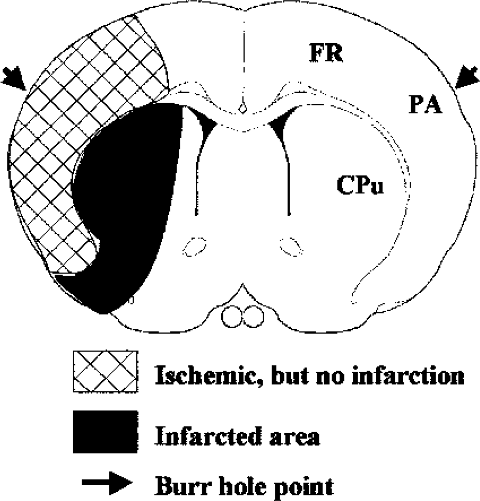
Verification of the focal ischemia/reperfusion model. Schematic diagram depicts regions from which tissues were sampled for the high-performance liquid chromatography with electrochemical detection analysis, ARP assay, and DNA repair assays, and the approximate distribution of infarction in the brain 72 hours after 1 hour of MCAO. The arrows point to the approximate locations of burr holes made for laser Doppler flowmetry measurement. FR, frontal cortex; PA, parietal cortex; Cpu, caudate putamen.
Induction of 8-oxodG in the brain after ischemia
One-hour MCAO significantly increased the 8oxodG/2-dG ratio in both ischemic frontal/parietal cortex and caudate-putamen within 15 minutes of reperfusion compared with the contralateral hemisphere of ischemic brains or with sham-operated control brains (Fig. 2). The levels of 8-oxodG within the frontal/parietal cortex remained significantly elevated up to 4 hours after reperfusion, and thereafter subsided to control levels. In contrast, the levels of 8-OHdG in the caudate-putamen were persistently increased throughout the time course of reperfusion (72 hours). There were no significant changes in the levels of 8-OHdG in the contralateral hemisphere at any time point studied.
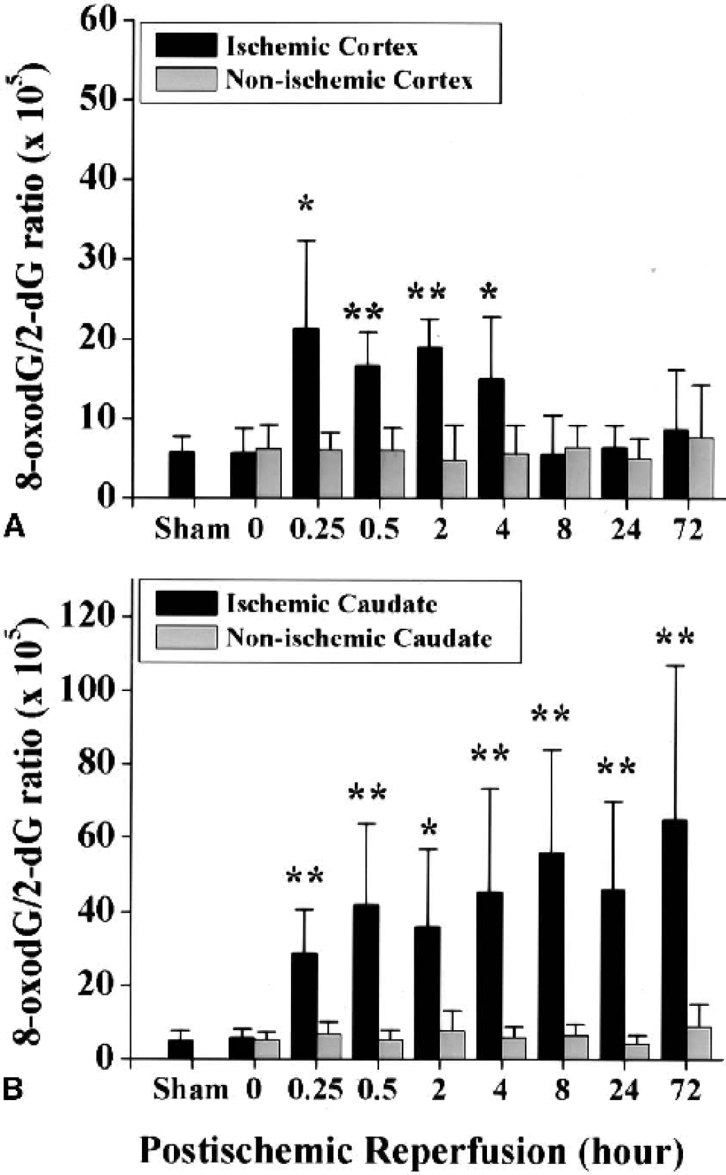
Induction of 8-oxodG in nuclear DNA after focal ischemia and reperfusion measured using high-performance liquid chromatography with electrochemical detection. Top graph, frontal-parietal cortex (noninfarcted region); bottom graph, caudate putamen (infarcted region). dG, deoxyguanosine. Data are mean ± SD (n = 6–8 per time point). ∗ P < 0.05; ∗∗ P < 0.01 versus contralateral nonischemic hemispheres.
Induction of AP sites in the brain after ischemia
The principle and specificity of the ARP assay for the detection of AP sites has previously been described (Asaeda et al., 1998; Ide et al., 1993; Kubo et al., 1992; Nagayama et al., 2000a). Using this method, we detected less than one AP site per 105 nucleotides in DNA samples prepared from control brains. Within 15 minutes after MCAO, levels of AP sites were markedly increased in the ischemic parietal cortex and caudate-putamen (Fig. 3a and Fig. 3b). After 2 hours or longer duration of reperfusion, the levels of AP sites in the cortex gradually subsided, but were still significantly higher than in the controls until 72 hours after ischemia. In contrast, the levels of AP sites remained significantly increased in the caudate-putamen throughout the 72-hour time frame studied.
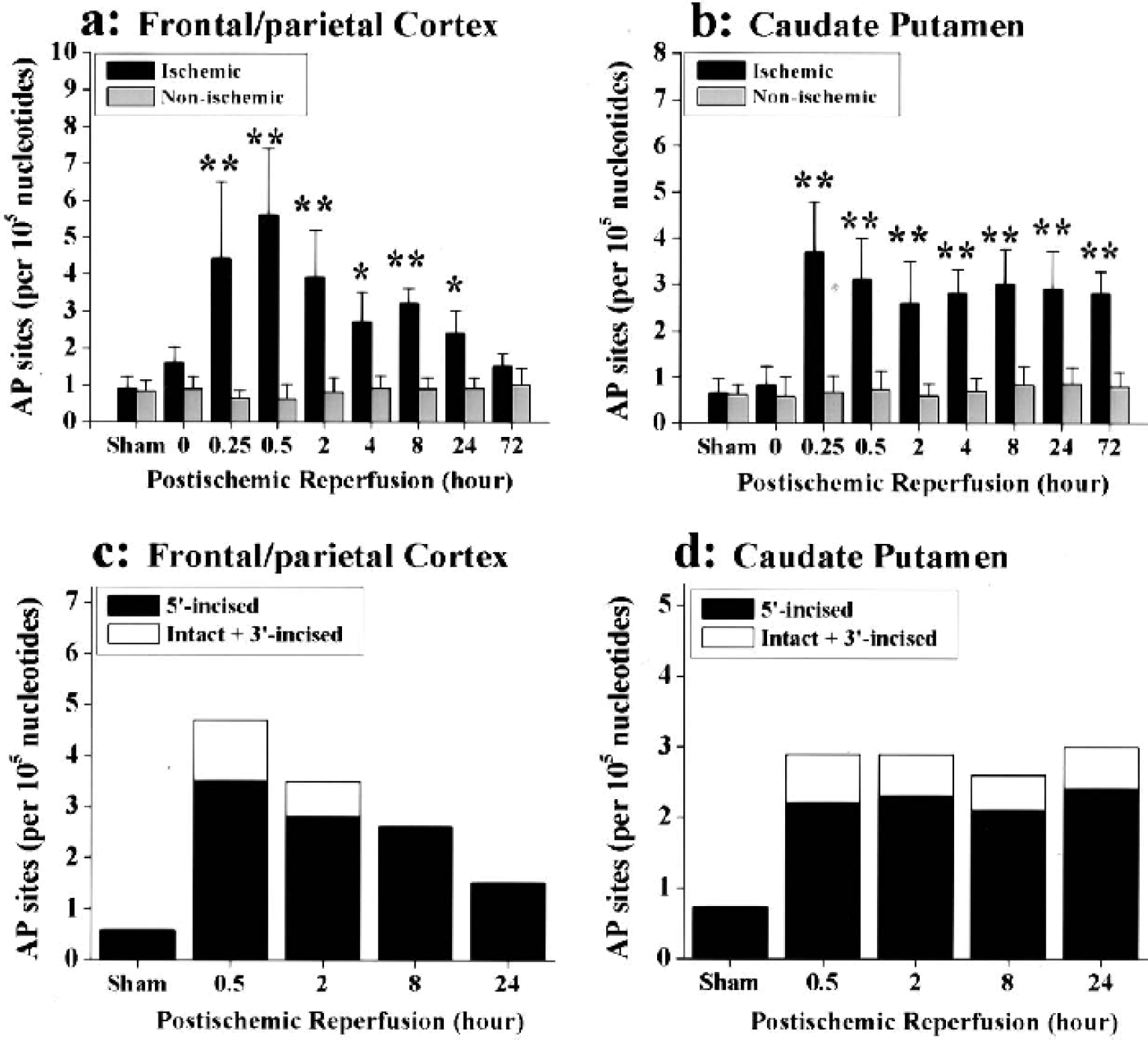
Induction of AP sites in nuclear DNA after focal ischemia and reperfusion measured using the ARP assay. (
Because it has been shown previously that different forms of AP sites are repaired with different efficiencies in cells under oxidative stress (Nakamura et al., 2000), in the present study we performed the AP-site cleavage assay (Nakamura and Swenberg, 1999) to determine the amount of AP sites in the 5′-incised or intact or 3′-incised forms in selective time points after ischemia or in sham controls. In this assay, Exo III was used as the class II AP endonuclease to identify 3′-cleavage, and putrescine was used to detect 5′-cleavage of AP sites. As shown in Fig. 3c and Fig. 3d, the ischemic insult induced the accumulation of AP sites in brain predominantly in the 5′-incised form at about twice the level of the combination of the intact and 3′-incised forms. Furthermore, the intact and 3′-incised AP sites were completely repaired in the cortex within 8 hours of reperfusion, whereas they were insignificantly repaired in the caudate. In contrast to the repair of the intact and 3′-incised AP sites, the 5′-incised AP sites were repaired slowly in the cortex; the amounts did not subside to control levels until 24 to 72 hours after ischemia. The 5′-incised AP sites were continuously accumulated in the caudate throughout the reperfusion, indicating that this type of lesion was particularly inefficiently repaired in this region after ischemia.
Alterations of DNA repair activity in the brain after ischemia
Alterations in overall BER activity
The 8-oxodG–dependent overall BER activity (which requires the actions of all BER steps) was examined in nuclear extracts obtained at 0.5, 2, 8, 24, and 72 hours after 1 hour of MCAO or sham operation. At earlier time points (0.5–2 hours), the DNA repair-synthesis activity was not significantly changed in either cortex or caudate-putamen after ischemia (Fig. 4). At 8 hours after ischemia and thereafter, however, significant increases in DNA repair-synthesis activity were detected in nuclear extracts from the frontal/parietal cortex. In contrast, the activity was significantly decreased in nuclear extracts from the caudate-putamen.
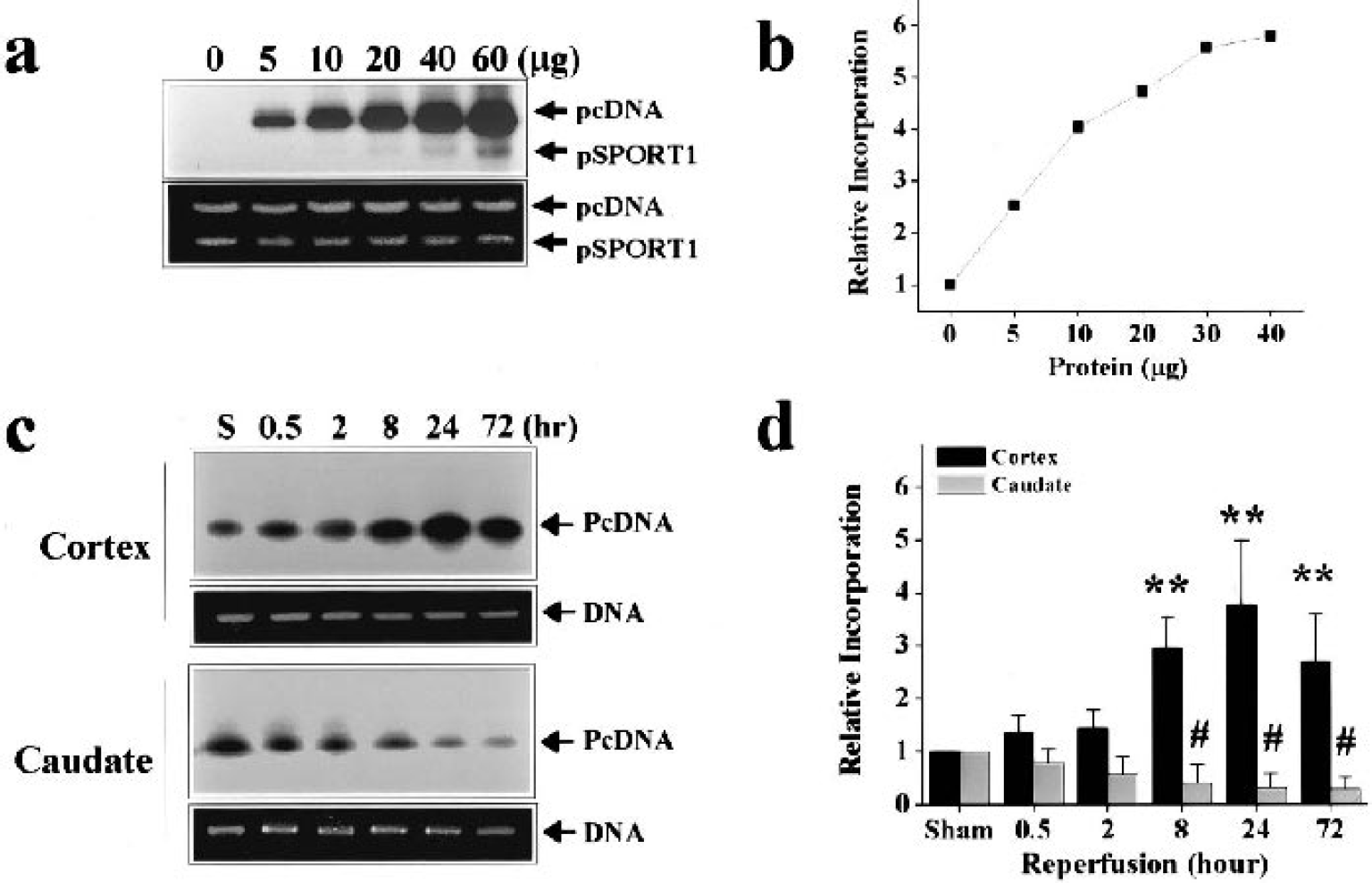
Alterations of 8-oxodG–initiated overall BER activity in nuclear extracts after focal ischemia and reperfusion. The DNA repair incorporation assay was performed using the 8-oxodG–containing pcDNA plasmids. (
Alterations in incision activity for base lesions
Results of previous studies indicated that incision of certain oxidative DNA base lesions such as 8-oxodG is the rate-limiting step in lesion-dependent BER conducted by neuronal cell extracts (Chen et al., 2000). To determine whether ischemia-induced alterations in overall BER activity in the brain were associated with the alterations in the incision activity for 8-oxodG or AP site, oligonucleotide incision assays were performed employing specific substrates containing precisely positioned 8-oxodG or AP site. In the cortex, the 8-oxodG-incision activity was unchanged in earlier time points (0.5–2 hours), but it was significantly increased at 8 to 72 hours after ischemia (Fig. 5a and Fig. 5b). In contrast, the AP site-incision activity was not significantly changed in the cortex throughout this time period (Fig. 5c and Fig. 5d). In the caudate-putamen, both 8-oxodG- and AP site-incision activities were decreased below control levels 8 to 72 hours after ischemia (Fig. 5a and Fig. 5d), consistent with the time course of decreases in overall BER activity in this region.
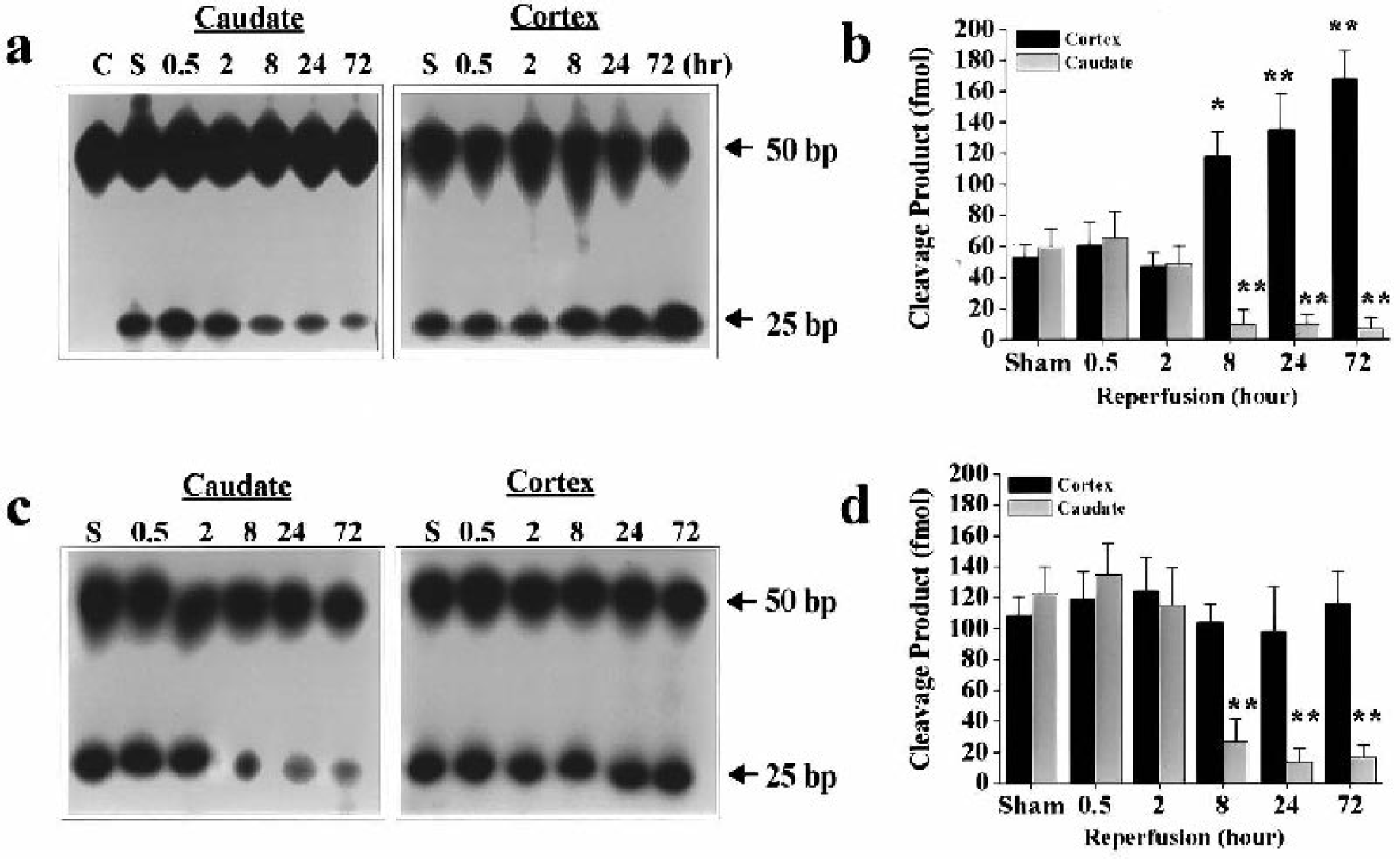
Alterations of base incision activity in brain nuclear extracts after focal ischemia and reperfusion. (

Alterations of DNA polymerase-β activity after focal ischemia and reperfusion, detected using the reconstituted cell-free BER system. (
Alterations in DNA polymerase-β activity and DNA ligase activity
DNA polymerase-β is the predominant DNA polymerase responsible for the “gap-filling” step in the BER pathway in the brain. To determine the role of DNA polymerase-β in the altered overall BER activity after ischemia, we examined its activity in nuclear extracts using a repair assay employing synthesized DNA duplex that contains a precisely positioned single-nucleotide nick (Fig. 6a and Fig. 6b). Titration assays showed that brain nuclear extracts resulted in protein concentration-dependent radiolabel of the repair substrate (Fig. 6c and Fig. 6d). Using this assay, we detected markedly increased DNA polymerase β-mediated nucleotide synthesis activity (increased formation of both 26- and 50-mer products) in the ischemic cortex, beginning at 2 hours after ischemia and lasting for at least 72 hours (Fig. 6e). In contrast, this nucleotide synthesis activity was significantly decreased in the caudate 2 hours after ischemia and thereafter (Fig. 6f).
The role of DNA polymerase-β in the induced BER activity occurring in the ischemic cortex was further confirmed by the observation that immunodepletion of DNA polymerase-β from cortical extracts before the assay diminished the nucleotide synthesis in the repair substrate (Fig. 6g).
To determine if the increased DNA polymerase-β activity in the ischemic cortex is associated with a comparable increase in the DNA ligase activity, we performed the standard oligonucleotide ligation assay using cortical extracts. No significant alteration in DNA ligase activity was detected at any time point after ischemia (Fig. 7). This result suggests that the increased formation of the 50-mer repair product resulting from ischemic cortical extracts (see Fig. 6e) was likely due to the increased dual activities of DNA polymerase-β, nucleotide synthesis, and dRP cleavage, and the latter is prerequisite for the subsequent action by DNA ligases.
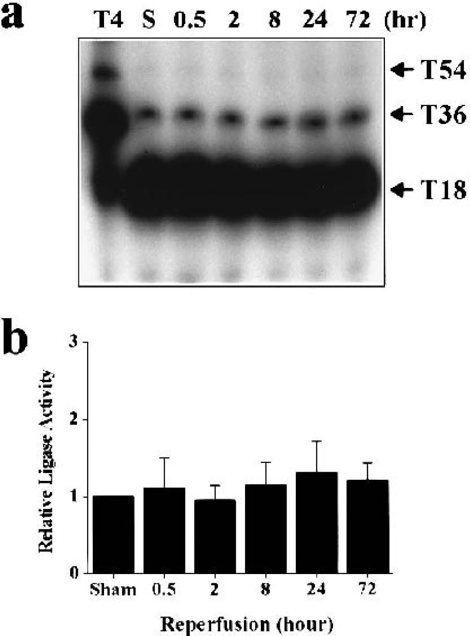
Induced BER activity in the ischemic cortex does not involve the alteration in DNA ligase activity. (
Alterations in BER enzyme expression in the brain after ischemia
Expression of essential BER enzymes
Western blot analysis was performed using nuclear protein extracts to determine whether the alterations of BER activity in the brain are associated with the changes in the expression of BER enzymes (Fig. 8). In the cortex, the levels of APE, DNA ligase I, DNA ligase III, and XRCC1 were not significantly changed after ischemia. The levels of OGG1 were significantly increased 8 to 72 hours after ischemia, consistent with the upregulation of 8-oxodG–incision activity observed in this region. The immunoreactivity for DNA polymerase-β was markedly increased beginning 8 hours after ischemia, and remained elevated throughout the time course. In the caudate-putamen, however, the levels for all BER enzymes tested showed marked decreases at 8 to 72 hours after ischemia (data not shown).
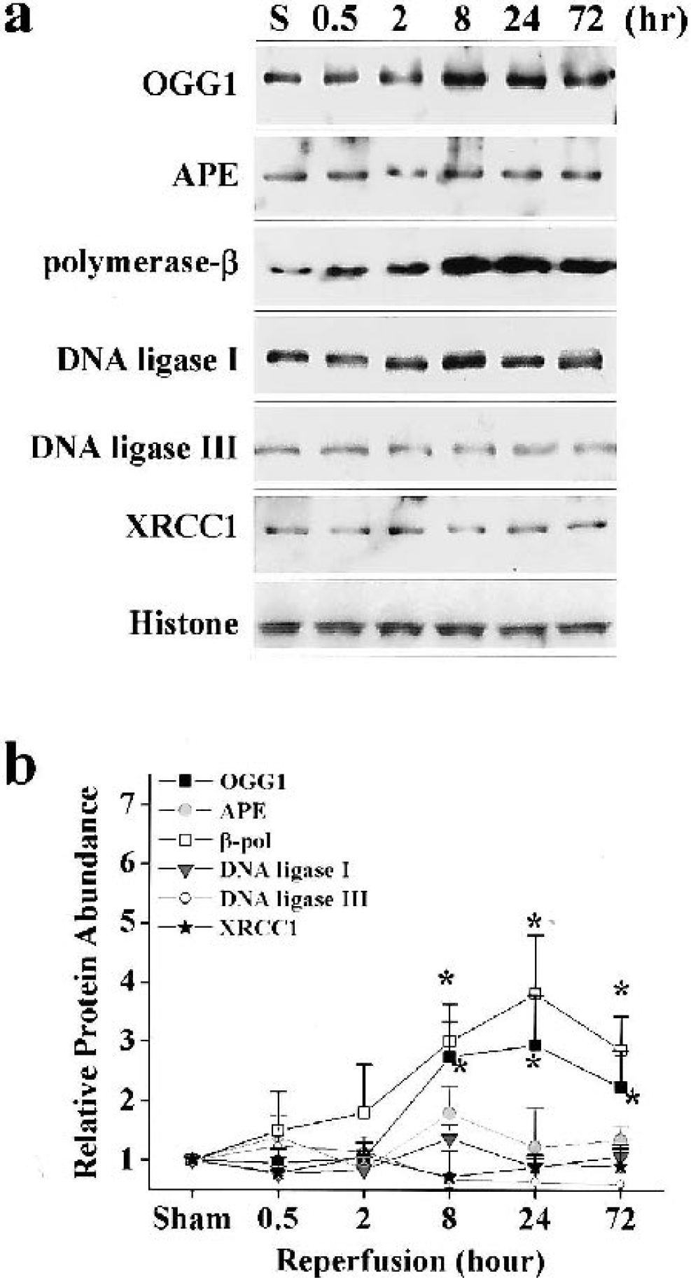
Ischemia-induced regulation of expression of essential BER enzymes in the frontal-parietal cortex. (
Induction of DNA polymerase-β gene expression
Because the increase in DNA polymerase-β activity has an essential role in the induction of overall BER activity in the cortex after ischemia, we further characterized the expression of DNA polymerase-β in the brain after MCAO. Northern blots confirmed the expression of DNA polymerase-β mRNA (∼1.5 kb) in the cortex and, compared with sham controls, the levels were increased at 4 (1.8-fold), 8 (3.1-fold), and 24 hours (2.9-fold) after ischemia (Fig. 9a). As shown by in situ hybridization (Fig. 9b), increased expression of β-pol mRNA was localized mainly in the frontal-parietal cortex after ischemia, whereas in the caudate the increased signals were limited to the border zone of infarction. The increased β-pol mRNA signals in the cortex showed a homogenous pattern at 8 hours after ischemia, whereas at 24 hours after ischemia there were patches of cortical areas where the mRNA signals were subsided to control levels. The patches of mRNA reduction likely reflected the scattered small infarction or cell loss in the cortex, which occurs rather frequently in this model. Examination of emulsion-coated sections confirmed the cellular localization of increased silver grains resulting from DNA polymerase-β mRNA (data not shown). In control experiments, sections hybridized with the sense cDNA probe showed a low-level background signal that was homogenous throughout the brain section.
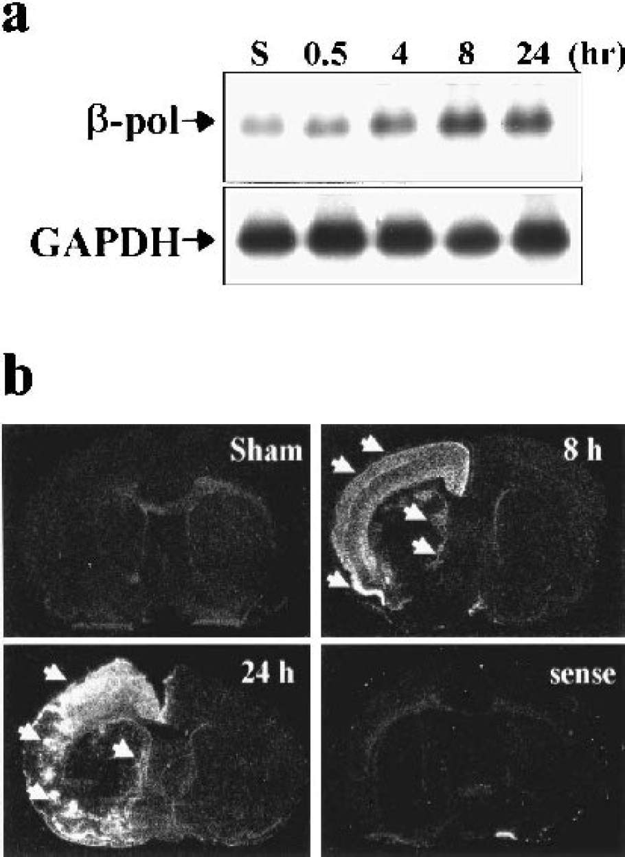
Induced DNA polymerase-β mRNA expression after focal ischemia and reperfusion. (
The cellular distribution of DNA polymerase-β was examined using immunohistochemistry. In normal nonischemic brains, neurons throughout most forebrain regions, including caudate-putamen, cortex, and thalamus, contained weak DNA polymerase-β immunoreactivity. Only the granular cell layer in the hippocampus and scattered neurons in the piriform cortex were highly DNA polymerase-β immunoreactive. Immunostaining in ischemic brains showed increased DNA polymerase-β immunoreactivity in regions affected by MCAO; this increase began to be detectable at 4 hours, but became apparent at 8 and 24 hours after ischemia. As shown at 24 hours after ischemia (Fig. 10a), increased DNA polymerase-β immunoreactivity with a clearly demonstrated nuclear localization was distributed throughout the frontal-parietal cortex and many cells in the infarct border zone in the caudate-putamen. In contrast, increased DNA polymerase-β immunoreactivity was not detected within the core of infarction.
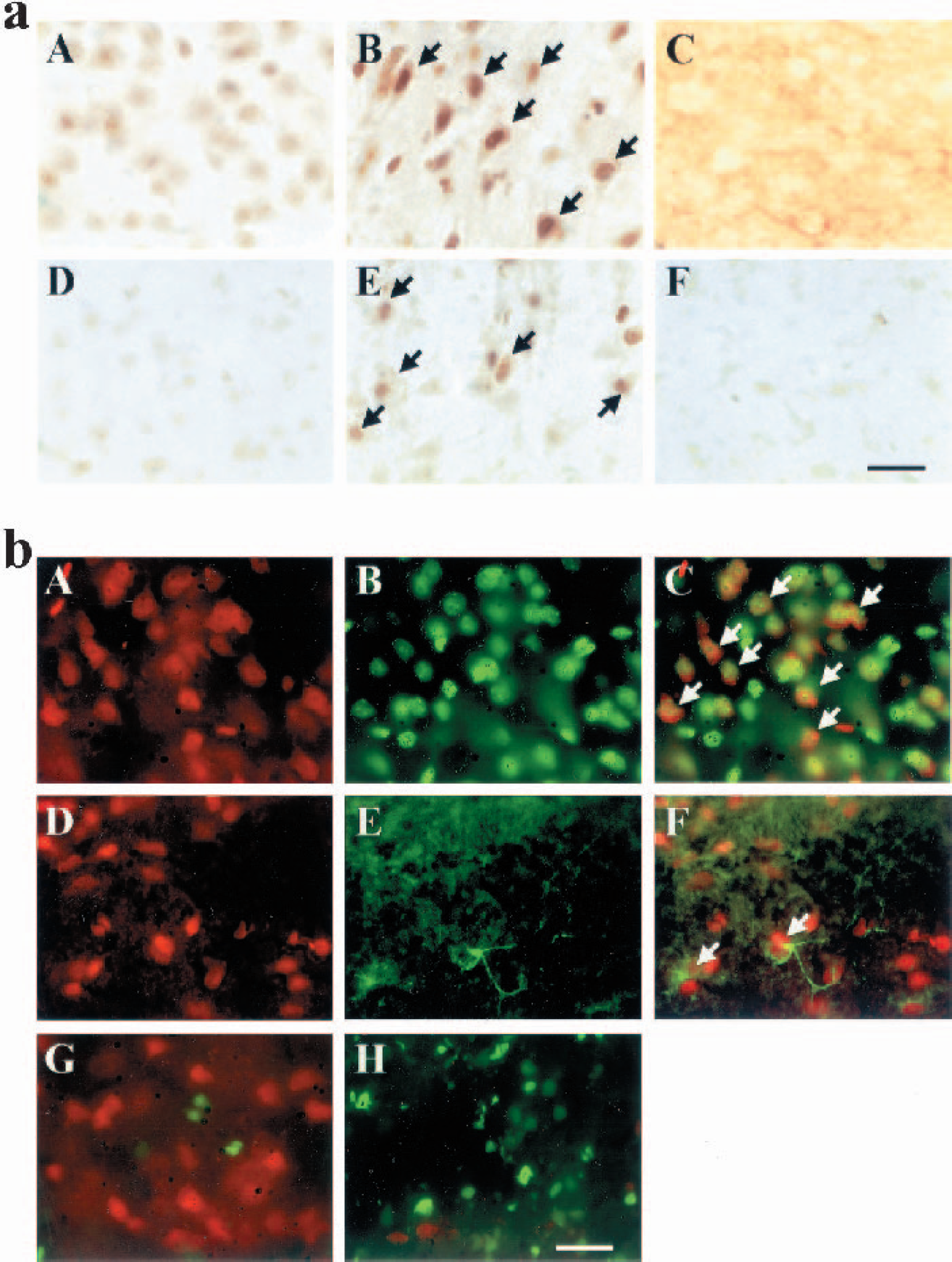
Characterization of DNA polymerase-β protein expression after ischemia. (
Double-label immunofluorescent staining performed at 24 hours after ischemia revealed that the increased DNA polymerase-β immunoreactivity was present predominantly in neurons, showing frequently co-localization with the neuronal marker NeuN (Fig. 10b). Increased DNA polymerase-β immunoreactivity was also detected in some activated astrocytes (GFAP immunoreactive), which were present mainly in the infarct border zone.
Double-label immunofluorescent staining of DNA polymerase-β and PANT (DNA polymerase I-mediated biotin-dATP nick translation) was performed in additional brains obtained 24 hours after ischemia to determine the relationship between DNA polymerase-β expression and the induction of DNA single-strand breaks, another form of oxidative DNA lesion repaired via the BER pathway (Chen et al., 1997). Positive staining for DNA strand breaks and DNA polymerase-β immunoreactivity was frequently observed within the same microscopic fields in the infarct border zone within the caudate or cortex. However, increased DNA polymerase-β immunoreactivity was found only in cells that did not show DNA single-strand breaks, whereas cells that contained DNA single-strand breaks showed no detectable DNA polymerase-β immunoreactivity (Fig. 10b).
DISCUSSION
Emerging evidence has suggested that induction of oxidative DNA damage plays an important role in mediating neuronal death in cerebral ischemia (Chen et al., 1997; Cui et al., 1999; Huang et al., 2000; Lin et al., 2000; Liu et al., 1996; Nagayama et al., 2000a). However, little is known about how the endogenous DNA repair mechanisms in the brain respond to ischemia and oxidative DNA damage. In the present study, we studied the regulation of nuclear DNA BER activity in a well-characterized rat model of transient focal cerebral ischemia, in parallel with the measurement of evolutional changes of endogenous oxidative DNA damage. The major observations resulting from this study are that (1) nuclear BER activity is markedly altered (up- or downregulated) in the postischemic brain in both reperfusion-duration–dependent and ischemic-region–dependent manners, and the alteration in BER activity is attributed to the altered expression and/or activity of essential BER enzymes including DNA polymerase-β and OGG1; and that 2) whereas a long-lasting activation of BER activity in the ischemic cortex is associated with the recovery of endogenously induced oxidative DNA lesions and, eventually, cell survival, a short-lasting BER activity in the ischemic caudate-putamen is associated with accumulation of DNA damage and subsequent cell death. These results strongly suggest that inducible nuclear BER activity constitutes an endogenous mechanism for neuroprotection against ischemia-induced oxidative stress and oxidative DNA damage.
The data presented here show that oxidative DNA damage, in the forms of base lesions and AP site, was markedly induced in the brain during the early stages of postischemic reperfusion. This is the first study to quantitatively measure 8-oxodG and AP site in focal cerebral ischemia and reperfusion. Within 15 minutes after the initiation of reperfusion, the amounts of both types of oxidative lesions were significantly increased in the caudate and cortex. Not surprisingly, the evolution of DNA damage after prolonged reperfusion was markedly different between these two brain regions: both types of DNA lesions were nearly fully recovered in the ischemic cortex, which eventually survived ischemic injury; however, the lesions were increasingly accumulated in the caudate, which was destined to develop infarction. This pattern of changes in the levels of 8-oxodG and AP site during reperfusion was compatible to those in DNA single-strand breaks, another marker of oxidative damage, previously reported in the same ischemia model (Chen et al., 1997). Therefore, these results are consistent with the notion that the reversibility of oxidative DNA damage after cerebral ischemia may be one of the important factors that determine the fate of ischemic tissues (Chen et al., 1997; Huang et al., 2000).
Based on the observation that large amounts of oxidative DNA damage were efficiently repaired in the ischemic cortex during postischemic reperfusion, we speculated that the DNA repair machinery might be activated in this region in order to match the increased demand imposed by the accumulating lesions. To test this hypothesis, DNA BER activity was measured in nuclear extracts using the in vitro DNA repair-synthesis assay, which uses repair substrates rich in 8-oxodG, the prevalent oxidative lesion known to be repaired endogenously via the BER pathway (Aspinwall et al., 1997; Chen et al., 2000; Demple and Harrison, 1994; Hilbert et al., 1997; Roldan-Arjona et al., 1997; Srivastava et al., 1998). We found that 8-oxodG-initiated DNA repair synthesis, reflecting the overall nuclear BER activity, was markedly and persistently increased (up to 72 hours) in the ischemic cortex during reperfusion. We also found that this increase in overall BER activity was attributed to the enhancement of both 8-oxodG glycosylase and DNA polymerase-β activities; however, AP endonuclease and DNA ligase activities were not changed in the cortex after ischemia, suggesting that they are most likely not involved in the activation of BER pathway. The induced 8-oxodG–incision activity was the result of the upregulation of OGG1 expression after ischemia because the time course of both events coincided (8–72 hours after ischemia). As the first step and indeed the rate-limiting step in the 8-oxodG–initiated BER pathway (Chen et al., 2000), the accelerated incision of 8-oxodG would efficiently provide repair substrates for the subsequent enzymatic actions of AP endonuclease and DNA polymerase-β, and thus is critical for the activation of the 8-oxodG–initiated BER pathway. This result confirmed the previous observation in a murine model of forebrain ischemia that upregulation of OGG1 contributes to the inducible repair of 8-oxodG in ischemic neurons (Lin et al., 2000).
We found that the induced gene expression and increased enzymatic activity of DNA polymerase-β is instrumental in ischemia-induced activation of the BER pathway. Because DNA polymerase-β functions downstream of lesion-specific DNA glycosylases and AP endonuclease, its activation should contribute to the inducible repair of various base lesions including 8-oxodG as well as AP site. In addition to its nucleotide synthesis activity, DNA polymerase-β also possesses the dRP-lyase activity, which is prerequisite for the subsequent “nick-sealing” step of BER by DNA ligases and has been suggested to play a particularly important role in mediating the protection by DNA polymerase-β against DNA damage-induced cytotoxicity (Sobol et al., 2000). The marked induction of DNA polymerase-β activity seen in this study (2–72 hours after ischemia) is attributable not only to the upregulation of gene expression (∼4 hours after ischemia for mRNA, ∼8 hours for protein), but likely involves a posttranslational activation of this enzyme (∼2 hours after ischemia, preceding gene overexpression). Although the precise mechanism by which DNA polymerase-β undergoes posttranslational activation is unclear, a recent study has suggested that the transcriptional cofactor p300 potently regulates DNA polymerase-β activity by acetylating it at lysine 72 (Hasan et al., 2002). However, the persistent increases in DNA polymerase-β activity in the cortex after ischemia may rely on the prolonged upregulation of the DNA polymerase-β gene. Several factors involved in ischemic injury, such as hypoxia and oxidative stress, could activate the transcriptional process of DNA polymerase-β by enhancing the DNA binding of ATF-1/CREB, the major promoter activator of the DNA polymerase-β gene (Narayan et al., 1996). Several recent studies have confirmed that ATF-1/CREB is upregulated in neurons surviving ischemia or oxidative stress (Mabuchi et al., 2001; Tanaka et al., 2000; Zaman et al., 2000).
The inducible expression of BER enzymes and upregulation of cellular BER activity in ischemic neurons may have important functional significance. Previous work in mammalian cells has defined a strong linkage between cellular BER capacity and the fate of cells that are subjected to oxidative stress or other genotoxic insults. At least four lines of evidence support a critical role of BER activity in promoting cell survival after genotoxic stresses and DNA damage. First, a number of studies have shown that cells deficient in essential BER enzymes, such as DNA polymerase-β or APE, show increased accumulation of oxidative DNA lesions and markedly decreased cell viability in response to oxidative stress or hypoxic injury (Clairmont and Sweasy, 1996; Horton et al., 2002; Kaina et al., 1998; Ochs et al., 1999; Sobol et al., 1996; Walker and Sikorska, 1994). Second, inducible expression of BER enzymes are involved in the induction of “adaptive response” in cells, whereby upregulation of APE or DNA polymerase-β induced by sublethal levels of oxidative stress offers protection against the cytotoxicity of lethal doses of oxidative stress or other DNA-damaging agents (Grosch et al., 1998; Horton et al., 2000; Ramana et al., 1998). Third, several studies using gene-transfection approaches to enhance BER capacity in mammalian cells show protection of cells against oxidative stress (Grosch et al., 1998; Hansen et al., 1998; Tomicic et al., 1997). For example, transfection of the human APE cDNA driven by an inducible promoter into CHO cells markedly reduced cytotoxicity of oxygen-derived free radicals (Grosch et al., 1998). Finally, Lin et al. (2000) showed that the upregulation of OGG1 activity in mouse brain during postischemic reperfusion was associated with the accelerated repair of 8-oxodG in functional genes, suggesting a protective role for the 8-oxodG–dependent BER pathway in ischemic injury. Taken together, these studies clearly show that an alteration in cellular BER activity can substantially affect the outcome of genotoxic insults and oxidative DNA damage. Therefore, based on the data obtained from this study, we suggest that the ischemia-induced upregulation of BER activity may play a role in promoting cell survival in the ischemic penumbra (such as the frontal/parietal cortex) by limiting the genotoxic effect of oxidative stress. However, to directly prove the functional role of BER activity in ischemic brain injury, one needs to specifically alter (upregulate or downregulate) BER activity in the brain, which will be an important goal for future studies.
Accordingly, failure to express BER enzymes in the ischemic caudate may contribute directly to the decreased BER activity and, consequently, the extremely poor recovery of oxidative lesions in this region during postischemic reperfusion. These results are consistent with the previous reports that the expression of the BER enzyme AP endogenous was markedly decreased in the brain after severe focal ischemia and thus may contribute to irreversible tissue injury (Fujimura et al., 1999; Kawase et al., 1999).
Another intriguing and novel finding of this study was that different forms of AP sites were repaired with different temporal profiles in the cortex after ischemia. The results of the AP site cleavage assay showed that while the intact and/or 3′-incised AP sites were quickly repaired (∼2 hours) in the ischemic cortex, the 5′-incised AP sites were not completely repaired until 24 to 72 hours after ischemia. These results are consistent with the recent observations by (Nakamura et al. (2000) that, in mammalian cells, the 5′-incised AP sites induced by ROS are particularly resistant to the BER process compared with the intact or 3′-incised AP sites. It has been proposed that different mechanisms may be involved in the formation of different AP sites under oxidative stress, and the 5′-incised AP sites induced by ROS may represent modified AP sites that cannot be efficiently released from the DNA backbone through β-elimination by DNA polymerase-β (Nakamura et al., 2000). Specific DNA glycosylases, such as OGG1, play a major role in the generation of AP sites during the excision of oxidative base lesions, which most likely introduce 3′-incised AP sites (Bjoras et al., 1997; Rosenquist et al., 1997). Furthermore, oxidative stress also induces labile ring-saturated pyrimidine adducts that subsequently result in intact AP sites (Breen and Murphy, 1995).
Shown previously in the in vitro AP-site repair assay, intact and 3′-incised AP sites can be readily repaired by class II AP endonuclease and the dRP-lyase activity possessed by DNA polymerase-β, respectively (Dianov et al., 1998; Fortini et al., 1999). However, the 5′-incised AP sites induced by ROS are structurally different from the above forms, in which ROS may also induce sugar lesions by direct proton extraction from the C-4′ or C-5′ position of deoxyribose (Balasubramanian et al., 1998; Nakamura and Swenberg, 1999). Such alterations render the AP sites refractory to β-elimination (excision of the sugar-phosphate residue) by preventing DNA polymerase-β from reaching the aldehydic moiety, and thus these sites are not efficiently repaired by the DNA polymerase-β-mediated short-patch BER pathway (Demple and Harrison, 1994; Klungland and Lindahl, 1997). However, these lesions can be efficiently repaired via the alternate BER pathway, the so-called long-patch BER pathway, which involves the synthesis of two to six (but most frequently four) nucleotides (Frosina et al., 1996; Klungland and Lindahl, 1997; Lu et al., 2001; Prasad et al., 2001). The long-patch pathway requires the action of Flap endonuclease-1 (FEN1), which excises a short 5′-dRp–containing oligomer (typically, 5′-dRp-trinucleotide), instead of the dRp group alone, downstream of the AP site. The nucleotide gap generated by FEN1 is then filled by strand displacement DNA synthesis by DNA polymerase-β (Dianov et al., 1999; Horton et al., 2000; Prasad et al., 2001). We have recently cloned the rat brain FEN1 gene and found that the expression levels of FEN1 as well as the long-patch BER activity are extremely low in adult brain (Lan, unpublished observations, 2002). Based on the observed temporal profile for the repair of 5′-incised AP sites in the ischemic cortex, we speculate that the activity of long-patch BER pathway might be induced in the cortex during postischemic reperfusion. Future studies are warranted to investigate the role of long-patch BER in the repair of modified AP site after ischemia.
The findings derived from this study have clear significance. We demonstrated that active repair of endogenous oxidative DNA lesions took place in the frontal/parietal cortex, a region that survived the insults of focal ischemia and reperfusion. We further demonstrated that the repair of endogenous oxidative lesions in the cortex was associated with induced cellular BER activity, which is attributed to the overexpression and activation of BER enzymes DNA polymerase-β and OGG1. The inducible BER activity observed in this study may represent a novel and important endogenous mechanism for neuroprotection. This mechanism, together with various cellular antioxidant systems (Chan, 2001), may prevent the accumulation of lethal oxidative lesions and thus promote cell survival after ischemia. Therefore, the future use of molecular or pharmacologic means to enhance the cellular capacity for DNA repair in the brain may have therapeutic implications in cerebral ischemia.
Footnotes
Acknowledgments
The authors thank Carol Culver for editorial assistance and Pat Strickler for secretarial support.