
Abstract
To investigate long-term adaptations after unilateral carotid artery ligation, the effect of forebrain ischemia on cerebral blood flow and ATP levels was determined at various times after ligation. Unilateral carotid artery ligation was performed in male Wistar rats 0, 3, or 7 days before forebrain ischemia. Laser-Doppler blood flow was monitored bilaterally over the parietal cortex and ATP was measured in the subadjacent cortex of both hemispheres at the end of a 10-minute episode of ischemia. In the 0-day group, forebrain ischemia reduced cortical blood flow to 12% ± 8% (mean ± SD) of preischemic values and lowered cortical ATP to 26% ± 35% of control levels in the ipsilateral hemisphere. Delaying the onset of forebrain ischemia for 3 days after carotid artery ligation significantly improved cortical blood flow (29% ± 12%, P < 0.05) and ATP levels (92% ± 11%, P < 0.05) in the ipsilateral hemisphere. Delaying forebrain ischemia for 7 days also significantly improved ipsilateral blood flow (36% ± 11%, P < 0.05) and ATP levels (81% ± 29%, P < 0.05) compared with the 0-day group. In the contralateral hemisphere, the reduction in blood flow and ATP levels was not significantly altered by delaying the onset of forebrain ischemia for 3 or 7 days. These results show that unilateral carotid artery ligation induces long-term vascular adaptations that improve the collateral circulation and preserve ATP levels during a subsequent episode of ischemia.
Keywords
In the rat, occlusion of one common carotid artery alone causes only minor alterations in cerebral blood flow in the ipsilateral hemisphere (Salford and Siesjö, 1974; De Ley et al., 1985; Coyle and Panzenbeck, 1990). However, when combined with systemic hypoxia, unilateral carotid artery ligation is associated with ischemic neuronal injury in the ipsilateral hemisphere of both adult and neonatal rat brain (Levine, 1960; Salford et al., 1973; Rice et al., 1981). Recently, we demonstrated that delaying the onset of hypoxia for 24 hours after carotid ligation prevented the depletion of ATP and markedly diminished the extent of ischemic injury in the brain of neonatal rats (Hylton et al., 1995). These results indicate that adaptations occur after carotid ligation that increase the tolerance of the brain to a subsequent episode of hypoxia. One possibility is an adaptation of the cerebral vasculature that improves blood flow during hypoxia, thus preserving energy metabolites and preventing ischemic injury. However, the long-term vascular adaptations after carotid artery ligation remain poorly understood.
Unilateral carotid artery ligation was reported to enlarge one of the major collateral vessels, the ipsilateral posterior communicating artery, in the adult rat (Coyle and Panzenbeck, 1990). Enlargement of this collateral vessel would be expected to improve blood flow in the ipsilateral hemisphere during a subsequent episode of hypoxia or ischemia. Indeed, improved collateral circulation has been shown in the ipsilateral hemisphere 6 weeks after unilateral carotid artery ligation (Coyle and Panzenbeck, 1990). However, it is not clear whether such adaptations in the collateral circulation can occur within the first few days after carotid artery ligation. Thus, the objective of the present study was to determine whether an adaptive improvement in the collateral circulation occurred during the first week after unilateral carotid artery ligation. In addition, we wished to determine whether any improvement in the collateral circulation during ischemia would be sufficient to preserve tissue levels of ATP. Preliminary results of this work have been reported previously (Bronner et al., 1997).
MATERIALS AND METHODS
Cortical blood flow during forebrain ischemia
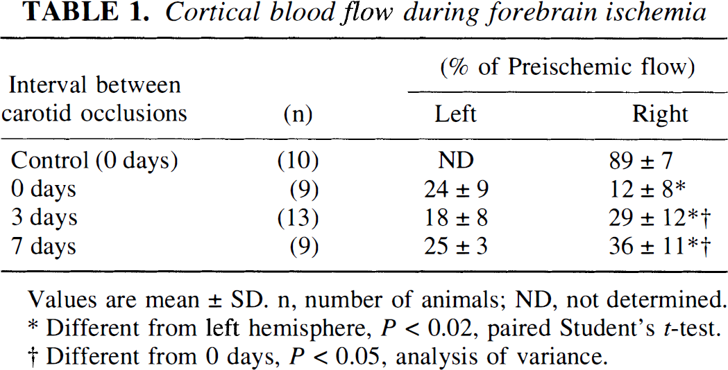
Values are mean ± SD. n, number of animals; ND, not determined.
Different from left hemisphere, P < 0.02, paired Student's t-test.
Different from 0 days, P < 0.05, analysis of variance.
Cortical ATP levels during forebrain ischemia
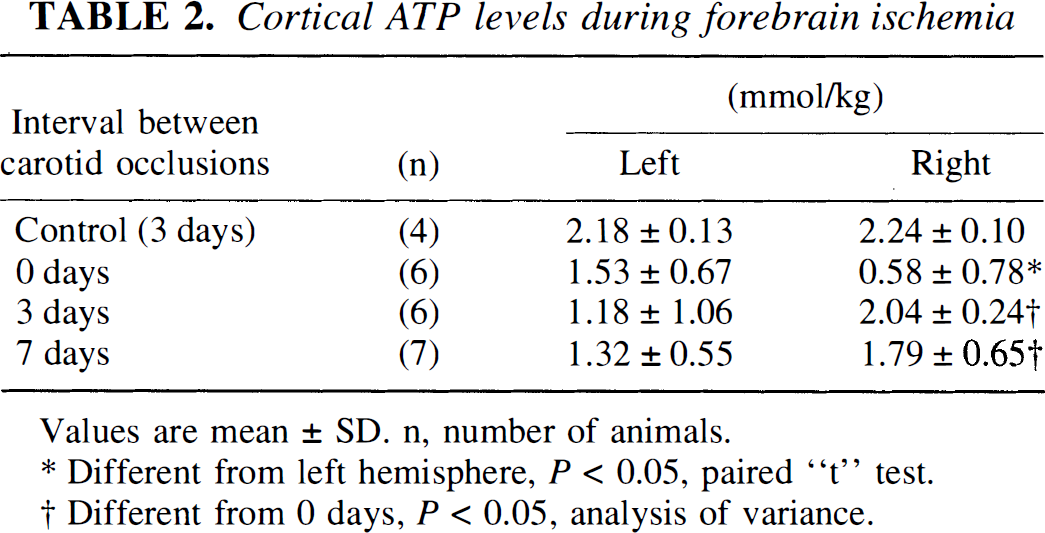
Values are mean ± SD. n, number of animals.
Different from left hemisphere, P < 0.05, paired “t” test.
Different from 0 days, P < 0.05, analysis of variance.
Differences in mean values of blood flow and ATP between groups were tested for statistical significance using analysis of variance, followed by Student's t-tests with Bonferroni corrections for multiple comparisons. Differences in mean values of flow and ATP between hemispheres were tested for statistical significance using a paired Student's t-test.
RESULTS
Unilateral carotid artery ligation alone had only a minor effect on ipsilateral cortical blood flow (Table 1, control) and had no effect on ipsilateral levels of ATP measured 3 days after ligation (Table 2, control). Carotid ligation caused ptosis in 23% of the animals, presumably the result of damage to the sympathetic nerve trunk during isolation of the carotid artery. However, there was no apparent difference in results between animals showing ptosis and those that did not.
Simultaneous occlusion of both carotid arteries, coupled with arterial hypotension, reduced cortical blood flow to 24 ± 9% and 12 ± 8% of preischemic values in the left and right hemispheres, respectively (Table 1, 0 days). Similarly, cortical ATP declined to 70 ± 31% and 26 ± 35% of control levels in the left and right hemispheres, respectively (Table 2, 0 days). The interhemispheric differences in cortical blood flow and ATP levels were statistically significant in the 0-day animals.
Delaying the onset of forebrain ischemia for 3 days after ligation of the carotid artery on the right side resulted in a 2.4-fold increase in cortical blood flow and a 3.5-fold increase in ATP levels in the ipsilateral hemisphere, relative to the values in 0-day animals (Tables 1 and 2). In the left (contralateral) hemisphere, the reductions in blood flow and ATP levels were not significantly different from those in the 0-day animals. At 3 days, cortical blood flow in the right (ipsilateral) hemisphere was significantly greater than that in left hemisphere, the reverse of the difference observed at 0 days. The interhemispheric difference in ATP levels at 3 days was not statistically significant.
Delaying the onset of forebrain ischemia for 7 days also significantly increased cortical blood flow and ATP levels in the right (ipsilateral) hemisphere, compared with the 0-day animals (Tables 1 and 2). However, there were no significant differences in blood flow and ATP levels between the 3- and 7-day animals. Similarly, blood flow and ATP levels in the left (contralateral) hemisphere at 7 days were not significantly different from those at 0 days. Again, the interhemispheric difference in cortical blood flow at 7 days was statistically significant, while the difference in ATP levels was not.
DISCUSSION
The present results show that unilateral carotid artery ligation in the rat induces cerebrovascular adaptations that improve collateral blood flow and ATP levels in the ipsilateral hemisphere during a subsequent episode of ischemia. The adaptive improvement in collateral circulation was evident by 3 days after ligation, a time by which the ipsilateral posterior communicating artery has been reported to be enlarged by 50% (Coyle and Panzenbeck, 1990). Thus, the adaptive improvement in collateral circulation during ischemia may be explained by an enlargement of this major collateral artery. However, there may also be adaptations in the ipsilateral microcirculation that contribute to the preservation of blood flow during ischemia. For example, enlargement of smaller anastomotic vessels between the distributions of the anterior cerebral and middle cerebral arteries has been shown after permanent occlusion of the middle cerebral artery in the rat (Coyle, 1984; Coyle and Heistad, 1991). Whatever the explanation, it is clear that unilateral carotid artery ligation induces cerebrovascular adaptations within 3 days, which improve the collateral circulation during a subsequent episode of ischemia.
The mechanisms coupling unilateral carotid artery ligation with adaptations in the cerebral vasculature remain unknown. Although carotid ligation causes an immediate drop of intravascular pressure distal to the ligation, the present results confirm previous studies showing only a minor reduction of blood flow in the ipsilateral hemisphere (Salford and Siesjö, 1974; De Ley et al., 1985). Presumably, rapid vasodilatation limits the decrease in blood flow after carotid ligation. The present results also show normal ATP levels in the ipsilateral hemisphere 3 days after carotid ligation. Thus, unilateral carotid artery ligation alone does not cause significant alterations in ipsilateral blood flow or energy state. However, carotid artery ligation markedly diminishes the circulatory reserve in the ipsilateral hemisphere, as evidenced by a suppression of the blood flow responses to hypoxia, hypercapnia, or hypotension (Salford and Siesjö, 1974; Sengupta et al., 1973; Sengupta et al., 1974). Interestingly, the blood flow response to hypercapnia was reported to recover gradually during the first 2 weeks after unilateral ligation, indicating an adaptive increase in the circulatory reserve similar to that observed in the present study (De Ley et al., 1985).
An unexpected finding in the present study was the asymmetric reduction of cortical blood flow and ATP levels in the two hemispheres after simultaneous occlusion of both carotid arteries. It is possible that interhemispheric differences in flow are caused by minor asymmetries in the vascular anatomy and are enhanced at levels of perfusion that are threshold for maintaining ATP. It has been noted that left and right posterior communicating arteries differ in size (mean difference, 26%) in Fischer 344 rats (Coyle and Panzenbeck, 1990). Furthermore, variations in the branching pattern of posterior communicating arteries were reported to occur twice as frequently on the right side than on the left side (Brown, 1966). Whatever the cause, flow differences between hemispheres should not affect the ipsilateral flow adaptations observed in the present investigation.
The significance of the present findings relates to the compensatory changes that occur in the brain when blood flow through a major vessel is reduced. These compensations are both immediate and slowly adapting. Immediate vasodilation and compensatory flow through existing collateral vessels serve to maintain blood flow at nearly normal levels after unilateral carotid artery occlusion. However, long-term changes are required to restore circulatory reserve and, thus, maintain blood flow during a superimposed episode of hypoxia or ischemia. Identifying the mechanism of these slowly adapting compensations may have therapeutic relevance to carotid stenosis in humans.