
Abstract
Proneuronal basic helix–loop–helix (bHLH) transcription factor, neurogenin 1 (Ngn1), regulates neuronal differentiation during development of the cerebral cortex. Akt mediates proneuronal bHLH protein function to promote neuronal differentiation. Here, we show that recombinant human erythropoietin (rhEPO) significantly increased Akt activity and Ngn1 mRNA levels in neural progenitor cells derived from the subventricular zone (SVZ) of adult rat, which was coincident with increases of neural progenitor cell proliferation, differentiation, and neurite outgrowth. Inhibition of Akt activity by the phosphatidylinositol 3-kinase/Akt (PI3K/Akt) inhibitor, LY294002, abolished rhEPO-increased Ngn1 mRNA levels and the effects of rhEPO on neural progenitor cells. In addition, reducing expression of endogenous Ngn1 by means of short-interfering RNA (siRNA) blocked rhEPO-enhanced neuronal differentiation and neurite outgrowth but not rhEPO-increased proliferation. Furthermore, treatment of stroke rat with rhEPO significantly increased Ngn1 mRNA levels in SVZ cells. These data suggest that rhEPO acts as an extracellular molecule that activates the PI3K/Akt pathway, which enhances adult neural progenitor cell proliferation, differentiation, and neurite outgrowth, and Ngn1 is required for Akt-mediated neuronal differentiation and neurite outgrowth.
Introduction
Proneuronal basic helix–loop–helix (bHLH) transcription factors including neurogenin 1 (Ngn1) regulate neurogenesis (Vojtek et al, 2003). Neurogenin 1 is expressed exclusively in the ventricular zone where neural progenitor cells reside during development of the cerebral cortex and promotes neural progenitor cell differentiation into neurons (Sun et al, 2001) Mice with targeted disruption of Ngn1 exhibit a loss of progenitor population (Bertrand et al, 2002). Neural progenitor cells in the subventricular zone (SVZ) of the adult rodent migrate towards the olfactory bulb where they differentiate into granule and periglomerular neurons (Moreno-Lopez et al, 2000). Olfactory progenitor cells express Ngn1 in the adult rodent (Cau et al, 2002).
Erythropoietin (EPO), a hematopoietic cytokine, is expressed in the developing central nervous system and in neural progenitor cells of adult rodent brain (Malhotra et al, 2004; Ribatti et al, 1999). Endogenous and exogenous EPO increases neurogenesis in the normal and stroke brain (Shingo et al, 2001; Wang et al, 2004). Erythropoietin binds to its receptor and activates intracellular signaling intermediates including phosphoinositide 3-kinase (PI3K) and the serine-threonine kinase Akt (Folli et al, 1996; Kaplan and Miller, 2000). The PI3K/Akt pathway regulates cell proliferation, survival, and migration (Deregibus et al, 2003). A recent study shows that Akt regulates activity of bHLH to promote neuronal differentiation (Vojtek et al, 2003). Whether the PI3K/Akt signaling pathway and Ngn1 are involved in EPO-enhanced neurogenesis, however, is not known.
Neural progenitor cells isolated from the adult SVZ exhibit neural stem cell characteristics in the presence of growth factors (Gritti et al, 1996; Reynolds and Weiss, 1992; Morshead et al, 1994). Blocking endogenous EPO decreases neurogenesis, while increasesing of EPO levels enhances neurogenesis in neural progenitor cells derived from the SVZ of adult rodent (Shingo et al, 2001). Accordingly, using neural progenitor cells isolated from the SVZ, the present study tested the hypothesis that EPO enhances neurogenesis by Ngn1, which is increased via activating the PI3K/Akt signaling pathway.
Materials and methods
All experimental procedures were approved by the institutional Animals Care and Use Committee of Henry Ford Hospital.
Animal Model
The middle cerebral artery (MCA) of male Wistar rats was occluded by placement of an embolus at the origin of the MCA (Zhang et al, 1997).
Neurosphere Culture
Subventricular zone neural progenitor cells were dissociated from normal (n = 6) male Wistar rats (3 to 4 months), as previously reported (Morshead et al, 2002; Chiasson et al, 1999). Subventricular zone cells contain actively proliferating progenitor cells and relatively quiescent neural stem cells (Doetsch et al, 1997; Morshead et al, 1998; Zhang et al, 2004). The cells were plated at a density of 2 × 10⁴ cells per milliliter in growth medium. Growth medium contains DMEM-F-12 medium (Invitrogen Corporation, Carlsbad, CA, USA), 20 ng/mL of epidermal growth factor (EGF, R&D System, Minneapolis, MN, USA), and basic fibroblast growth factor (bFGF, R&D system, Minneapolis, MN, USA). DMEM-F-12 medium contains
Proliferation and Differentiation Assays
For proliferation, single cells at a density of 10 cells per microliter were incubated in the growth medium for 7 days and bromodeoxyuridine (BrdU, 30 μg/mL) was added 18 h before the termination of incubation. The total number of neurospheres and the percentage of BrdU-positive cells were measured. For differentiation, neurospheres were cultured in the differentiation medium for 10 days. The percentage of TuJ1 and GFAP-positive cells were measured.
Experimental Protocol
(1) To examine the effects of recombinant human EPO (rhEPO) on neurogenesis and Ngn1, neural progenitor cells were incubated in the presence of rhEPO (10 U/mL, epoietin α; AMGEN, Thousand Oaks, CA, USA). Neurogenesis and Ngn1 mRNA levels were measured. (2) To examine whether Akt activation is involved in the effects of rhEPO on neurogenesis, neural progenitor cells were incubated in the presence of rhEPO (10 U/mL) with or without LY294002 (a selective PI3K inhibitor, 10 μmol/L; Calbiochem, San Diego, CA, USA) or Wortmannin (a potent and specific PI3K inhibitor, 2 μmol/L, Sigma-Aldrich, St Louis, MO, USA). Neurogenesis and Akt activation were measured. (3) To examine the role of Ngn1 on rhEPO enhanced neurogenesis, neural progenitor cells were transfected with rat Ngn1 short-interfering RNA (siRNA) cassettes and incubated with rhEPO. Neurogenesis and Ngn1 levels were measured. (4) To examine the effect of rhEPO on Ngn1 mRNA levels in vivo, stroke rats were treated with saline (n = 3) or rhEPO (n = 3) at a dose of 5,000 units/kg daily, intraperitoneally (i.p.) for 7 days starting at 24 h after stroke. These rats were killed 7 days after stroke for measurements of Ngn1 mRNA levels.
Rat Neurogenin 1 Short-Interfering RNA Synthesis and Transfection
Rat Ngn1 siRNA cassettes were designed according to rat Ngn1 sequence in the gene bank (U67777) using siRNA target finder (GenScript Corp. Piscataway, NJ, USA). The selected sequences were chemically synthesized and the cassettes were constructed by PCR, which consists of a 505 bp human Hi promoter and terminator sequence flanking a DNA insert encoding a small hairpin RNA (GenScript Corp. Piscataway, NJ, USA). A BLAST search against rat genome was performed for the specificity of all target sequences and the scrambled sequences. All cassettes were labeled at 5’ end with Cy3 for control of transfection efficiency (Figure 2). Neural progenitor cells were transfected using FuGENE 6 Transfection Reagent (Roche Applied Science, Indianapolis, IN, USA) following the manufactor's instructions. The total amount of siRNA per transfection was kept constant to 0.5 μg/mL. Neurogenin 1 mRNA and protein levels were measured 48 or 72 h after transfection.
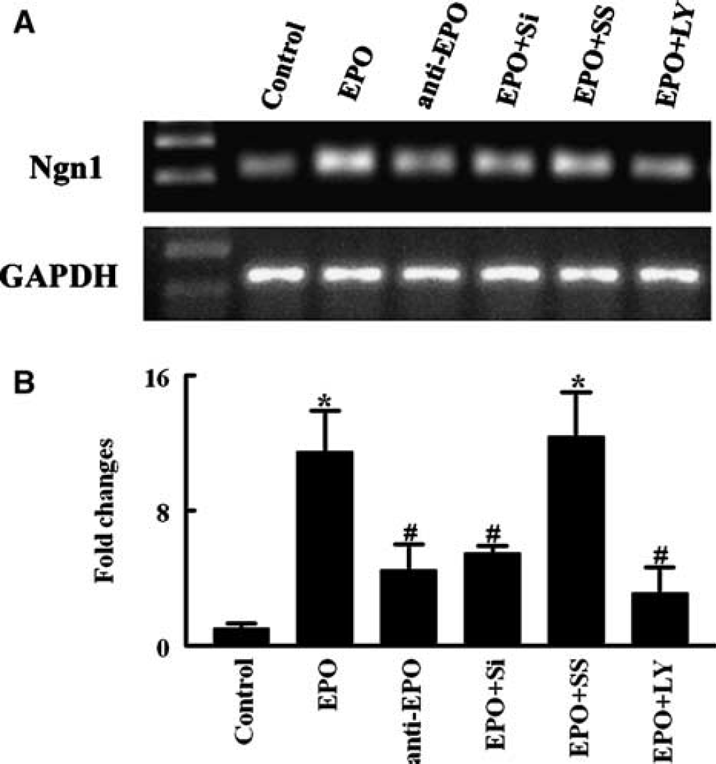
Recombinant human erythropoietin (rhEPO) upregulates neurogenin 1 (Ngn1) expression in neural progenitor cells. Panels
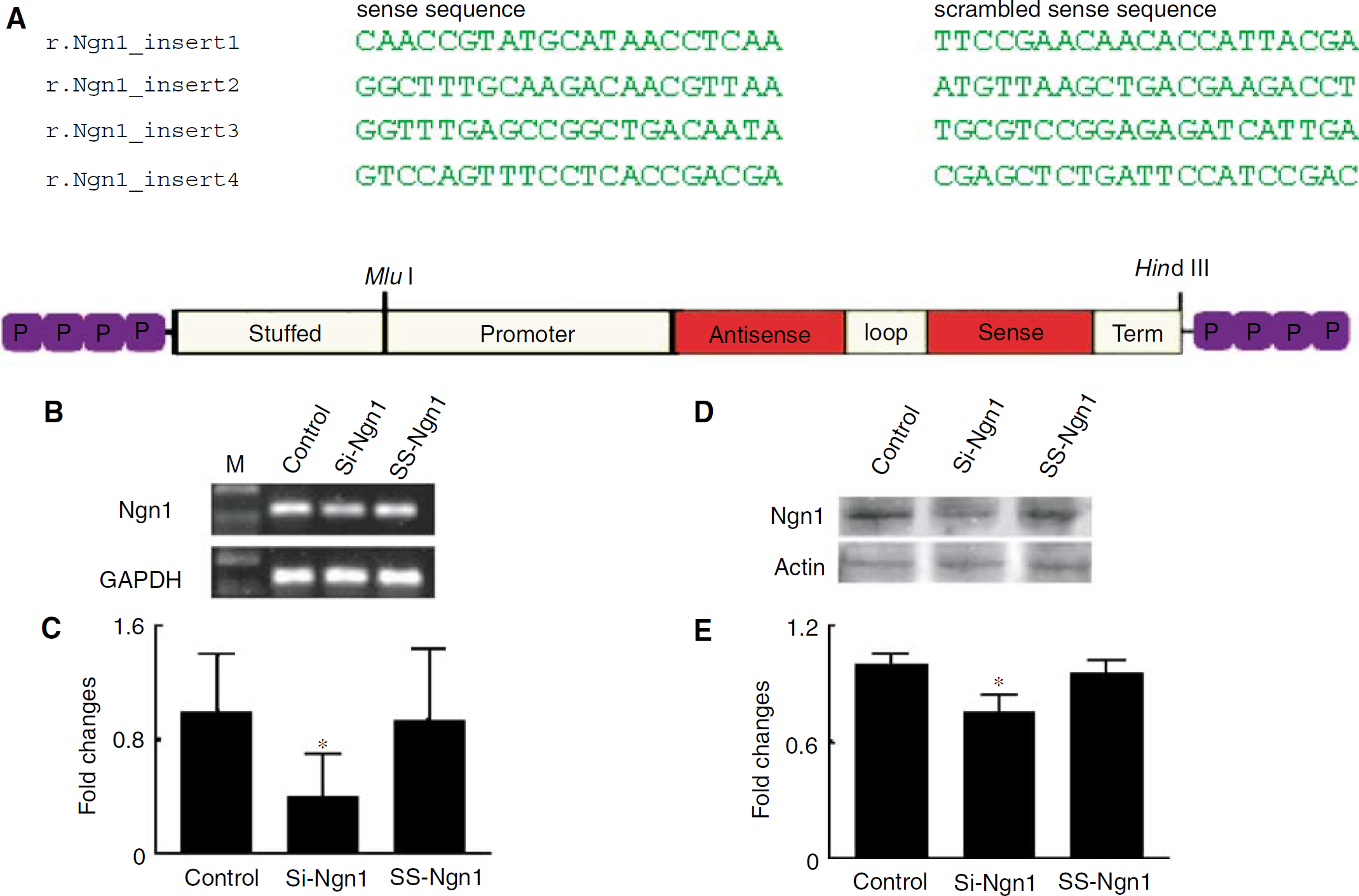
Neurogenin 1 (Ngn1) suppression by short-interfering RNA (siRNA). (
Real-Time RT-PCR
Quantitative PCR was performed using SYBR Green real-time PCR method. Total RNA was isolated from neurosphere cultures using the Stratagene Absolutely RNA MicroRNA isolation kit (Stratagene, La Jolla, CA, USA). Quantitative RT-PCR was performed on an ABI 7000 PCR instrument (Applied Biosystems, Foster City, CA, USA) using 3-stage program parameters provided by the manufacturer, as follows: 2 mins at 50°C, 10 mins at 95°C, and then 40 cycles of 15 secs at 95°C, and 1 min at 60°C. Specificity of the produced amplification product was confirmed by examination of dissociation reaction plots. A distinct single peak indicated that a single DNA sequence was amplified during PCR. PCR products were run on 2% agarose gels to confirm that correct molecular sizes were present. Each sample was tested in triplicate using quantitative RT-PCR, and samples obtained from three independent experiments were used for analysis of relative gene expression data using the 2−ΔΔCT method (Livak and Schmittgen, 2001). The following primers for real-time PCR were designed using Primer Express software (ABI): glyceraldehyde-3-phosphate dehyrogenase (GAPDH, FWD: AGA GAG AGG CCC TCA GTT GCT, REV: TTG TGA GGG AGA TGC TCA GTG T), Ngn1 (FWD: CAG TAG TCC CTC GGC TTC AG, REV: AAG CAG GGT GTC GTA TGG AG).
Immunohistochemistry and Quantification
Single immunofluorescent staining of cultured cells was performed, as previously described (Zhang et al, 2001; Zhang et al, 2003). The following primary antibodies were used in the present study: mouse anti-BrdU (1:1,000; Boehringer Mannheim, Indianapolis, IN, USA), mouse anti-β-tubulin III (TuJ-1, 1:1,000; Covance the Development Services Company, MI, USA), rabbit antiglial fibrillary acidic protein (GFAP, 1:500; Dako Cytomation California Inc., Carpinteria, CA, USA). Cultured cells were fixed in 4% paraformaldehyde for 15 to 20 mins at room temperature. Nonspecific binding sites were blocked with 5% normal goat serum for 30 mins at room temperature. The cells were then incubated with the primary antibodies listed above and with CY3-conjugated secondary antibodies. Nuclei were counterstained with 4', 6'-diamidino-2-phenylindole (DAPI) (Vector Laboratories, Burlingame, CA, USA). The number of BrdU, TuJ1, and GFAP-positive cells and total DAPI cell number were counted and the percentage of each cell type was determined.
Western Blot Analysis
Western blots were performed according to published methods (Bradford, 1976). Briefly, lysates from neural progenitor cells were sonicated for 10 secs and centrifuged at 14,000g for 20 mins. Protein concentration in the supernatants of cell extract was determined using a BCA protein assay kit (Pierce, Rockford, IL, USA). Equal amounts of proteins were loaded on 10% SDS-polyacrylamide gel. After electrophoresis, the proteins were transferred to nitrocellulose membranes and the blots were subsequently probed with the following antibodies: phosphospecific Akt (Ser473), Akt (Cell Signaling Technology Inc., Beverly, MA, USA) and Ngn1 (Chemicon Internation Inc., Temecula, CA, USA). For detection, horseradish peroxidase (HRP)-conjugated secondary antibodies were used (1:2,000) followed by enhanced chemiluminescence (ECL) development (Amersham, Buckinghamshire, UK). Normalization of results was ensured by running parallel Western blots with phosphorylation-independent antibodies or β-actin antibody. The optical density was quantified using an image processing and analysis program (Scion image, Ederick, MA, USA).
Assay of Akt Kinase Activity
Akt kinase activity of the cultured cells was determined using a commercial Akt kinase activity kit according to the manufacture's instructions (Cell Signaling Technology Inc., MA, USA). Briefly, the cultured cells were lysed and the protein concentration of the supernatants was determined using the Bio-Rad protein assay reagent. The proteins (250 μg) were incubated with gentle rocking at 4°C overnight with immobilized anti-Akt antibody cross-linked to agarose hydrazide beads. The immunoprecipitated products were resuspended in 40 μL of kinase assay buffer containing 1 μg of GSK-3α fusion protein (Cell Signaling Technology Inc., MA, USA). Reaction products were resolved by 12% SDS-PAGE followed by Western blotting (Hayakama et al, 1999) with an anti-phospho-GSK-3α/β antibody, as described previously (Hayakawa et al, 2000).
Measurements of Neurite Outgrowth
To analyze neurite outgrowth, TuJ1-positive cells were digitized using a ×20 objective (Zeiss) via the MCID computer imaging analysis system (Imaging Research, St Catharines, Canada). Neurite outgrowth was quantified using a software program developed in our laboratory, which includes measurements of the number, length, and diameter of branches. Sixty TuJ1-positive cells per group were measured and all measurements were performed by experimenters masked to the treatment of each culture condition.
Statistical Analysis
One-way analysis of variance (ANOVA) followed by the Student–Newman–Keuls test was used. The data were presented as means ± s.d. A value of P < 0.05 was taken as significant.
Results
Neurogenin 1 Promotes Erythropoietin-Increased Neuronal Differentiation and Neurite Outgrowth
Neurogenin 1 promotes neural progenitor cell differentiation into neurons (Vojtek et al, 2003). We first measured Ngn1 mRNA levels of neural progenitor cells. Real-time RT-PCR revealed that neural progenitor cells expressed Ngn1 and treatment of neural progenitor cells with rhEPO significantly increased Ngn1 mRNA level compared with control group (Figure 1). To examine the effect of Ngn1 on neurogenesis in neural progenitor cells, we constructed siRNA cassettes against rat Ngn1 (siRNA-Ngn1). Real-time RT-PCR and Western blot analysis showed a significant blockage of Ngn1 mRNA and proteins on extracts from neural progenitor cells 48 and 72 h after transfection with siRNA-Ngn1 cassettes compared with extracts from scrambled cassettes (Figure 2), indicating that the siRNA-Ngn1 cassettes are effective in reducing endogenous Ngn1 levels. We then examined the effects of blocking Ngn1 on rhEPO-enhanced neural progenitor cell proliferation and differentiation. Neural progenitor cells were transfected by siRNA-Ngn1 and scrambled cassettes in the presence of rhEPO for 48 h. After that, the cells were transferred to differentiation medium and the number of TuJ1-positive cells was counted. Treatment with rhEPO alone significantly increased the number of TuJ1-positive cells compared with the number in the control group (Figure 3). However, cells receiving siRNA-Ngn1 cassettes with rhEPO showed a significant reduction of the number of TuJ1-positive cells, whereas scrambled cassettes did not reduce the number of TuJ1-positive cells compared with the rhEPO alone group (Figure 3). In the presence of siRNA-Ngn1 cassettes, neural progenitor cells treated with rhEPO did not decrease the number of BrdU-positive cells compared with the number in the rhEPO alone group (Figure 4). Real-time RT-PCR showed that siRNA-Ngn1 but not scrambled cassettes blocked rhEPO-increased Ngn1 mRNA (Figure 1). These data suggest that endogenous Ngn1 is a requirement for EPO-enhanced neural differentiation but not proliferation.
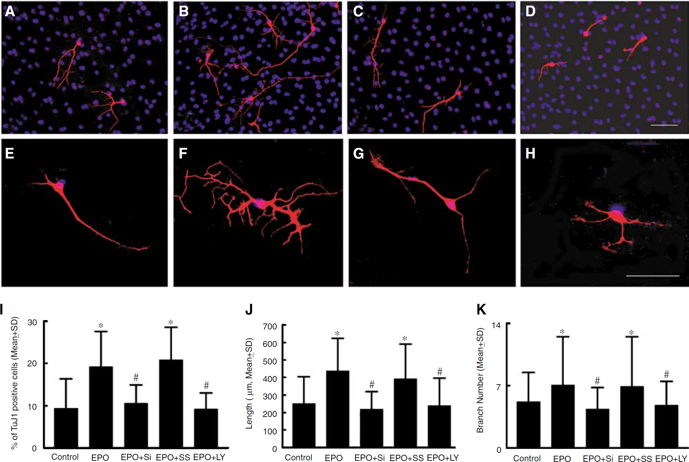
Recombinant human erythropoietin (rhEPO) enhances neuronal differentiation and neurite outgrowth. Recombinant human EPO enhances neuronal differentiation and elicits a more elaborate neurite growth pattern than control. Inhibition of neurogenin 1 (Ngn1) by short-interfering RNAs (siRNAs) and LY294002 reduced TuJ1-positive cell number and neurite growth. (
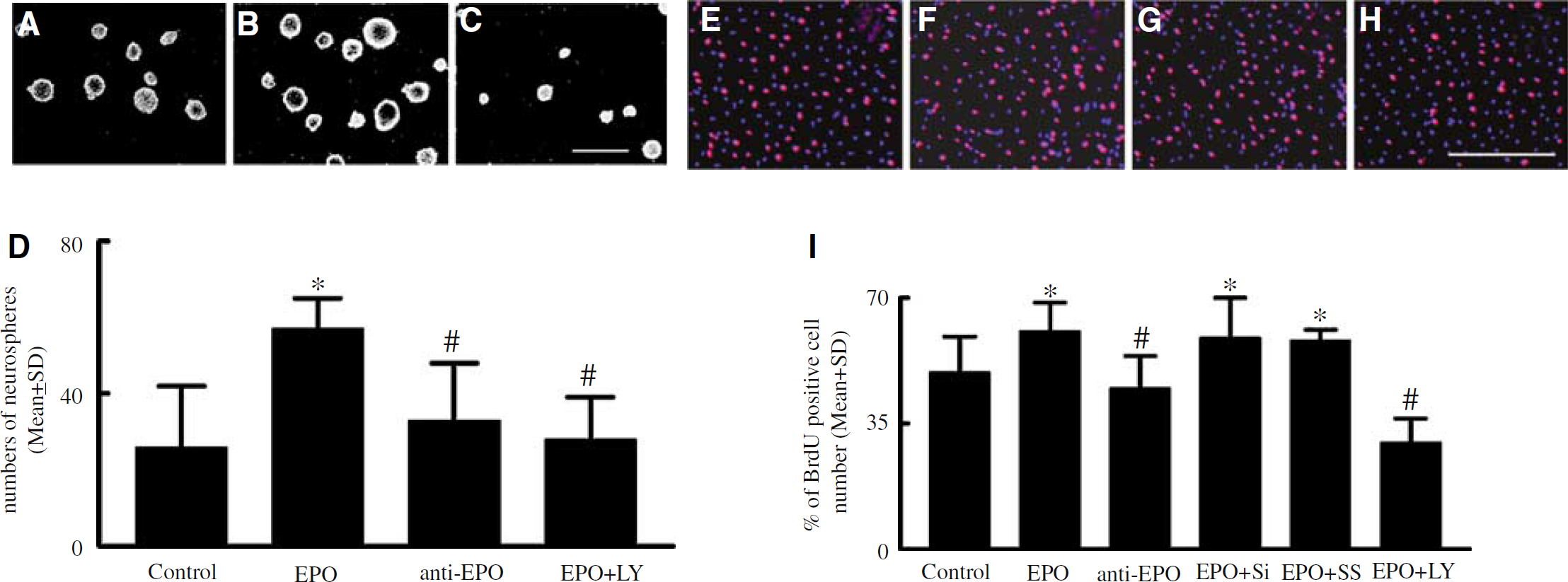
Recombinant human erythropoietin (rhEPO) increases neural progenitor cells proliferation. (
Neurons develop neurites during their maturation (Sayas et al, 2002). We also examined the effects of Ngn1 on neurite outgrowth. Neurite outgrowth was quantified by measuring the length and number of branches extending from TuJ1-positive cell soma. TuJ1-positive cells exhibited a few short branches in the vehicle treated group (Figure 3; Table 1). However, TuJ1-positive cells showed complex patterns when neural progenitor cells were incubated with rhEPO alone. Quantitative analysis revealed that treatment with rhEPO significantly increased the number and length of branches (Figure 3; Table 1). Blocking endogenous Ngn1 by siRNA-Ngn1 cassettes significantly inhibited rhEPO-increased number and length of branches compared with rhEPO alone and rhEPO with scramble cassette groups (Figure 3; Table 1). These data suggest that in addition to enhancement of neurogenesis, rhEPO increases neurite outgrowth and Ngn1 mediates rhEPO-enhanced neurite outgrowth.
Effect of rhEPO on main branch neurite length and number
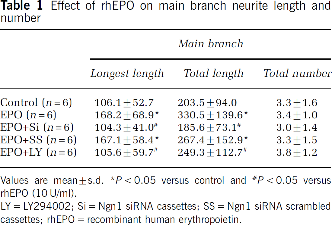
Values are mean ± s.d. *P < 0.05 versus control and <#P < 0.05 versus rhEPO (10 U/ml).
LY = LY294002; Si = Ngn1 siRNA cassettes; SS = Ngn1 siRNA scrambled cassettes; rhEPO = recombinant human erythropoietin.
The Phosphatidylinositol 3-Kinase/Akt Signaling Pathway Mediates Erythropoietin-Increased Neural Progenitor Cell Proliferation and Differentiation and Regulates Neurogenin 1 Levels
Akt regulates proneuronal bHLH genes (Vojtek et al, 2003). To examine whether rhEPO activates Akt, we performed Western blotting on extracts from neural progenitor cells treated with rhEPO. Incubation of neural progenitor cells with rhEPO significantly increased Akt phosphorylation compared with the control group (Figure 5). To determine the ability of activated Akt to phosphorylate its downstream targets, phosphorylation of GSK3α/β, a well-characterized Akt substrate, was measured. Erythropoietin-activated Akt significantly increased serine phosphorylation of GSK3α/β (Figure 5). In parallel, incubation of neural progenitor cells with rhEPO significantly increased numbers of BrdU and TuJ1 immunoreactive cells (Figures 3 and 4), which is consistent with our previous findings that rhEPO enhances neurogenesis in neural progenitor cells (Shingo et al, 2001; Wang et al, 2004). To examine whether activated Akt mediates rhEPO-increased neurogenesis, neural progenitor cells were incubated with rhEPO in the presence of the PI3K-Akt inhibitor LY294002 or vehicle control. In the presence of LY294002, numbers of BrdU and TuJ1-positive cells significantly reduced (Figures 3 and 4). In addition, inhibition of PI3K/Akt by LY294002 blocked Akt activation and serine phosphorylation of GSK3α/β (Figure 5). Erythropoietin-induced phosphorylation of Akt was also abolished by Wortmanin (data not shown). Thus, these data suggest that Akt activation likely mediates rhEPO-enhanced neurogenesis.
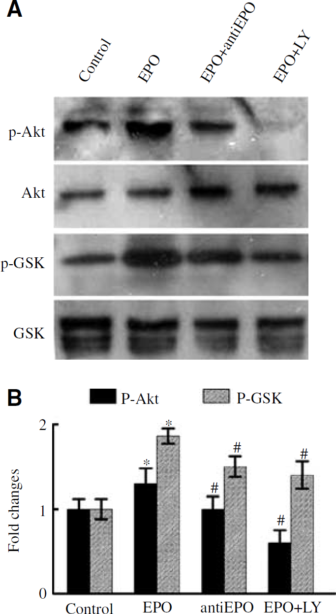
Effects of recombinant human erythropoietin (rhEPO) and LY 294002 on phosphorylation of Akt, GSK3α/β. Western blot analysis (
To examine whether blocking PI3K/Akt affects Ngn1, neuronal progenitor cells were treated with rhEPO and LY294002 for 48 h and total RNA were extracted from these cells. Inhibition of PI3K/Akt significantly attenuated rhEPO-increased Ngn1 mRNA levels (Figure 1), suggesting that the PI3K/Akt signaling pathway regulates Ngn1 expression.
Erythropoietin Increases Neurogenin 1 Levels of Neural Progenitor Cells In Vivo
Treatment of stroke with rhEPO enhances neurogenesis and improves functional recovery (Shingo et al, 2001; Wang et al, 2004). To examine whether administration of rhEPO affects Ngn1, rhEPO was administered 24 h after stroke. Ngn1 mRNA levels in the SVZ were measured. Treatment with rhEPO significantly (P < 0.05) increased Ngn1 mRNA levels compared with stroke-only rats (Figure 6).
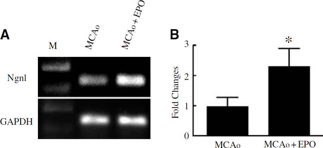
Erythropoietin upregulated neurogenin 1 (Ngn1) expression in stroke brain. Quantitative measurements of real-time RT-PCR show that Ngn1 mRNA was significantly higher in the recombinant human erythropoietin (rhEPO)-treated stroke group than the stroke-only group (
Discussion
The present study shows that EPO activates Akt, and blocking PI3K/Akt by the PI3K/Akt pathway inhibitor, LY294002, abolishes EPO-induced neuronal progenitor cell proliferation, differentiation, and neurite outgrowth. In addition, silencing endogenous Ngn1 by siRNA-Ngn1 cassettes blocks EPO-enhanced neuronal differentiation and neurite outgrowth but not proliferation. Furthermore, the effects of Ngn1 on neural progenitor cells were attenuated by inhibition of PI3K/Akt. These data indicate that the PI3K/Akt pathway mediates EPO-enhanced neurogenesis and Ngn1 is required for Akt-mediated neuronal differentiation in the adult neural progenitor cells.
The PI3K/Akt Pathway Mediates Erythropoietin-Enhanced Neurogenesis
A previous study suggests that EPO regulates neurogenesis via EPO stimulation of STAT5 phosphorylation and NF-κB translocation (Shingo et al, 2001). However, signal pathways that mediate EPO-enhanced neurogenesis have not been elucidated. The PI3K/Akt pathway mediates neurogenesis during Xenopus development (Vojtek et al, 2003; Peng et al, 2004). GSK3β is involved in neurite outgrowth (Goold and Gordon-Weeks, 2001). Growth factors such as platelet-derived growth factor activate PI3K/Akt (Auger et al, 1989; Varticovski et al, 1989). We found that EPO activated Akt and its downstream target GSK3α/β and inhibition of PI3K/Akt by LY294002 blocked EPO-induced activation of Akt and GSK3α/β, which was associated with abrogation of neural progenitor cell proliferation, differentiation, and neurite outgrowth. These data are consistent with findings that the PI3K/Akt pathway regulates EPO-stimulated expansion of hematopoietic progenitor cells (Joosten et al, 2004).
Therefore, our data suggest that EPO acts as an extracellular signaling molecule that activates the PI3K/Akt pathway, which mediates neural progenitor cell proliferation, differentiation, and neurite outgrowth.
Neurogenin 1 is Required for Akt-Mediated Neural Progenitor Cell Differentiation
Proneuronal bHLH transcription factors including Mash1, neurogenin-1, and 2 regulate neurogenesis (Sun et al, 2001). During development of the cerebral cortex, Ngn1 is expressed exclusively in the ventricular zone where neural progenitor cells reside and promotes neural progenitor cell differentiation into neurons (Kim et al, 2004). Mice with targeted disruption of Ngn1 exhibit a loss of progenitor population (Bertrand et al, 2002). In the adult rodent, the ventricular zone of the lateral ventricle is replaced by an ependymal layer and the SVZ containing neural progenitor cells generates neurons and glial cells throughout animal life (Morshead et al, 1998). Here, we found that adult neural progenitor cells expressed Ngn1 and treatment with EPO increased Ngn1 levels, both in cultured neural progenitor cells and in the SVZ cells of animals. These data are in agreement with observations that EPO increases another proneuronal bHLH transcription factor, Mash1, suggesting that EPO may regulate Ngn1. Consistent with a role for Ngn1 in promoting neural progenitor cell differentiation into neurons but not proliferation (Sun et al, 2001), we found that blocking endogenous Ngn1 by silencing Ngn1 RNA abolished EPO-increased neural progenitor cell differentiation and neurite outgrowth, but not EPO-increased proliferation. These data suggest that Ngn1 is required for EPO-enhanced neuronal differentiation and neurite outgrowth. Although we do not examine the effects of Ngn1 on neural progenitor cells derived from the dentate gyrus, Ngn1 may have neurogenic effects in the dentate gyrus since Ngn2 expresses in the dentate gyrus (Raineteau et al, 2004).
A few studies have investigated signal transduction pathways that regulate proneuronal bHLH transcriptional activity in neurogenesis (Kim et al, 2004). By silence of endogenous Akt, Vojtek et al (2003) showed that Akt regulates the assembly and activity of bHLH–coactivator complexes to promote neuronal differentiation. We found that blocking the PI3K/Akt pathway downregulated Ngn1 expression in the presence of EPO, suggesting that Akt may have direct effects on Ngn1 expression, consistent with previous observations of Akt in regulating neuronal differentiation at the level of transcription. Collectively, our data suggest that EPO-like other growth factors binds to its receptor and activates intracellular PI3K/Akt signaling pathway, which mediates adult neural progenitor cell proliferation, differentiation, and neurite outgrowth and the proneuronal bHLH protein Ngn1 is required for Akt-mediated neuronal differentiation and neurite outgrowth.