
Abstract
The synthesis of silver nanoparticles of varying size has been achieved using different molar concentrations of NaOH while the effect of changing the temperature has been studied. AgNO3, gelatine, glucose and NaOH are used as a silver precursor, stabilizer, reducing agent and accelerator respectively. The synthesized nanoparticles have been characterized by a FESEM study, X-ray diffractometry, Raman spectroscopy and UV-vis spectroscopy. The colloidal sols of the silver nanoparticles in a biopolymer gelatine show strong surface plasmon resonance absorption peaks. The visible photoluminescence emission from the synthesized silver nanocrystals has been recorded within the wavelength range of 400–600 nm under UV excitation. The synthesized nanoparticles may be extremely useful in making biosensor devices as well as for other applications.
1. Introduction
Silver nanoparticles (Ag-NPs) have been widely used during the past few years in various applications, such as biomedicine, biosensors, catalysis, pharmaceuticals and photonics [1, 2]. A variety of methods exists to synthesize Ag-NPs, including the microemulsion technique, γ-ray, UV or microwave irradiation and spray pyrolysis [3–6]. Polymers have also been used as matrices in nanocomposites or as stabilizers to provide stability for the metal nanoparticles against oxidation, agglomeration and precipitation [7]. The preparation of Ag-NPs have been reported by several researchers giving well-dispersed Ag-NPs [8] and using various polymers, such as polyethylene glycol [9], poly methyl methacrylate (PMM) [10], poly vinyl alcohol (PVA) [11], polyaniline [12] and polyacrylonitrile (PAN) [13]. Natural polymers, such as natural rubber [14], polysaccharides [15], cellulose [16], chitosan [17] and starch [18], have been used as matrices or stabilizers for the preparation of metallic nanoparticles because of their non-toxicity and biocompatibility. The use of a polymer as the stabilizing agent and the OH– ion as an accelerator in the synthesis of metallic nanoparticles was reported by Kwon et al. [19]. Gelatine is a natural biopolymer, extracted from the partial hydrolysis of collagen and which has good biocompatibility and biodegradability. In recent years, it has been widely used in wound dressings and as a drug carrier and tissue scaffold [20].
We report here the synthesis of silver nanoparticles by a simple chemical process using gelatine as a capping agent and glucose as a reducing agent with NaOH as an accelerator.
2. Experiment
2.1 Synthesis
All of the chemicals and reagents in this work are of analytical grade from Merck, India, and have been used without further purification. 1 g of gelatine is added to an aqueous solution of AgNO3 (10 ml, 0.2M) in a flask. Different molar concentrations of an aqueous solution of NaOH (10 ml) are added to an AgNO3 solution to prepare various samples, viz. 0.5M (S1), 1.0M (S2) and 5.0M (S3), at a room temperature of 25 °C. To each of these solutions is added 10 ml of an aqueous solution of glucose (1 M) each. The suspension immediately turns brown in all of the samples, indicating the formation of silver nanoparticles. The reaction continued for 15 min. The obtained suspensions are centrifuged at 15,000 rpm for 20 min. Furthermore, samples S4, S5 and S6 have been prepared, keeping the molar concentration (5M) of NaOH constant and increasing the temperature to 30 oC, 60 oC and 100 oC respectively.
For the morphological analysis of the prepared nanostructured samples, field emission scanning electron microscopy (FESEM, Carl Zeiss Ultra 55 model) of the centrifuged samples was performed. The average particle size of the prepared Ag nanoparticles was determined using the Sigma Scan Pro software.
The structural characterization of the synthesized Ag-NPs was done using powder X-ray diffraction (PXRD) (Philips, Analytical X-Pert Pro diffractometer) and the XRD pattern was analysed.
Room temperature Raman spectroscopy was performed using a LABRAM-HR800 laser Raman spectrometer with 514 nm and 633 nm laser radiation. To avoid the laser heating the sample, the incident power was kept at a low value of 1.99 mW.
UV-vis spectral analysis was done using a double-beam spectrophotometer (Hitachi, U-3010) with the samples dispersed in distilled water and kept in a quartz cuvette with a path length of 10 mm. The photoluminescence emission spectra from the samples (dispersed in distilled water) were recorded by a spectrofluorometer (LS 55, Perkin Elmer) at room temperature using a four sided polished quartz cuvette with a path length of 10 mm.
3. Results and discussion
The chemical reaction for the formation of Ag nanoparticles is:


In the dispersion of the silver ions in the gelatine matrix (Eq. 1), the gelatine reacts with Ag+ ions to form a stable gelatinous complex [Ag(gel)]+ which further reacts with OH− ions to form Ag-NPs by the reduction of silver ions through the oxidation to gluconic acid (Eq. 2)[21].
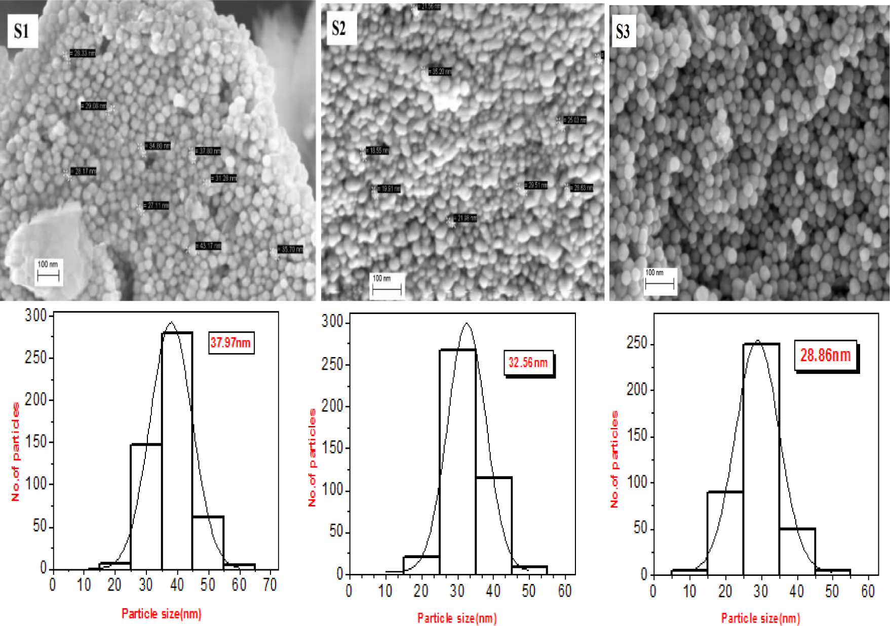
The FESEM micrographs demonstrate the formation of Ag-NPs at different molar concentrations of NaOH. Figure 1 and Figure 2 show the typical FESEM images and the corresponding particle size distribution of the prepared Ag-NPs.
FE

FESEM images of Ag-NPs and their particle size distribution at temperatures 30 °C (S4), 60 °C (S5) and 100 °C (S6).
Figure 3 shows the powder X-ray diffraction analysis and the typical XRD pattern for sample S3. The XRD peaks at 2θ values of 37°, 43°, 64° and 77° can be attributed to the (111), (200), (220) and (311) crystallographic planes, respectively, of the face-centred cubic (fcc) structure of silver nanocrystals (JCPDS Card No. 04-0783). The other peaks relating to any crystalline impurity (e.g., Ag2O) are absent. The particle size measured using Scherer's formula compares well with the average values calculated from the FESEM micrographs. The size of the silver nanocrystallites estimated from the FWHM of the (111) peak, using the Scherrer formula, has been found to be less than 40 nm.

pattern of Ag-NPs.
Figure 4 demonstrates how the morphology of Ag-NPs affects the surface enhanced Raman spectra (SERS). Following Choi et al. [22], we would expect that the samples with a decreasing particle size - which could be produced by using different NaOH concentrations [Figure 4(a)] - to provide good enhancement with a single strong plasmon resonance which is slightly blue shifted, whereas the samples synthesized at different temperatures [Figure 4(b)] provide strong plasmon resonance without any shift. In our study, we observed slight blue shifts to confirm that there were changes in particle size with the changing NaOH concentration, whereas no such changes occurred with varying the temperature. The Raman peaks in the range of 500–1000 cm−1 are due to the glass substrate holding the samples.
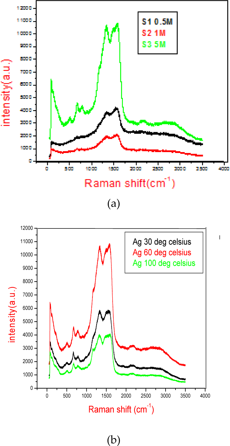
Raman spectra of Ag-NPs with 514 nm excitation wavelength: (a) for different molar concentrations; (b) for different temperatures.
In Figure 5, we find that the addition of 0.5 M (S1) NaOH led to the broadening of the SPR peak at 405 nm. In sample S2, with a greater molar concentration of NaOH of 1.0 M, the absorbance intensity increased due to an increase in the silver concentration and the absorbance peak was observed to be blue shifted to 403 nm. In sample S3, with a NaOH molar concentration of 5.0 M, the further addition of NaOH resulted in a blue shift of λmax to 395 nm. Such observations were due to a decrease in the particle size of Ag-NPs. These results are in good agreement with the results obtained using field emission scanning electron microscopy (FESEM) images of Ag-NPs and their particle size distribution.

UV-vis absorbance spectra of Ag-NPs prepared at different molar concentrations of NaOH.
The UV-vis absorption spectra of samples prepared at different temperatures while keeping the concentration of NaOH constant are shown in Figure 6. The obtained Ag-NPs show the characteristic surface plasmon resonance (SPR) band for silver nanoparticles without any blue shift of absorption peaks at varying temperatures.

UV-visible spectra of Ag-NPs prepared at different temperatures.
Tables 1 and 2 show in tabular form the observations regarding the absorbance properties of the synthesized Ag nanoparticles.
Absorbance characteristics of Ag-NPs prepared with different molar concentrations of NaOH at room temperature (25° C).

Absorbance characteristics of Ag-NPs prepared at different temperatures with the concentration of NaOH kept constant at 5 M.

The photoluminescence spectra obtained from the synthesized silver nanoparticles are shown in Figure 7(a). The PL emission has been obtained within the visible range, from 400 to 600 nm, with peak positions at 504, 496 and 478 nm for S1, S2 and S3 samples respectively. The PL emission peaks have been found to be red shifted by ~ 100 nm, 90 nm and 80 nm, respectively, from their corresponding UV-vis absorption peaks. Figure 7(b) shows that there is no change in the position of SPR (λmax=497 nm) for samples S4 to S6. Thus, we conclude that there is no change in particle size with the variation of the temperature, although the reaction rate increases with an increase of the temperature.

PL emission spectra of: (a) samples S1, S2 and S3; (b) samples S4, S5 and S6.
As the samples were prepared separately under different conditions, the difference in PL emission peaks could be attributed to different states of agglomeration and growth in particle size in cases of the synthesis of sample S3 when compared with samples S4, S5 and S6.
The peak at 750 nm could be attributed to the presence of surface defects and defect states.
The visible luminescence of Ag nanoparticles is due to the excitation of electrons from occupied d bands into states above the Fermi level. Subsequent relaxation by the electron-phonon scattering process leads to an energy loss and, finally, the photoluminescent radiative recombination of an electron from an occupied sp band with the hole takes place. The optical properties of silver nanoparticles depend on both interband and intraband transitions between electronic states.
4. Conclusions
We have demonstrated a simple green synthesis route to prepare high purity Ag-NPs with a glucose reduction of aqueous AgNO3 at different molar concentrations of NaOH used as a reaction accelerator. Nanoparticle agglomeration has been controlled with the addition of gelatine as a stabilizing agent. Gelatine is a biopolymer which is biocompatible and biodegradable. The structural characterization of the samples was performed using XRD, Raman spectra and FESEM observations, which led us to infer that the synthesized nanoparticles were of facecentredcubic (fcc) structure and smaller than 40 nm in size. The UV-visible spectroscopy has shown a shift of the SPR peak, depending upon the changing average particle size of the Ag-NPs, synthesized at different molar concentrations of NaOH. Visible photoluminescence emission has been observed from the synthesized silver nanoparticles within the 400 - 600 nm wavelength range, with peak emissions between 478 and 504 nm. Such nanoparticles may find wide applications in biosensor devices, since the gelatine capping may help their ready attachment with biological systems.
Footnotes
5. Acknowledgements
The authors would like to express their gratitude to Dr. P. Kumbhakar for his valuable support. They also express their sincere thanks to Mr. R. Kuladeep, Mr. R. P. Saida and Mr. M. Chiranjeevi for their technical support.