
Abstract
We demonstrate the synthesis of silver nanoparticles by a potentially benign species of bryophilous Rhizoctonia in two different media. The first medium supports fungal growth and the up-regulation of nitrate reductase, while the second medium supports fungal growth and the repression of nitrate reductase. For both media, the resulting silver nanoparticles were ca. 25–50nm and were subglobose to broadly ellipsoidal in shape. The optical analysis of the silver nanoparticles from both media demonstrated plasmon resonance at 415nm, confirming their metallic properties. The liquid colour change typically observed for extracellular silver nanoparticle formation was absent in both media. The silver nanoparticles in the two different media displayed different chemical associations; fewer associated chemicals were found with the media, which supported the up-regulation of nitrate reductase. Another difference included plate-like silver nanoparticle conglomerations, which were only encountered on hyphae from the medium that repressed nitrate reductase. There was also a noticeable difference in the capping agent formations between each media. The Rhizoctonia isolate examined in this study is suitable for large-scale industrial applications because it does not produce spores and would have minimal impact on air quality.
1. Introduction
Nanoparticles (NPs) are defined as having one dimension 100nm or less in size and due to their large surface area they tend to react differently than larger particles of the same composition, allowing them to be utilized in novel applications. Silver NPs are utilized for their optical, antibacterial, electrochemical and catalytic properties [1, 2]. Unfortunately chemical synthesis of silver NPs utilizes environmentally toxic or biologically hazardous reducing agents [3], so there has been a search for greener production alternatives [1, 2, 4–6].
Many microorganisms, plant extracts and fungi have been shown to produce NPs through biological pathways [1–4, 6, 7]. Synthesis of silver NPs has been investigated utilizing many ubiquitous fungal species including Trichoderma, Fusarium and Penicillium [8–10]. Recent research regarding the use of fungi has generally investigated potential redox systems using silver nitrate as the source of silver ions [8, 10]. Several enzymes, α-NADPH-dependent reductases and nitrate-dependent reductases, were implicated in silver NP synthesis for Fusarium oxysporum [2, 9]. Nitrate reductase was also suggested to initiate NP formation in a Penicillium species [10]. Research has shown that nitrate can induce nitrate reductase, while ammonium and glutamine can cause nitrate reductase repression in fungi [11, 12]. If nitrate reductase were responsible for silver NP formation, then repression of the enzyme would either inhibit NP formation or cause NP formation by another pathway.
Unfortunately, most fungal species that were recently studied (Fusarium oxysporum, Aspergillus and Verticillium species) are plant or human pathogens or fungi that have a negative impact on indoor air quality and cause sick building syndrome due to the release of fungal spores, which would limit or prohibit their large scale production [9, 10, 13]. Fungi that produce sterile mycelium (no spores) would have little impact on air quality and also represent another avenue for a greener production alternative.
Bryophytes are known to uptake and retain metal particles from their environment and many bryophytes have been shown to be metal-tolerant [14, 15]. Bryophyte extracts from Fissidens minutes (moss) [16], Anthoceros (hornwort) [17] and Riccia (thalloid liverwort) [18] species have demonstrated the ability to produce silver NPs. Bryophyte fungal endophytes offer another possible avenue for NP formation since the endophytes would also have to contend with the uptake and retention of the metal particles. To our knowledge, no bryophyte fungal endophyte has been evaluated for the synthesis of silver NPs.
Our objective is to determine if an endophytic bryophilous fungus with sterile mycelium can synthesize metallic silver NPs in two different media; one that promotes and one that represses nitrate reductase. We will evaluate the subsequent NPs on their morphological characteristics, chemical composition and optical properties.
2. Experiment
2.1 Fungal Isolate, Growth and Synthesis of AgNPs
Isolate A3 (Rhizoctonia sp.) from Conocephalum conicum (thalloid liverwort) was assayed for its ability to convert aqueous silver ions to silver NPs. The isolate was selected based on its previous ability to tolerate matric-induced water stress, an indication that it is a bryophyte endophyte and has sterile mycelium. A culture of the isolate was deposited in the American Type Culture Collection.
The AgNO3+CN and Ag2SO4+CN media is consistent with the carbon/nitrogen ratio of Spezieller Nährstoffarmer agar, while utilizing the same concentration of silver ions as reported by other researchers [8, 10, 13]. The AgNO3+CN media supports fungal growth and supports the expression of nitrate reductase, the enzyme reported to be implicated in the production of silver NPs. Nitrate is an inducer of nitrate reductase and the carbon/nitrogen ratio supports better growth than a non-carbon containing medium. The Ag2SO4+CN media also supports fungal growth, but the nitrogen source (L-glutamine) represses the production of nitrate reductase, whereby silver NPs would need to be produced by other means.
An agar plug of actively growing mycelium was antiseptically placed into an autoclaved Erlenmeyer flask that contained 300ml of initial growth medium (8g Nutrient Broth [Difco] + 4g Yeast extract [Difco] + 10g dextrose [L−1 distilled water]) and placed on shake culture (123rpm at ca. 25°C) for 14 days. Under a laminar flow hood, fungal colonies were gravimetrically separated from the initial growth medium using autoclaved Whatman #1 filter paper. Approximately 20ml of autoclaved water was used to wash the excess growth medium from around the fungal mycelia. After three consecutive washes, sterile forceps were used to transfer ca. 5.0g (wet weight) of the washed isolate to 4 sterile Erlenmeyer flasks, each containing one of the following media: AgNO3 medium consisting of a fixed carbon/nitrogen (CN) ratio to support fungal growth and up-regulation of nitrate reductase (AgNO3+CN: 1.0mM AgNO3 + 0.83 g KNO3 + 0.2g glucose + 0.2 g of dextrose, L−1 distilled water) or Ag2SO4 medium, which consisted of the same fixed CN ratio to support fungal growth and L-glutamine to induce the repression of nitrate reductase (Ag2SO4+CN: 1.0mM Ag2SO4 + 1.0g L-glutamine + 0.2g glucose + 0.2g of dextrose, L−1 distilled water). The inoculated Erlenmeyer flasks were returned to shake culture for 96 hours at room temperature, at which time the production and characteristics of the silver NP formations were assayed.
2.1.1 Centrifuge procedure for AgNP isolation
Two cultures, one cultured in Ag2SO4+CN and one cultured in AgNO3+CN, were sonicated at 10W ultrasound with a cell disruptor to disrupt the mycelia, while another non-sonicated set was used as a control. The control test tubes were placed in a Clay-Adams centrifuge and spun at 2800rpm. The supernatants were collected and set aside (silver ion containing solution). Sterile water (10ml) was added to each control test tube containing the mycelium, shaken vigorously by hand for 30 seconds and spun at 15000rpm (30,996 g) for 10 minutes in a Beckman Model J2–21 centrifuge. The supernatant was collected in a separate clean test tube (unaltered wash solution).
The sonicated cultures were spun at 15000rpm (30,996g) for 10 minutes in a Beckman Model J2–21 centrifuge. The supernatant was collected in a separate clean test tube (silver ion containing solution). To each pellet, 10ml of sterile water was added, followed by vigorous hand shaking for 30 seconds to re-suspend the pellet. The test tubes containing the re-suspended pellets were centrifuged for a second time at the same settings. The supernatant was collected in a separate clean test tube (sonicated wash solution). The pellet was re-suspended in 10ml of sterile water (pellet solution) and all samples were assayed.
2.1.2 Media evaluation
The Rhizoctonia isolate was grown in an autoclaved Erlenmeyer flask containing 100ml of 1.0g KNO3 + 0.2g glucose + 0.2g of dextrose and L−1 distilled water and in a separate autoclaved Erlenmeyer flask containing 100ml of 1. g L-glutamine + 0.2g glucose + 0.2g of dextrose and L−1 distilled water for three days at 25°C. An aliquot (1ml) of each medium was added to separate 1.5ml centrifuge tubes and spun at 14000rpms for 2 min. to clear the medium of fungal fragments. A 500μl aliquot of each centrifuged medium was added to a separate 15ml plastic tube that contained 1500μl of the above nitrate-containing medium. Controls were 500μl of the virgin medium + 1500μl of the above nitrate medium. After 24 hours at 25°C the nitrate concentrations were determined colourimetrically using the Griess diazotization reaction and zinc as a catalyst.
2.1.3 Analysis
Imaging was carried out using a Vega TC SBII SEM equipped for simultaneous elemental analysis with an X-ray detector (X-MAX from Oxford Instrument) for Energy-Dispersive X-ray spectroscopy (EDX). Particle size was measured using Vega TC software. The optical transmission was done using a spectrograph USB2000 from Ocean Optics coupled with a Xenon light source.
3. Results and discussion
3.1 Media evaluation
Both media promoted good fungal growth for the Rhizoctonia isolate, but greater growth occurred on L-glutamine containing medium [Figure 1]. When the L-glutamine containing medium was evaluated for its ability to repress nitrate reductase, the results indicated that the nitrate concentration in the tube containing the fungal L-glutamine supernatant was comparable to the L-glutamine control tube. The nitrate evaluation medium demonstrated a noticeable reduction in colour intensity, thereby indicating a reduction in nitrate concentration, for the tube containing the fungal nitrate supernatant in comparison to its control tube.
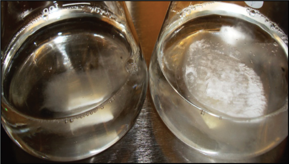
Isolate A3 growth on nitrate (left) and L-glutamine (right) evaluation media after 4 days at 25°C.
3.2 Silver NP characteristics
The bryophilous Rhizoctonia isolate was capable of silver NP production in both the AgNO3+CN and Ag2SO4+CN media. In both media, silver NPs were external to the fungal hyphae as visualized with SEM images and were concentrated near the hyphal surface and to a lesser extent, the NPs were unattached to the hyphal surface [Figure 2].

SEM image of bryophilous isolate A3 (Rhizoctonia sp.) mycelium and subsequently produced silver NPs when subjected to silver ions.
No significant change in the culture supernatant colour was observed in both media. This was in contrast to the appearance of a brown colour change in some previous fungal studies [8, 9], which was attributed to extracellular silver NP formation. The lack of supernatant colour change could have occurred due to the biosorption capacity of fungal mycelium to bind extracellular metals to their cell wall components [19, 20]. It is hypothesized that extracellular enzymatic NP formation occurred in the AgNO3+CN medium, as reported by [8, 9], followed by extracellular precipitation and subsequent NP binding to the hyphal cell wall. Our optical analysis of the various supernatants support this as the most likely conclusion since plasmon resonance was only detected in the wash solutions that were obtained after vigorous hand shaking of the washed fungal mycelia in distilled water.
Figure 3 demonstrates subglobose to broadly ellipsoidal silver NPs, ca. 25–50nm in width, embedded within a matrix, which were produced in the Ag2SO4+CN medium, while Figure 4 demonstrates the morphology of the silver NPs produced in the AgNO3+CN medium. They are of similar size and shape, but appear to have a different capping formation.

Silver NPs from Isolate A3 Ag2SO4+CN wash solution demonstrated that they are made up of individual NPs that appear to be embedded in a matrix.

Silver NPs from Isolate A3 AgNO3 +CN wash solution demonstrated that they are made up of individual NPs embedded within a matrix.
Larger silver particles within isolate A3 unaltered wash solutions where visualized as conglomerations that were on average 50 to 250nm. A conglomerate configuration [Figure 5] that was only visualized in the Ag2SO4+CN medium suggested their formation was created by nucleation centres on the hyphal cell wall.

Plate-like silver conglomerations that appear to be site of nucleation on Rhizoctonia isolate mycelium grown in Ag2SO4+CN medium.
3.2 Elemental analysis of A3 (Rhizoctonia sp.) wash solutions
The elemental analysis clearly demonstrated that the bright objects in the SEM images are silver NPs and no silver was detected outside of them. The following elements were also routinely detected: carbons (C), chlorine (Cl), nitrogen (N), oxygen (O), phosphorous (P), potassium (K) and sulphur (S). After both sonicated wash solutions were annealed at 200°C for 3 hours in air, the EDX analysis did not indicate chlorine in any of the targeted NPs for the AgNO3+CN wash solution. Chlorine was still detected by EDX analysis after the same annealing time for the targeted NPs in the Ag2SO4+CN wash solution. For both unaltered samples and both wash solution samples, silver was always present when chlorine and sulphur were detected. Nitrogen, phosphorus and excessive carbon were also present for many silver-containing NPs [Table 1].
Energy-dispersive X-ray spectroscopy analysis of isolate A3 silver NPs.
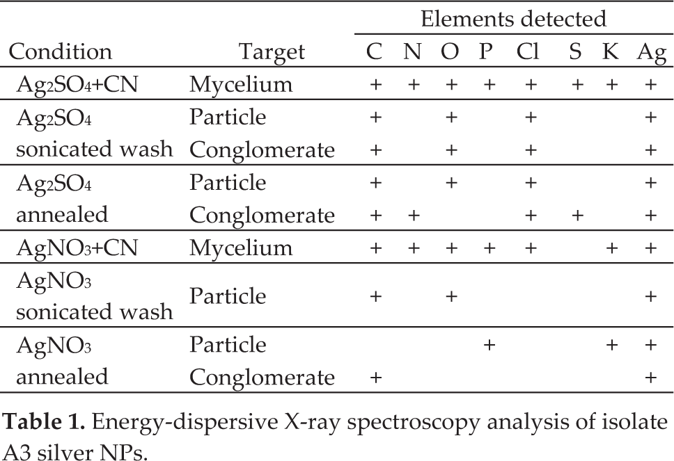
From the EDX analysis, we concluded that silver NPs produced using the AgNO3+CN medium produced silver NPs with less associated chemicals than by using the Ag2SO4+CN medium.
3.3 Optical transmission properties of A3 (Rhizoctonia sp.) solutions
The metallic state is characterized with charge carriers mobile enough to respond to electromagnetic fields. The measurement of this response is widely used for contactless detection of free charge carriers. In the case of NPs, their size and shape lead to formation of characteristic resonances, or absorption bands, which happen to be in the near-UV and visible spectral range for silver and gold NPs [22, 23]. Therefore, near-UV and visible light is a fine and precise probe for the detection of free charge carriers (resp. metallic state) in silver NPs and it has been widely used in the literature [6–9, 21–23]. Even a surface capping layer or an embedding dielectric matrix is not an obstacle to the detection of characteristic absorption bands, resp. the presence of metallic state.
Optical transmission measurements of the A3 unaltered wash solution from both media demonstrated a broad absorption band from 325nm to 600nm, with maximum absorption at 480nm [Figure 6]. Isolate A3 sonicated wash solutions from both media demonstrated a narrower absorption band, which shifted to shorter wavelengths with maximum absorption at 415nm. Such characteristic absorption bands for metallic silver NPs were not observed in any of the silver ion containing solutions for both media.

UV-vis absorption spectra for A3 unaltered supernatant wash and sonicated wash solutions after 96 hours.
The position of the narrower absorption bands in both A3 sonicated wash solutions were in agreement with the expected wavelengths [22, 23] for silver NPs within the size range of 25–50nm, which is also in agreement with the SEM images shown in Figures 2–5. The A3 unaltered wash solution obtained from both media contained larger NPs leading to maximum absorption at longer wavelengths, while the A3 sonicated wash solution from both media shows absorption peaking at shorter wavelengths. The silver NPs in the unaltered wash solutions from both media have a broad size distribution, as evidenced by the broad absorption extending in the UV range. These experimental observations demonstrate that the bio-synthesized silver NPs from both media contain enough mobile charge carriers to generate the characteristic plasmon resonance.
4. Conclusions
We have unequivocally demonstrated that a bryophilous Rhizoctonia isolate (A3) from Conocephalum conicum could convert silver ions into metallic silver NPs in a medium that supported and in a medium that repressed nitrate reductase. The subglobose 25–50nm silver NPs from both media were shown to be pure enough to demonstrate surface plasmon resonance at 415nm. In both test media, fungal produced silver NPs were clearly embedded in a capping agent and were shown to be attached to the hyphal surface. We have observed that the silver NPs had different associated chemicals depending upon the growth medium. Sonification and centrifugation isolation steps modified the UV-vis spectral absorption bands that demonstrated that the sonification process broke-up the larger conglomerations. The data also suggested that hyphal nucleation sites could be involved in the formation of some of the silver NPs in the Ag2SO4+CN medium, while the silver NP formation in the AgNO3+CN medium occurred by extracellular enzymatic initiation and precipitation, followed by hyphal biosorption. However, further research would be needed to understand the exact sequence and mechanisms involved. The successful demonstration of biosynthesis of metallic silver NPs motivates us to search for their application as antimicrobial agent. At this stage of our research, we found anecdotal evidence pointing toward the viability of this application. The unambiguous demonstration of this application would be a task requiring rigorous tests, which is beyond the scope of this work.
Footnotes
5. Acknowledgments
This work was made possible by a Lock Haven University Faculty Professional Development grant for promoting undergraduate research and by NSF grant #0923047. The authors would like to express their gratitude to Dr. Andrew Miller for technical support for the identification of the isolate.