
Abstract
Even with the prevalence of wounds, the medical technology for efficiently managing skin damage is still primitive. The disruption of any of the numerous healing processes can lead to problems in the time-sensitive healing actions of the dermal and epidermal layers. Bacterial infection is one of the major obstacles to proper wound healing as it poses a danger of causing long-term negative effects. Keeping the wound free of bacteria is imperative to the proper and hasty repair of dermal wounds. Silver has been widely used to treat wounds for its bactericidal properties. Although the mechanism of silver's antibacterial action is not fully understood, it exhibits a significant antimicrobial efficacy against a wide spectrum of bacterial species. A number of different approaches to the mechanism are reported and presented in this review. Silver nanoparticles (AgNPs) have been reported to exhibit enhanced antibacterial activity due to their increased surface-area-to-volume ratio. AgNPs are capable of various modifications, significantly broadening the therapeutic properties of the material as a result. This review explores the different aspects of silver and silver nanoparticles, and their antibacterial properties, which can be applied in the field of wound treatments.
1. Introduction
The process of evolution leading to life on dry land may have been significantly influenced by the development of skin, acting as a barrier preventing water loss [1]. Water is essential to all life forms on earth and the slightest change in the water level of any organism can bring harm to its host. Thus, the skin's role in the maintenance of a healthy body water is vital to survival on land. The skin, the largest external organ in the human body, accounts for about 16% of the adult human body [2]. The composition of the skin is intricate, involving multi-faceted layers responsible for various functions. There are two major components of the skin: the epidermis and the dermis. The epidermis, covered with keratin, is responsible for the physical, biochemical/chemical and adaptive immunological barrier functions [3]. Skin damage is such a common occurrence in daily life that its effects are passed over as negligible; however, in reality, it poses a threat to the security of the entire body, especially in unhygienic areas. The human body can experience a variety of injuries, including penetration, burns, and blunt trauma [4]. Some of these forms of damage can turn into chronic wounds, which cause long-term agony and complications in many patients, as a result of the inability of the body to properly heal itself. Dermal reparation is an elaborate process entailing the precise synchronization of countless components of the skin to fix damaged tissues, restore protective barrier function, and re-establish the homeostatic equilibrium state of the wound area [4]. The steps of wound repair can be categorized into haemostasis, inflammation, proliferation, and remodelling, with each phase involving the collaborative work of many tissues and cells [5]. Keratinocytes, fibroblasts, endothelial cells, macrophages, and platelets cells are among the essential cells involved in wound repair [6]. Haemostasis is characterized as the release of blood components into the injury site, allowing the platelets to come into contact with the exposed collagen and other components of the extracellular matrix. This release then triggers the release of clotting factors, essential growth factors, and cytokines such as platelet-derived growth factor (PDGF) and transforming growth factor (TGF-β), from the platelets [4]. During haemostasis, platelets adhere to the wound site, where they aggregate. Aggregation is then followed by coagulation cascade activation, which results in the formation of platelet clots in a protein mesh composed of cross-linked fibrin [7]. The inflammation stage involves neutrophils entering the injured area and beginning phagocytosis, in order to remove unwanted substances, bacteria, and unhealthy tissue. Macrophages also contribute to the phagocytosis process by further releasing PDGF and TGF-β during inflammation [4]. The sheer number of growth factors and cytokines involved in the overall wound healing task, including fibroblast growth factor, vascular endothelial growth factor, granulocyte-macrophage colony-stimulating factor, platelet-derived growth factor, connective tissue growth factor, interleukin, and tumour necrosis factor-alpha, indicates the complexity and fragility of the process [6]. The cytokines and growth factors released during the haemostasis and inflammation phases act as chemical signals that control matrix production, cell migration, differentiation, and enzyme expression. Through these chemical signals, new blood supplies and connective tissue cells are summoned into the damaged area. After the inflammation phase, fibroblasts migrate to the wound site and initiate the proliferative phase by releasing fresh extracellular matrices. The released matrices are then cross-linked and reorganized during the remodelling stage. Each step of the process must be carried out appropriately in a timely manner, in order for the wounded area to retrieve its homeostatic equilibrium. Any disruptions in the process may lead to undesirable conditions that may present more serious problems at the wound site. Thus, it is critical for the health of the patient that the processes involved in the healing of dermal damage are not disrupted.
The bacterial infection of wounds is one of the major concerns in wound treatment. Chronic wounds, a major cause of morbidity, are an example of a condition resulting from bacterial infection [8]. The clinical spectrum of wound colonization and infection describes the stages of bacterial interaction with a wound: contamination, colonization, local infection/critical colonization, the spreading of invasive infection, and septicaemia [8]. Each stage of infection produces various effects on the injury site, making it difficult to understand the interaction of bacteria with the disruption of wound healing. Biofilms—self-secreted extracellular polysaccharide matrices—provide protection for bacteria, through benefits such as increased infection rates, substrate accessibility, metabolic efficiency, and resilience towards environmental stress, along with inhibitors of agents that remove or terminate bacteria [9]. Thus, the presence of biofilms further complicates the interaction between dermal injury and bacteria. Chronic wounds and delayed wound repair are often characterized by elongated inflammation, faulty re-epithelialization, and poor matrix remodelling [8]. A proliferation of microorganisms at the injury site can disrupt the healing processes in a variety of ways. Thus, preventative measures for inhibiting the growth of harmful microorganisms can be an effective and straightforward course of action to help remove all the possible disruptions that bacteria may produce.
Silver is often utilized as an antibacterial agent, in order to provide a sanitary environment for the wound healing process. Silver was considered, historically, as one of the most frequently used antibacterial substances before the invention of antibiotics [10]. Silver was used in sutures to prevent postoperative inflammation and infections when treating battle wounds during the First World War [11]. The use of antibiotics, metal ions, enzymes, and quaternary ammonium compounds can exhibit negative side effects: antibiotic resistance, environmental damage, and high costs [12]. Such downsides have encouraged the scientific community to look to other modes of bactericidal activity. The multiplicity of silver's bactericidal mechanisms gives it a wide range of effective applications in the inhibition of bacterial growth. Utilizing broad-spectrum antimicrobial silver in wound treatment, to prevent the initial growth of any bacteria, may contribute to the advancement of wound treatment systems by providing an ideal sanitary environment for maximum wound repair. The introduction of nanoparticles has allowed the scientific community to enhance the antibacterial properties of silver. The increased surface area of the nanoparticles in turn induces an increased rate of interaction between the test subjects and the ionic silver. The increase in bactericidal efficiency lowers the minimum inhibitory concentration, meaning that a lower concentration of the material may be incorporated into wound healing systems to exert the same degree of effect. A lower concentration of silver nanoparticles will reduce the possible interference of biological and chemical wound healing processes from the AgNPs themselves. Additionally, the modification capability of silver nanoparticles further enhances its utilitarian spectrum in skin reparation. This review paper explores recent studies to provide an overview of the development of technologies utilizing silver's antibacterial activity in wound care.
2. Silver in Wound Treatment
The mechanism of silver's antibacterial activity has been widely studied, in order to further understand its underlying process. Before now, it was already understood that silver's mode of bactericidal action involves numerous pathways. Silver atoms attach themselves to the thiol groups of enzymes involved in vital biological processes, including ion transport and transmembrane energy generation [13]. Silver catalyzes oxidation reactions between the oxygen and hydrogen atoms of the thiol, producing disulfide bonds and inhibiting bacterial growth [14]. Changes in the biologically vital molecules of cells lead to changes in the cellular structure, which can cause malfunctions and ultimately celldeath. Another mechanism of the antibacterial activity of silver is the disruption of bacterial cell respiration. Silver ions are capable of deactivating 30S ribosomal subunits. The deactivation of the ribosome complex results in an interference in the translation of proteins, a crucial part of bacterial reproduction [15]. Succinyl-coenzyme A synthase is a crucial component of the TCA cycle and catalyzes the reaction that yields succinate from succinyl-CoA, while simultaneously phosphorylating ADP to produce ATP [16]. Silver ions are able to deactivate Succinyl-coenzyme A synthase, rendering a certain ratio of the enzymes useless. Fructose biphosphate adolase, which is deactivated by the presence of silver ions, acts as the catalyst in the fructose-1,6,-bisphosphate cleavage reaction that yields glyceraldehyde 3-phosphate and dihydroxyacetone phosphate [16]. MalK, a maltose transporter, is another protein greatly affected by silver ions. After treating cells with 900 ppb Ag+ solution, a decrease in their expression of 30S ribosomal subunit protein, succinyl coenzyme A synthetase, maltose transporter, and fructose biphosphate adolase was observed, supporting silver's ability to disrupt multiple processes involved in cell respiration [15]. Ag+ ions have been reported to enter cells and denature DNA molecules by disrupting the hydrogen bonds between purine and pyrimidine base pairs [13]. Utilizing its antimicrobial properties, silver has been increasingly integrated into many wound treatments for burns, traumatic wounds, diabetic ulcers, and chronic wounds. The lack of a normal protection barrier, due to the presence of a wound, may lead to an increase in the chance of infection and further complications [17]. There is a wide variety of commercially available wound dressings containing silver, which act as a temporary protection barrier and provide ideal conditions for effective repair [18]. The antibacterial activity of silver ions is commonly tested on Gram-positive Staphylococcus aureus (S. aureus) and Gram-negative Escherichia coli (E. coli). Silver sulfadiazine (AgSD) is considered the gold standard in terms of the antibacterial activity of silver ions [19]. One comprehensive study of AgSD, regarding the mechanism of its action on burn wound infections, investigated the binding of silver ions and sulfadiazine (SD) to the bacterial cells, the dissociation of AgSD, reactions between AgSD and biologically significant components, and the synergistic effects of sodium sulfadiazine and AgSD. The results clearly indicate the lack of SD cell penetration, meaning that this is unlikely to be a component in the antibacterial activity of AgSD [17]. Thus, it can be inferred that the silver ions from the AgSD are solely responsible for its ability to inhibit bacterial growth. Additionally, the level of silver cellular binding of AgSD was compared to that of various silver salts, to investigate the dissociation rates that eventually yield the antibacterial silver ions. AgSD and AgNO3 showed maximum silver binding, while other silver compounds showed minimal uptake, due to their slow dissociation rates. In a mechanistic study, E. coli and S. aureus cultures were exposed to a 10 μg/ml AgNO3 solution by adding the solution directly into the medium [20]. The cells were then studied via transmission electron microscopy (TEM) and X-ray microanalysis. Feng et al. observed significant structural changes in both bacterial species. Within the AgNO3 treated group, electron-light regions were observed in the middle section of the cells. Particles of condensed substances and electron granules – aggregates of high-electron-density mass consisted of silver and protein depositions observed in TEM and SEM -were present throughout the cell structures in both cell types, and severe cell wall damage was seen in many E. coli cells. The X-ray microanalysis results affirmed the presence of silver, sulfur and phosphorus within the cells. The presence of phosphorus may be a result of damage to the DNA molecules. From these results, it can be inferred that the silver ions inflict a significant amount of structural damage to the cells, possibly enough to be considered lethal. The antimicrobial activity of silver is often utilized in wound care systems with multiple assets. One study of partial-thickness burn wound patients clinically explored the wound-healing properties of Askina Calgitrol Ag®, an alginate silver wound dressing, compared with those of 1% silver sulfadiazine, a substance commonly used as the standard for antimicrobial properties [21]. Sixty-five patients were randomly divided into two experiment groups:35 patients were treated with 1% AgSD and the remaining patients were treated with Askina Calgitrol Ag®. The experimental group exhibited improved efficacies in the following parameters: healing time, nursing time, pain score, and number of wound dressing changes. All determined parameter values were significantly lower in the Askina Clagitrol Ag® group. From these results, it can be inferred that the alginate silver wound dressing is much more effective in treating burn victims than the simple AgSD solution, most likely due to the presence of the wound exudate-like environment provided by the alginate-based product. Furthermore, the biopolymer matrix containing silver ions may afford an improved method of silver ion production at the wound site, which may lead to additional increases in the level of interaction between the silver ions and the microbes. The shorter healing time and fewer dressing changes exhibited in the experiments could also help significantly reduce the cost of wound care, which is beginning to become a major economic problem worldwide.
Even with the clinically tested effectiveness of the silver ions, the development and application of such chemotherapy should proceed with caution, as previously unforeseen side effects may present themselves under certain conditions. The limited efficiency of silver ion release, the confined number of silver species, the inability of silver ions to reach the wound bed, and the rapid consumption of silver ions may necessitate higher concentrations of silver compounds within the wound treatment systems, which could be lethal to healthy host cells [19]. Thus, other modes of silver ion delivery were sought, in order to increase the efficiency of the wound healing treatments.
3. Silver Nanoparticles
The utilization of silver nanoparticles to increase the surface-to-volume ratio has been reported to be successful in enhancing the bactericidal efficiency of the silver ions. The surface-to-volume ratio improvement is accompanied by an increase in contact area with the bacteria. In order for the silver to exhibit its antibacterial activity, it needs to be in its ionic form. Thus, the increase in the contact area of the nanoparticles is able to generate more silver ions able to interact with the bacteria, so as to damage them via its multiple pathways [22]. For the reasons provided, silver nanoparticles have been garnering a great deal of attention in the field of wound care systems.
Silver nanoparticles take similar approaches to killing bacteria. The nano scale of the particles is their major advantage in the improvement of their efficiency. The antibacterial activity of the silver nanoparticles, and its mechanism of action, was studied by analysing the membrane permeability, structural morphology, and growth of E. coli [23]. Li's group gathered data proving an increase in the leakage of reducing saccharides and proteins. The detected concentration of leaked reducing sugars increased from 30 μg/mg to 102.5 μg/mg in two hours, and the leaked protein concentration increased from 5.89 μg/mg to 11.96 μg/mg in four hours. The rise in the level of the leaked molecules seems to be due to an increase in the membrane permeability. The membrane permeability of the bacteria is dictated by the outer lipopolysaccharide (LPS) membrane. The altered outer LPS membrane structure exhibited increased permeability in E. coli [24]. Li's group found that the silver nanoparticles were inhibiting the activity of respiratory chain dehydrogenases in a concentration-dependent manner. The AgNP-treated bacterial cells were significantly deformed and fragmented. In addition, many of the cells exhibited large gaps in the cellular structure observed via TEM. Figure 1 represents the AgNP's function in bacterial inhibition and the improvement of silver ion delivery to the membrane and cytoplasm of the bacteria cells. Xiu et al. suggest that silver nanoparticles may contribute in the growth control of higher-order organisms [25].

Schematic representation of the improved silver ion delivery capability of silver nanoparticles formed with natural ligands. The pH difference between the cytoplasm and the membrane of the bacterial components enhances the delivery of the active substances. Reprinted with permission from Xiu, Z.-M., Q.-B. Zhang, H. L. Puppala, V. L. Colvin & P. J. J. Alvarez (2012) Negligible Particle-Specific Antibacterial Activity of Silver Nanoparticles. Nano Lett, 12, 4271–4275. Copyright © 2012 American Chemical Society.
The antibacterial activities of silver nanoparticles are influenced by the morphology and size of the nanoparticles. Morones et al. utilized high-angled annular dark-field microscopy (HAADF) and TEM to investigate the antibacterial activity of silver nanoparticles, in the size range of 1–100 nm, on Gram-negative bacteria [26]. Luria broth plates treated with various concentrations (0, 25, 50, 75, 100 μg/ml) of nano-silver were prepared and introduced to 10 ml of a prepared E. coli bacterial culture. The interrelation of the nano-particulates and E. coli was analysed by measuring the optical density at 595 nm. The silver nanoparticles' electrochemical nature was analysed by using stripping voltammetry. The TEM confirmed that the sizes of nanoparticles were between 1–10 nm. The majority of particles with diameters from 1 to 10 nm were octahedral, decahedral, or multiple-twinned icosahedral in shape. The study of the effects of different concentrations of AgNPs on bacterial growth demonstrated that concentrations above 75 μg/ml inhibited the growth. Further TEM analysis confirmed the presence of silver nanoparticles in the cell membrane and inside the bacteria. The HAADF image shows that the smaller-sized nanoparticles (5 nm) produced enhanced bactericidal efficacy, thus supporting the size dependence of the activity of silver nanoparticles. Panacek et al. synthesized silver nanoparticles varying in size, via a one-step reaction (Tollens process) [27]. In this method, different reducing agents (saccharides, glucose, galactose, maltose and lactose) were used. The concentration of AgNO3 (10−3 mol L−1) and reducing agents (0.01 mol L−1)were fixed, and varying amounts of ammonia were used from 0.2 to 0.005 mol L−1. A NaOH solution was used to initiate the reduction of [Ag(NH3)2]+. The silver colloid particles ranged in size from 25 to 100 nm, on average. This size variation was dependent on the ammonia concentration and on the type of reducing agent in the reaction system. The ammonia concentration was significant in defining the size of silver particles, since an increase in the ammonia concentration increased the particle size. The size transition from tens to hundreds of nanometres took place when the ammonia concentration was at 0.035 mol L−1. Additionally, the structures of saccharides influence the size of AgNPs. Disaccharides (maltose) were efficient in the reduction of [Ag(NH3)2]+ and produced monodispersed particles with an average size of 25 nm. These sizes were efficient (1.69 μg/mL of Ag) in inhibiting the bacterial growth against both Gram-positive and Gram-negative bacteria, including Staphylococcus aureus. The synthesized particles were characterized by DLS, TEM and UV-visible absorption spectrometry. Silver colloid nanoparticles with an average size of 25 nm were produced by reducing [Ag(NH3)2]+ with maltose. Through antimicrobial and bactericidal assays, it was found that 25-nm silver colloid particles demonstrated noticeable antimicrobial and bactericidal activity against multi-resistant bacteria species, including methicillin-resistantStaphylococcus aureus (MRSA). Martínez-Castañón et al. investigated the controlled formation of AgNPs at varying sizes(7, 29 and 89 nm) [28]. In this article, gallic acid was used to reduce and stabilize the AgNPs. Through TEM analysis, the silver nanoparticles were determined to be spherical at sizes 7 and 29 nm, and pseudo-spherical at 89 nm. The pH adjustment of the reactions dictated the size of the silver nanoparticles produced. The reduction at pH 11 and 10 resulted in 7-and 29-nm silver nanoparticles, respectively. The reaction mixture was irradiated with UV light (254 nm, 25 W) for 30 minutes, in order to yield 89-nm AgNPs. The solution was then heated for 30 minutes at 80°C. The antibacterial activities of the silver nanoparticles weretested against E. coli and S. aureus for comparison. Seven-nanometre AgNPs showed the greatest antibacterial activity against S. aureus and E. coli. Because of their miniscule nanoparticle size (7 nm) and high surface area, silver nanoparticles with 7 nm were able to easily reach the nuclear content of the test subjects, thereby killing the bacteria more efficiently. Espinosa-Cristóbal et al. investigated the antibacterial activity of AgNPs against Streptococcus mutans, a microorganism associated with dental caries [29]. In this study, 8.4-, 16.1- and 98-nmsilver nanoparticles were prepared using an adaptation of the synthesis method reported by Martínez-Castañón et al. The morphology of theS. mutans before and after their treatment with the AgNPs was surveyed via atomic force microscopy (AFM). The AgNPs determined to have an average diameter of 8.4 nm showed improved antibacterial activity compared with those of the larger nanoparticles (16.1 and 98 nm). Espinosa-Cristóbal et al. suggest that smaller nanoparticles release more Ag+ ions, which leads to an increase in their antibacterial effect. Lu et al. have also reported the size-dependent nature of the antibacterial activities of silver nanoparticles against anaerobic microbes [30]. The antibacterial activities of the differently sized AgNPs, synthesized by reduction, were evaluated by colony counting assays, the growth inhibition curve method, and corresponding minimum inhibitory concentration (MIC) measurements, against five anaerobic oral pathogenic bacteria (Actinobacillus actinomycetemcomitans, Fusobacterium nuceatum, Streptococcus mutans, Streptococcus sanguis and Streptococcus mitis) and one aerobic bacteria (E. coli). All three sizes were synthesized differently: the 5-nm AgNPs were synthesized by adding AgNO3 (0.5 mL, 0.2 M) to poly(N-vinyl-2-pyrrolidone) (PVP, MW & 55000), and stirring in a round-bottom flask which contained 5 mL of deionized water. After 30 minutes, a 0.1 M NaBH4 solution was added to reduce the Ag+.TEM images revealed the size of the AgNPs as 5 nm. Similarly, 15-nm AgNPs were synthesized by adding 0.5 mL AgNO3 (0.05 M) to a flask containing PVP (0.25 g) and 2- [4-(2-Hydroxyethyl)-1-piperazinyl]ethanesulfonic acid (HEPES) buffer (25 mL, 100 mM, pH 7.4). After 30 minutes, NaBH4 (0.1 M) was added to reduce the silver ions. Larger AgNPs (55 nm) were prepared under a higher temperature, using PEG as a reducing agent. The antibacterial effects of the particles against anaerobic bacteria were lower than those against aerobic bacteria. Thus, it can be inferred that bacteria requiring oxygen for their survival are more susceptible to AgNPs than those bacterial species that survive independently of O2 presence. The 5-nm AgNPs presented the best antibacterial activity against E. coli and F. nuceatum. Considering the higher antimicrobial properties and lower toxicity of AgNPs, compared to those of Ag compounds such as AgNO3 and silver sulfadiazine, they could be used for the treatment of periodontal pathogens with greater benefits. Recently, Agnihotri et al. reported the size-specific (5, 7, 10, 15, 20, 30, 50, 63, 85 and 100 nm) antibacterial efficacy of silver nanoparticles [31]. These particles were synthesized using NaBH4 as the primary reductant and trisodium citrate as the co-reducing agent, under two different thermal conditions. In stage I, metal salts were reduced using NaBH4 and the reaction mixture was maintained at 60°C. During this stage, reduction occurs, and a large number of AgNPs are formed. In stage II, the formed AgNPs undergo a growth process involving further reduction, with the trisodium citrate and at a higher temperature (90°C). However, it was suggested that the trisodium citrate remained passive during stage I, as it prevented AgNPs from agglomerating. Furthermore, the results suggested that NaBH4 was important for the formation of smaller-sized particles (5–20 nm), whereas trisodium citrate was responsible for the yield of bigger nanoparticles (60–100 nm), as shown in Figure 2.

The schematic representation of the two-step synthesis of size-dependent silver nanoparticles, using NaBH4 and sodium citrate reduction under two different thermal conditions. Agnihotri, S., S. Mukherji & S. Mukherji (2014) Size-controlled silver nanoparticles synthesized over the range 5–100 nm using the same protocol and their antibacterial efficacy. RSC Advances, 4, 3974–3983. Reproduced by permission of the Royal Society of Chemistry (RSC), on behalf of the Centre National de la Recherche Scientifique (CNRS) and the RSC.
From the results, the antibacterial efficacies of the silver nanoparticles with diameters smaller than 10 nm were significantly enhanced, as revealed through disc diffusion tests, corresponding MIC/minimal bactericidal concentration (MBC) values, and delayed bacterial growth kinetics. The 5-nm AgNPs (the smallest) demonstrated the quickest bactericidal activity on every test strain (E. coli MTCC 443, E. coli MTCC 739, B. subtilis MTCC 441, and S. aureus NCIM5021) compared to the 7-nm and 10-nm AgNPs at similar bacterial concentrations. In conclusion, the silver nanoparticles synthesized in a size-controlled manner exhibited broad-spectrum antibacterial activity in a size- and dose-dependent manner. Song et al. studied the shape-dependent bactericidal qualities of AgNPs [32]. Three different shapes of AgNPs were prepared. Typically, particles were prepared from equimolar concentrations (0.001 M) of AgNO3 and sodium citrate, under boiling conditions and in the presence of silver seed solution (3 ml). Spherically shaped AgNPs were obtained using a citrate reduction method. The rod-shaped and truncated triangular AgNPs were obtained via a seeded growth method. Both the particle types were grown together by adding 5 ml of silver seeds to a solution containing AgNO3 (5 ml, 0.01 M), ascorbic acid (0.1 M, 10 ml) and CTAB (146 ml, 0.1 M). Particle growth was accelerated by adding NaOH (1 ml, 1 M). After aging, the particles were purified by centrifugation to separate the truncated triangular and rod-shaped AgNPs. To measure the killing kinetics of nano-silver, agar plates were inoculated with 100 μl of bacterial suspensions and treated with various AgNP amounts, ranging from 1 to 100 μg, in order to determine the susceptibility of bacteria to silver. The truncated triangular nanoparticles, with a total silver content of 10μg, led to the complete hindrance of microbial growth. The improvement in antibacterial activity of the truncated triangular nanoparticles was based on the {111} lattice structure of the truncated triangular nanoplate. The truncated triangular nanoplates exhibited greater silver reactivity properties, due to their high-atom-density facets (such as {111}), whereas the spherical and rod-shaped nanoparticles contained a greater level of {100} facets compared to {111} facets. Therefore, it can be inferred that the high-atom-density of the truncated triangular silver nanoparticles' {111} lattice system enhances its antibacterial efficacy. The shape dependence of the bactericidal qualities of the AgNPs was supported by the results of the explored study. Furthermore, the differences in the bactericidal activity of differently shaped AgNPs were confirmed by EFTEM (Figure 3). The EFTEM pictures demonstrated alterations of the cell membrane that resulted in lysis. This shows that triangular nanoparticles displayer better bactericidal activity compared to spherical and rod-shaped nanoparticles, suggesting that the {111} lattice plane of triangular nanoparticles was in better contact with the bacteria cell wall, and thus able to cause more damage.

EFTEM images of E. coli cells. (A) Untreated E. coli, with visible flagella. (B) E. coli grown on agar plates, supplemented with Ag+ (AgNO3). Arrows indicate partially damaged membranes. These cells are viable. (C) E. coli treated with triangular silver nanoplates. Silver nanoparticles appear as dark irregular pits on the cell surface. (D) E. coli treated with spherical silver nanoparticles. (E) Enlarged image of part of the bacterial cell membrane, treated with triangular silver nanoparticles. The cell membrane is damaged in multiple locations. Pal S. et al. (2007) Does the Antibacterial Activity of Silver Nanoparticles Depend on the Shape of the Nanoparticle? A Study of the Gram-Negative Bacterium Escherichia coli.Appl Environ Microbiol, 73, 1712–1720.
One of the major benefits of the development process of nanoparticles is its capacity for modification. By varying the synthesis method and conditions, certain nanoparticle properties can be controlled as desired [31, 33, 34]. The size, composition, structure and other physical parameters of the silver nanoparticles significantly affect the properties of AgNPs. Interestingly, the antibacterial efficiency of silver can be tuned by changing its valency. Only a few reports have shown that the higher the valency, the greater the antibacterial activity. Djokic prepared high-valent silver oxysalts and studied their antimicrobial activity against Pseudomonas aeruginosa, Staphyloccus aureus and Candida albicans microorganisms [35]. This study suggested that oxidized silver oxysalts produce a greater rate of antibacterial activity compared to that of its lower-valent salts. Based on this concept, and taking advantage of the nanosized silver and the high surface-to-volume ratio of nanoparticles, novel silver complexes were developed as nanoparticles. Song et al. prepared nanosized high-valent silver-chlorhexidine complexes (Ag(III)CHX) using a reverse microemulsion system. In general, all AgNPs have Ag in a zero oxidation state; however, nanocrystalline Ag(III)CHX is structurally and chemically different from the conventional AgNPs. Furthermore, Ag(III)CHX exhibited higher antibacterial activity against various Gram-positive and Gram-negative bacteria species.
A silver(I) chlorhexidine complex was synthesized in an in situ precipitation of the complex, from a weakly acidic AgNO3 solution and chlorhexidine (CHX). Ag(II)CHX was synthesized by the oxidation of Ag(I), followed by the complexation of the oxidized metal with chlorhexidine, as shown in Figure 4.
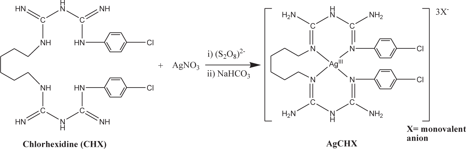
Reaction scheme of the silver chlorhexidine synthesis
The antibacterial activities of the novel complexes were assessed in vitro on four gram-positive (Enterococcus faecalis, Propionibacterium acnes, Staphylococcus epidermidis, Staphylococcus aureus) and four gram-negative species (Pseudomonas aeruginosa, Klebsiella pneumonia, Citrobacter freundii, Acinetobacter calcoaceticus). The developed silver complexes were far more efficient in killing the tested bacteria and did so at a much faster rate when compared to AgSD and AgNO3. These improved bactericidal efficacies of the polydiguanide complexes might be a result of the higher availability of silver ions in the medium as a result of the reduction in the deactivation of the silver ions. Modifying the valency of the silver also seems to affect the antimicrobial activity of the nanoparticles [35]. In an attempt to prepare trivalent silver polydiguanide complex (Ag(III)CHX) as nanoparticles, a reverse micro-emulsion method was utilized as shown in Figure 5.A g(III)CHX nanoparticles were prepared using a microemulsion system comprising water, dioctyl sulfosuccinate sodium salt(AOT) and heptane. The formation of AgNPs can be divided into four main steps: first, the oxidation of Ag+ to Ag3+; then, the stabilization of Ag3+ by complexation with CHX molecules, followed by the neutralization of pH with sodium bicarbonate (NaHCO3); and finally, nucleation and particle growth. The nanoparticles were analysed through transmission electron microscope analysis, X-ray photoelectron spectroscopy, and zeta potential measurements. The water in a micro-emulsion system provides a limited micro-environment for the formation of nanoparticles and plays a crucial part in tuning the dimensions of the Ag(III)CHX nanoparticles. The MIC, MBC, and IC50 values were measured using agar dilution and broth dilution methods on a variety of Gram-positive and negative bacteria species. The antimicrobial properties of the synthesized Ag(III)CHX were much greater than those of AgNO3, AgSD and CHX alone. The most significant difference between the levels of bactericidal efficiency was exhibited on the Acinteobacter calcoaceticus, where the Ag(III)CHX outperformed AgNO3 by more than ten-fold. Although the mechanisms of the enhancement are poorly understood, the study clearly presents the increased antimicrobial strength of nanocrystalline silver's high valency. An in vitro study of the concentration-dependent cellular responses induced by silver nanoparticles was performed on human fibrosarcoma (HT-1080) and human skin/carcinoma(A431) cells [37]. The morphology studies showed that exposure to AgNP concentrations ranging from 6.25 to 50 μg/mL caused the cells to become less polyhedral, more fusiform, shrunken and rounded. The IC50 values for the HT-1080 and A431, determined by XTT assays, were 10.6 and 11.6 μg/mL, respectively. Cells treated with concentrations around half the IC50 exhibited oxidative stress through significant decreases in the levels of glutathione and superoxide dismutase, and an escalation in the level of lipid peroxidation. The DNA fragmentation observed in the AgNP-treated group suggested the apoptosis of the AgNP-treated cells.

The synthesis sequence for silver chlorhexidine nanoparticles in a reverse microemulsion. Reprinted with permission from Pal, S., E. J. Yoon, Y. K. Tak, E. C. Choi & J. M. Song (2009) Synthesis of Highly Antibacterial Nanocrystalline Trivalent Silver Polydiguanide. J Am Chem Soc, 131, 16147–55. Copyright © 2009 American Chemical Society.
Silver nanoparticles are often incorporated into wound treatment systems to provide clinically desirable properties and an ideal environment for rapid and effective healing. One silver nanoparticle-based dressing, Acticoat™Flex 3, was tested on a fibroblast cell culture and a partial-thickness burn patient to study its effects [38]. In this study, the in vitro experiments showed that the AgNPs reduced mitochondrial activity without causing skin cell lysis or compromising the nuclear integrity of the skin cells, according to the data obtained through cellular staining techniques. The reduced mitochondrial activity in the studied area of interest suggests a decrease in the presence of bacteria, thus supporting the antibacterial activity of the AgNPs. Skin biopsies of AgNP-treated patients were analysed via TEM and inductively coupled plasma mass spectrometry (ICP-MS). The results led to the conclusion that AgNPs are released as aggregates and localized in the cytoplasm of fibroblasts, have different distributions within the upper and lower dermis, and do not cause any noticeable cell death. The ability of AgNPs to reduce the growth of bacteria at the area of interest without causing any serious damage to the skin cells, as presented in this article, sets the basis of silver's clinical safety and antibacterial activity. Tian et al. investigated the in vivo wound-healing properties of topically delivered AgNPs, and observed swift healing and enhanced scarring appearance in a dose-dependent manner [39]. The samples of nanosilver were coated on a wound dressing applied to the area of interest. In the thermal injury mouse model from this study, the average of the healing times of animals inoculated with AgNPs was much shorter than those of the negative control group and AgSD treatment group. In addition, the appearances of the healed areas of the AgNP-treated groups were the most similar to that of normal skin, whereas the silver sulfadiazine groups exhibited hypertrophic scarring. Tian's group also presented results suggesting improved antibacterial properties, compared to those of antibiotics, through quantitative polymerase chain reaction (PCR), immunohistochemistry, and proteomic studies of the wound sites of the animals. The wound dressing coated with nano-silver was significantly more efficient in hindering bacterial growth at the wound site than the silver sulfadiazine (AgSD) solution. The microorganism culture from the wound swabs exhibited bacterial culture growth after three days in the AgSD-treated group and after seven days in the AgNP-treated group. In a separate study, silver nanoparticles were impregnated into bacterial cellulose to provide it with antibacterial properties [40]. Bacterial cellulose, an ideal wound dressing material because it provides a moist environment at the site, does not contain any bactericidal activity. Thus, silver nanoparticles were prepared, analysed, and impregnated inside the bacterial cellulose fibre by soaking the bacterial cellulose membranes in 0.001 M of aAgNO3 solution and then reducing the silver ion-saturated bacterial cellulose pellicles with various concentrations of aqueous NaBH4. The freeze-dried silver nanoparticles-infused bacterial cellulose was examined for its release of silver ions, swelling, and antimicrobial activities. The results from the disc diffusion method supported the successful infusion of the AgNPs into the bacterial cellulose through the presentation of an inhibition zone, which the bacterial cellulose alone did not exhibit. In terms of the bacterial growth reduction of the silver nanoparticles-infused bacterial cellulose, 99.7% and 99.9% reductions were observed through colony forming count methods in E. coli and S. aureus, respectively. Meanwhile, the pure bacterial cellulose groups exhibited 34.6% and 40.7% increase. This study shows that AgNPs can be incorporated into biological components without altering their properties (most importantly, the clinically advantageous properties). Tang et al. synthesized silver dendrimer nanocomposites with diameters ranging from 9.5, 7.6 to 16.2 to 65 nm [41]. The synthesized nanocomposites were used to prepare antibacterial cotton fabrics as possible wound treatment utensils. The silver-dendrimer nanocomposites were synthesized using the following protocol: G2 dendrimer (1.9 mM) and methanol (30 ml) were first put into a conical flask and stirred vigorously, to form a dendrimer-methanol solution (10% w/w). To adjust the pH for better protonation, oxalic acid (0.07 mol) was added, followed by an AgNO3 (1.9–0.96mM) solution. The reaction mixture was stirred for two hours in the dark at room temperature. Finally, 10 ml of a NaBH4 (0.95–0.048Mm) aqueous solution was added to the above solution to reduce the metal ions. The reaction mixture was stirred for another hour to yield the silver nanocomposite solution. The antibacterial cotton fabric was prepared by soaking the cotton fabric in the antibacterial agent solution (Ag-dendrimer nanocomposite+H2O, pH8.0) for five minutes, stirring at room temperature, then washing the cotton with deionized water and air-drying the cotton. The antibacterial activity of the cotton fabric containing dendrimer-encapsulated silver nanoparticles was tested against S. aureus and E. coli through the inhibition zone method. The authors' results suggest that the antibacterial activity of the silver nanocomposite cotton fabrics was dependent on the size of the silver nanoparticles: the smaller the AgNPs, the larger the zone of inhibition. The application of poly(amidoamine) (PAMAM) dendrimers has been explored in numerous ways, including in gene transfection, the delivery of drugs, and bacterial growth inhibition. Its wide range of uses stems from the fact that PAMAM possesses exclusive properties, such as large molecular size, well-defined molecular structure and narrow size distribution. Strydom et al. recently studied the PAMAM dendrimer with sulfadiazine (SD), for which its silver nanoparticle complex was synthesized [42]. The novel dendrimer complex showed high antibacterial activity and improved water solubility compared with the gold standard for the topical treatment of burn wounds: silver sulfadiazine (AgSD). The nanoparticle complex was prepared by utilizing a microfluidization method; the particle size was characterized by DLS and the morphology determined by TEM. To prepare the AgSD nanoparticles, the authors followed a method previously used for silver-dendrimer complex. In this method, 10ml of AgNO3 solution was added to the PAMAM dendrimer (10 ml) and irradiated with UV light at different time intervals [43]. To obtain a sulfadiazine-dendrimer (SD-dendrimer) complex, a method developed by Prieto et al. was followed. To synthesize the SD-dendrimer complex, the dendrimer and SD were first dissolved in methanol at different molar ratios. Then, these solutions were incubated for 45 minutes at room temperature. The methanol was removed to obtain a solid product, which was then dissolved in a buffer solution and centrifuged to obtain the soluble SD-dendrimer complex. In this method, equimolar solutions of Ag/G4.0,G5.0-PAMAM-NH2 complex and SD/G3.5, G4.5-PAMAM-COOH complex were mixed, and the silver sulfadiazine complex was produced as a precipitate. This method was scaled up using a modified microfluidizer process. Structural analysis was performed using X-Ray powder diffraction and Raman spectroscopy. The antibacterial efficacy was determined against commonly found burn wound bacteria:S. aureus, S. epidermis, P. auroginosa and E. coli. The authors also studied the drug release kinetics of the material and observed that the synthesized antibiotic nanoparticles exhibited greater release rates, compared to those of commercially available silver sulfadiazine (AgSD) products. The efficient release rates of the dendrimer-coated AgSD nanoparticles may be responsible for the greater antibacterial activity in the burn wound bacterial culture exhibited by the novel nanomaterial. Silver is mostly known for its indirect contribution to wound care, by aiding in the removal and preventing the presence of unwanted materials at the wound site; however, recent studies have been shedding light on the unprecedented properties that contribute advantageously to damage repair. Liu et al. investigated how the AgNPs influence specific cells during the wound healing process [44]. The cellular response of keratinocytes and fibroblasts (the cell types involved in dermal contraction and epidermal re-epithelialization), from an excisional wound model in rodents, were studied in this article. The wound contraction rate was determined by measuring the wound site area over time. The wound closure process of the AgNP-treated group displayed an accelerated rate. A histological study of the epithelial tongue growth, using an immunohistochemistry stain to measure cell division, confirmed a significant increase in cell proliferation in the AgNP-treated groups. The authors concluded that the AgNPs promoted re-epithelialization through increased keratinocyte migration and proliferation, and wound contraction, by driving the differentiation of fibroblasts into myofibroblasts. These mechanistic explanations of the AgNPs' wound healing promotion illuminate possible methods for further improving wound care efficiency, by shortening the healing time of the wound site. AgNPs were reported to affect both local and systemic inflammation, as shown in Figure 6(A) [39]. Tian et al. also studied burn injury sites via proteomic analyses, to monitor the expression levels of major acute-phase proteins: haemopexin (Hpx), haptoglobin (Hpg), and serum amyloid protein component P (SAP). The plasma glycoproteins—Hpx and Hpg—bind to haemoglobin and free haeme, which are released during intravascular haemolysis to defend against oxidative damage. Figure 6(B) exhibits the silver nanoparticles' (ND) ability to normalize the expression levels of the acute-phase proteins to the levels exhibited by normal skin, faster than untreated and silver sulfadiazine- (SSD-)treated skin. The levels of cytokines involved in the dermal injury repair process (including IFN-γ, VEGF, TGF-β1, IL-6, and IL-10) were affected by the presence of AgNPs [39]. Cytokine modulation of the silver nanoparticles was investigated using quantitative real-time PCR. The IFN-γ, VEGF, and IL-10levels of the AgNP-treated groups were higher than those exhibited in the AgSD-treated groups, while IL-6 mRNA levels of the AgNP group was significantly lower. The cytokine expression modulation of the silver nanoparticles was dose-dependent. The changes in the levels of the different cytokines suggested a method of controlling specific molecules, which could lead to a greater understanding of wound healing and its dose-dependent interaction with foreign materials. Nadworny et al. reported that nano-silver reduced inflammation when tested on a porcine model of contact dermatitis [45]. The area of interest was treated with silver nanoparticle dressings, 0.5% silver nitrate, or saline solutions. Among the experiment groups, the skin treated with silver nano dressing was the closest to healthy skin, according to the erythema and oedema examinations and histological data. The authors concluded that the decreased inflammation was associated with a rise in the degree of inflammatory cell apoptosis and a decrease in the expression of pro-inflammatory cytokines and gelatinase activity. Silver nanoparticles were also successful in reducing inflammation in different forms. Nanocrystalline silver was reported to significantly reduce the colonic inflammation caused by ulcerative colitis in mice [46]. The intracolonically or orally administered silver nanoparticles significantly restrained the expression of tumour necrosis factor-alpha (TNF-α), matrix metalloproteinase-9 (MMP-9), interleukin-1beta (IL-1β), and IL-12. In another study, different concentrations of nanocrystalline silver (NCS) were intravesically administered to rats as a possible treatment for interstitial cystitis [47]. Boucher et al. aimed to assess the anti-inflammatory qualities of AgNPs by studying the bladders and urine of silver nanoparticle-treated rats. The urine of the subjects was collected throughout the treatment period and the bladder was obtained from the rats after the four-hour treatment period. The histamine levels of the NCS-treated rats were reduced compared to those of the PBS-treated rats. The histamine and TNF-α levels released from the bladder explants were lower in the rats treated with NCS. These studies support silver nanoparticles' ability to reduce inflammation: a crucial aspect in encouraging wound healing via a more direct approach.

(A) Neutrophil immunohistochemical microscopy images of ND (silver nanoparticles), SSD (silver sulfadiazine), and negative control groups, using naphthol ASD chloroacetate esterase. (B) Images monitoring the expression of proteins and serum markers, in burn injuries. The proteins were analysed using 2D electrophoresis and MALDI-TOF MS protein identification. Tian, J., K. K. Wong, C. M. Ho, C. N. Lok, W. Y. Yu, C. M. Che, J. F. Chiu & P. K. Tam (2007) Topical delivery of silver nanoparticles promotes wound healing. ChemMedChem, 2, 129–36. Reprinted with permission from ChemMedChem.
4. Modified Silver and Silver Nanoparticles
One of the major benefits of the development process of nanoparticles is its capacity for modification. By varying the synthesis method and conditions, certain properties of the nanoparticles can be controlled as desired [31, 33, 34]. The size, composition, structure and other physical parameters of the silver nanoparticles significantly affect the properties of AgNPs. Furthermore, the conjugation of AgNPs is possible, depending on the composition of the nanocrystalline silver, which makes AgNPs much more versatile.
Song et al. developed multifunctional composites comprising the dual properties of antibacterial activity and wound healing [48]. In this work, a bi-valent silver polydiguanide was conjugated to histatin-1 (HST-1): a known potent wound-healing polypeptide. HST-1 is known to be a major factor in wound closure. Its induction of wound closure stems from its ability to be actively internalized by epithelial cells, to enhance epithelial migration using the extracellular signal-regulated kinases 1/2 (ERK1/2) pathway [49]. Solid-phase peptide synthesis, utilizing Fmoc chemistry with an automatic peptide synthesizer, was carried out to yield the linear peptide HST-1. The product was then purified using RP-HPLC and characterized using mass spectrometry (MS). Ag(II)CHX, known for its enhanced bactericidal activity, was synthesized using a previously established procedure. The formed Ag(II)CHX was then dissolved in DMSO and conjugated with HST-1 at different ratios. The reaction mixtures were incubated at 2–8°C for one hour. The various peptide-metal complex solutions were then stored at 2–8°C for one month, before being tested for their antibacterial activity and used in cell-spreading assays. Circular dichroism spectroscopy was used to confirm the formation of the HSt-1- [Ag(II)CHX] complex. The antibacterial properties of the novel polypeptide complex were measured by calculating its MICs and MBCs on four strains of Gram-negative bacteria (Pseudomonas aeruginosa, Klebsiella pneumonia, Citrobacter freundii and Acinetobacter calcoaceticus) and four strains of Gram-positive bacteria (Propionibacterium acnes, Staphylococcus epidermidis, Staphylococcus aureus and Enterococcus faecalis). The MICs and MBCs of Ag(II)CHX were compared to those of the Hst-1- [Ag(II)CHX]. The MICs and MBCs of the Hst-1- [Ag(II)CHX] were slightly higher than those of the Ag(II)CHX, but seemed to retain a large part of the starting material's antibacterial activity. Furthermore, the wound-healing properties of the synthesized complex were studied using cell-spreading assays, as shown in Figure 7. The in vitro cell-spreading assay was prepared by preparing and culturing 3T3-L1 cells, with a cell-free gap in the centre. The samples were treated with the different substances to test their cell migration-enhancing abilities. The relative cell-free gaps (RGt) were calculated at different time intervals (0, 12, and 24 hours), in order to compare the wound-healing properties of the novel polypeptide silver complex. As shown in Figure 7, the Ag(II)CHX-treated samples did not exhibit much cell migration promotion, while both the HST-1- and HST-1- [Ag(II)CHX]-treated cells exhibited much smaller cell-free gap areas, compared to that of the control after 12 hours. The promotion of cell migration, shown in the materials consisting of polypeptide or polypeptide-conjugated complexes, show that the silver nanoparticles can be conjugated with desired properties, in order to aid in the treatment of wounds.

(A) Neutrophil immunohistochemical microscopy images of ND (silver nanoparticles), SSD (silver sulfadiazine), and negative control groups, using naphthol ASD chloroacetate esterase. (B) Images monitoring the expression of proteins and serum markers, in burn injuries. The proteins were analysed using 2D electrophoresis and MALDI-TOF MS protein identification. Tian, J., K. K. Wong, C. M. Ho, C. N. Lok, W. Y. Yu, C. M. Che, J. F. Chiu & P. K. Tam (2007) Topical delivery of silver nanoparticles promotes wound healing. ChemMedChem, 2, 129–36. Reprinted with permission from ChemMedChem.
5. Conclusion
In various different forms, silver is capable of aiding the wound healing process, mainly through its antibacterial activity. The broad spectrum of the silver ion's antibacterial activity—a result of the multiplicity of the bactericidal mechanism—has proven itself to be effective on a variety of bacteria species in multitudinous studies. Although silver has already been incorporated into commercially available products, the advancement of such wound care would have positive implications for the health and economy of humanity, especially if the less developed countries were granted an increased access to such medical necessities. Through the development of nanoparticles, and their modification at the molecular and nano level, the application efficiency of wound treatments containing silver has been improving noticeably. These developments may be able to significantly reduce the time required for the wound area to reach its normal homeostatic equilibrium, while reducing the risk of unwanted complications and improving the physical appearance of the scar by reducing hypertrophic scarring. While seemingly effective and safe, the safety of such materials should always be investigated thoroughly. By proceeding with punctiliousness, and studying its versatility and its limits, silver may be used to provide the medical community with a new generation of highly effective and multi-dimensional wound treatment systems.
Footnotes
6. Acknowledgements
This research work was supported by the National Research Foundation of Korea (NRF) Grantof the Ministry of Education, Science and Technology (MEST) (2015RlA2AlA05001842). We are also grateful to the Research Institute of Pharmaceutical Sciences at Seoul National University for providing some experimental equipment.