
Abstract
The utility of point of care ultrasound training during medical school is becoming more and more evident. At the Loma Linda University School of Medicine, we have formally integrated ultrasound education into the curriculum of all four years. Exposure begins in the first few months of Year 1 and takes form in a variety of educational mediums through Year 4. Whether students receive training through mandatory sessions during physical diagnosis courses or extracurricular workshops provided through the Ultrasound Interest Group–-the experience equips learners of at all different skill levels with the confidence to apply what they have learned to patient care. The successful integration of ultrasound training into the medical curriculum can be attributed to progressive administration, devoted faculty and eager students. The perspective of medical students during the integration process is described in this paper.
Introduction
Utilization of point-of-care ultrasound (POCUS) has increased over the past decade due to the distinct advantage of providing physicians with immediate information about their patients and aiding them in the diagnosis and management of patients. 1 Medical specialties that are currently utilizing POCUS include emergency medicine, internal medicine, obstetrics and gynecology, critical care, cardiology, and anesthesia. 2 This has led to an increased need for POCUS training during residency and ultimately in the undergraduate medical curriculum. 3
A recent national survey of all medical school programs showed that many are in favor of integrating ultrasound into curriculum. 1 The survey also showed that while many schools have integrated ultrasound education into the curriculum, the results are highly variable, ranging from full vertical integration to an optional component. 1 In the preclinical years, some of what is taught includes the physics of ultrasound and basic ultrasound image acquisition. 3 Many schools have chosen to introduce the use of ultrasound in anatomy laboratories, which has been shown to improve students’ ability to learn gross anatomy.3–5 Additionally, it has been to shown to improve students’ accuracy with physical examination and their ability to learn and understand gross anatomy better as well as physiology when an ultrasound curriculum is implemented.2,6,7
At the Loma Linda University School of Medicine (LLUSM), we have formally integrated POCUS training into all four years of undergraduate medical curricula. A recent study comparing ultrasound proficiency between Year 1 students who received formal training with those who had not suggested that a vertical curriculum is necessary to allow students to improve image acquisition skills and prevent loss of knowledge. 8 Other studies have also supported the idea of a vertical ultrasound component in the curriculum for undergraduate medical education.3,5 LLUSM has implemented a longitudinal curriculum with a four-year stepwise approach by introducing ultrasound initially into Year 1 and annually expanded into the subsequent medical school years. The main pitfall that was encountered was not having enough ultrasound equipment. This was overcome by the School of Medicine by making financial investments each year to buy more equipment. The following is written by LLUSM medical students with their perspectives on integration of ultrasound during their medical training.
Year 1 Student Experience
Exposure to POCUS begins in the first few months of Year 1 and is integrated within the Physical Diagnosis (PDX) course by organ system. Training sessions include a hands-on laboratory where students learn focused history taking, physical examination maneuvers, and associated ultrasound techniques. Ultrasound learning is required in Year 1. In preparation for the ultrasound portion of these sessions, students are given a brief handout introducing each ultrasound viewing window that will be taught, along with a short prequiz to ensure that students have familiarized themselves with the anatomy of the different viewing windows. During the laboratory session, the faculty will review the basic knobology, discuss patient positioning and comfort during the examination, and demonstrate the relevant ultrasound views. Following the demonstration, students are given the opportunity to practice the various viewing windows on each other under faculty guidance. Students interested in additional practice may make an appointment to utilize the ultrasound machines anytime during business hours.
In the 2015–2016 academic year, 72% (126/176) of Year 1 students are active members of the school's Ultrasound Interest Group (USIG). The group is designed to encourage extracurricular collaboration between student teachers experienced in ultrasound and student learners interested in further mastery. The campus USIG is responsible for both arranging peer-taught training sessions that correspond to the Year 1 PDX laboratory and providing a student-written and faculty-reviewed e-book (lluultrasound.org) of advanced ultrasound technique and interpretation. At the end of the first year, Year 1 students are tested over their familiarity of the fundamentals of ultrasound through a 22-point Ultrasound Objective Structured Clinical Examination (Fig. 1) that tests the scanning technique and assesses knowledge of anatomical structures. 8

Ultrasound Objective Structured Clinical Examination performed at the end of the first year.
Implementation of POCUS in the first year has been the result of collaboration between integrated PDX laboratories and extracurricular student-led USIG activities. In the PDX laboratory, juxtaposing ultrasound with the physical examination is a practical way of reminding all students of the anatomy of the organ system and the relationships on a live human body. For instance, deep epigastric palpation of the aorta becomes more meaningful when an ultrasound probe is placed on the abdomen and the aorta is actually visualized centimeters from the probe tip. Four peer-taught USIG sessions are offered to the first-year students, even though they are held voluntarily in the evening. Across the four training sessions, an average of 53.3% of the 2015–2016 Year 1 class took advantage of these sessions. In the past year, qualitative postevent surveys show that students are extremely satisfied with the peer-taught sessions and find them valuable to their education. Some of the benefits cited include hands-on time with the equipment, practical applications of their education, and a good supplement to cadaver dissection. Finally, and perhaps most significantly, ultrasound provides a way to safely interact with the material that Year 1 students have been learning in the classroom. There are few opportunities for hands-on learning during the first year of medical school, but ultrasound helps to fill the need.
Year 2 Student Experience
Year 2 medical students are at the core of the Loma Linda University USIG; they produce pivotal momentum for the application of bedside ultrasound on real patients in clerkships. Year 2 students obtain the most diverse ultrasound training of all four undergraduate years. Currently, students are given the flexibility to involve themselves in various activities throughout the year to maintain and build upon the skills they acquired in first year. They serve as elected officers of USIG, volunteer as teachers for first-year PDX laboratories, and participate in LLUSM's renowned Annual Ultrasound Symposium.
USIG is the largest student-run organization on campus at LLUSM, and it is the only interest group with a formal educational curriculum. Students from all four classes are involved in USIG. However, second-year students have traditionally held the coveted officer positions. After successfully completing and excelling in the basic ultrasound techniques of first year, a primary team of second-year officers are elected from their peers to govern USIG, to organize and administer all first-year training sessions, and to sufficiently prepare all first- and second-year students for the bedside ultrasound techniques they will be expected to deliver come the start of clerkships in third year. When second-year students are placed in these positions of leadership, they are able to refine their skills, to grow their confidence in teaching, and to demonstrate profound self-directed learning.
Year 2 students frequently volunteer as models for ultrasound symposiums and training sessions. After having benefited from such sessions in their first year of medical school, the students recognize their value and generously reciprocate, allowing novice students and residents to scan their own anatomy. Most volunteers have learned that this an excellent learning tactic as they can often assist the trainees themselves after a few successful scans.
Finally, Year 2 students are instrumental in orchestrating the annual Ultrafest event, a free, student-run ultrasound symposium for medical students featuring various modalities in POCUS, averaging 200 student participants from across California medical schools. LLUSM is currently a leader, alongside programs such as UCI, UCLA, UCSD, USC, and Stanford, in finding ways to close the knowledge gap and produce graduates who provide the highest quality of patient care. In preparation for Ultrafest each year, the USIG recruits faculty physicians from the state of California with expertise in bedside ultrasonography to join LLUSM in its mission. The faculty guide students in small groups through a series of short, 50-minute hands-on sessions throughout the daylong event. Training modules have historically featured echocardiography, obstetrics (second–third trimester), eFAST scan, ultrasound-guided procedures (central line, peripheral line, thoracentesis, lumbar puncture, and fine-needle aspiration), musculoskeletal, otorhinolaryngology, and advanced case simulations, featuring the Simulation Center in Centennial Complex at LLUSM. The Simulation Center is located on the fourth floor of the Centennial Complex and is maintained by a dedicated simulation staff. Students have full access to ultrasound equipment and phantoms housed in the Simulation Center. Ultrafest is the crowning achievement of the LLU USIG organization because it provides a unique venue in which medical students receive nearly one-on-one training from experts in their respective fields.
Although there is much flexibility for ultrasound educational experiences during Year 2, there remains an outstanding need in the LLUSM ultrasound curriculum for the required training activities during this year. Students who choose not to engage in the optional events currently run the risk of forgetting the techniques they learned during Year 1. A proposal of requiring learning core clinical ultrasound applications (cardiac, lung, abdomen, and procedures) is being considered at the time of this writing to bridge the transition from Year 1 to Year 3 curriculum, without imposing unreasonable burdens on the Year 2 student schedule.
Year 3 Student Experience
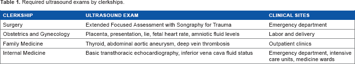
For Year 3 medical students, an ultrasound curriculum had been integrated into four core clerkships, namely, Family Medicine, Internal Medicine, Surgery, and Obstetrics and Gynecology. The format is similar in each clerkship. Formal didactics take form in short video lectures. Following didactics, students participate in bedside training sessions, which are taught in a peer-to-peer fashion on live normal models. Table 1 summarizes ultrasound examinations taught during the different clerkships. Abnormal scans are introduced to students with PowerPoint slides, showing images and videos of the pathology. Studies have shown that peer-to-peer ultrasound training can be very effective.9,10 Faculty are present for oversight and act as an expert reference. Several online resources, such as SonoSim ultrasound simulators and Blue Phantom simulation equipment, are available for optional self-directed learning.
Required ultrasound exams by clerkships.
The students have found that the emergency department (ED), intensive care unit (ICU), and obstetrics (OB) wards were exceptional clinical environments to learn bedside ultrasound due to availability of machines, frequent indications, and abundant attending and resident ultrasound expertise. They also frequently see the team make clinical decisions based on POCUS findings. Students find great joy confirming physical examination findings with ultrasound, as well as often being the first to make critical findings that greatly impact patient care.
However, ultrasound utilization may become difficult for students in the outpatient setting when the residents and attending physicians have little or no bedside ultrasound training. To address this issue, when working with attendings and residents who are not trained, students are instructed to only perform ultrasound examinations on patients who have had formal radiologic examinations to compare their findings with formal tests. This creates a self-directed learning environment, which enables the students to learn pathology with lessened risks of false findings.
Year 4 Student Experience
Unlike the first three years of medical training in which ultrasound occupies a place in the LLUSM medical curriculum, ultrasound learning in the fourth year is student dependent. Involvement in the USIG remains an option where fourth-year cabinet positions focus on curriculum development and integration of the third-year clerkships. Currently, the School of Medicine has a goal of aligning the goals of both faculty and students when formulating a curriculum. To accomplish this, each clerkship director is paired with at least one fourth-year cabinet member from the USIG who is interested in that specialty for residency. This student is responsible for partnering with the clerkship director to develop and improve upon a curriculum for that clerkship. They will also be responsible for coordinating the instruction of this material during each clerkship block. This benefits the clerkship by providing them with motivated students as a source of work and material, and benefits the students by giving them opportunities for personal growth, valuable experience for their residency application, and additionally close partnering relationships with the clerkship directors in their desired field.
For students who desire, there is also an excellent fourth-year ultrasound elective. Students choosing this option spend two weeks scanning patients in the high-volume ER and gaining experience in such areas as FAST, abdominal, and lower extremity scanning, while the second two weeks are spent in the ICU where cardiac and pulmonary ultrasound is paramount.
While the opportunities for formal ultrasound instruction may be fewer in the fourth year, this is a time where the students really can begin to reap the benefits of three years’ prior instruction. For example, ER and ICU are required senior rotations where the applications for ultrasound are numerous. In addition, students doing electives in such areas as obstetrics, internal medicine, family medicine, surgery, and many others have found ultrasound useful. Not only does ultrasound application in these settings solidify previous knowledge and skill, but it also enhances patient care and also tends to benefit students on evaluations as they are able to offer a knowledge base and know-how that might not have been previously realized by residents and faculty. In a similar way, prior ultrasound training has been noted to benefit some students who choose to do away or audition rotations, offering an area where they can shine over other fellow, yet competing, students from programs with less ultrasound exposure.
Conclusion
There are several keys to the success of ultrasound at LLUSM, a few of which are shared here in brief.
First and foremost are several championing faculty and administrators of the School of Medicine. The contributions of Dr. Vi Am Dinh cannot be overstated. Not only is he the founding faculty advisor of the student USIG, but he is also an inspirational visionary for the future of ultrasound in medicine and its importance in medical student training. Likewise, the LLUSM curriculum administrators deserve much recognition for their innovative willingness to accept ultrasound as part of its formal curriculum while collaborating with students in the formulation of the ultrasound curriculum.
Another secret to the success of POCUS training at LLUSM is the USIG's priority on recruitment. Through research and their own personal testimony, USIG leaders have been able to share with their fellow students the value of ultrasound and numerous possibilities for additional teaching, instruction, research, and leadership that the interest group offers. As a result, within its third year of existence, the interest group was the largest and the most active interest group on campus. This large population of student members has been central to the success of POCUS training on campus as it provides a source of Funding (via small member dues) for expansion, strong student support for policy change in the School of Medicine, and importantly a vast pool of new ideas with the manpower for their development and materialization. Regarding acquisition of resources, Funding for the ultrasound equipment was made by the School of Medicine and donations from Loma Linda Alumni. The use of ultrasound in the clinical setting by students is purchased and owned by the medical center. Currently, students do not have their own handheld devices.
Finally, the partnership between the USIG and the LLUSM administration has enabled the successful integration of all POCUS training programs available at LLU. Frequently, one of the pitfalls to the realization of new ideas in medical education is identifying and recruiting the energy and workforce to realize them. By uniting with the USIG, LLUSM has a surplus of both at its disposal. The USIG members also benefit from this arrangement as it creates invaluable research and leadership opportunities that few, if any, other interest groups or student organizations can offer.
Author Contributions
Conceived and designed the experiments: JYF, CK, RK, JM, AS, DSU, LAV, ZJ and VAD. Analyzed the data: JYF, CK, RK, JM, AS, DSU, LAV, ZJ and VAD. Wrote the first draft of the manuscript: JYF, CK, RK, JM, AS, DSU, L AV, ZJ and VAD. Contributed to the writing of the manuscript: JYF, CK, RK, JM, AS, DSU, LAV, ZJ and VAD. Agree with manuscript results and conclusions: JYF, CK, RK, JM, AS, DSU, LAV, ZJ and VAD. Jointly developed the structure and arguments for the paper: JYF, CK, RK, JM, AS, DSU, LAV, ZJ and VAD. Made critical revisions and approved final version: JYF, CK, RK, JM, AS, DSU, LAV, ZJ and VAD. All authors reviewed and approved of the final manuscript.