
Abstract
BACKGROUND:
Family with sequence similarity 83 member A (FAM83A) can promote tumor cell proliferation and facilitate epidermal growth factor tyrosine kinase inhibitor resistance in some malignant tumors, but its role in lung cancer has not been directly explored.
OBJECTIVE:
We investigated FAM83A expression in lung adenocarcinoma (LUAD) and its significance in clinicopathologic characteristics and prognosis of the disease.
PATIENTS AND METHODS:
We analyzed the mRNA expression of FAM83A in LUAD and normal (or adjacent) lung tissues from Oncomine database firstly. Then, we detected FAM83A protein expression in five paired fresh LUAD and adjacent lung tissue specimens from patients in our hospital by Western blotting. In addtion, FAM83A expression in 86 paraffin-embedded archived LUAD samples was evaluated by Immunohistochemistry, and the correlations between FAM83A expression and clinicopathologic characteristics and prognosis of the patients were analyzed.
RESULTS:
Oncomine data analysis manifested that FAM83A mRNA expression was increased in LUAD. Western blotting revealed higher FAM83A expression in fresh LUAD tissues than in the adjacent lung tissues (
CONCLUSION:
FAM83A is upregulated in advanced LUAD and is related to unfavorible prognosis. FAM83A might be a novel diagnostic and prognositic biomarker for LUAD.
Introduction
Lung cancer ranks the first in morbidity of malignant tumors and yields a high mortality worldwide [1]. In recent years, progress in identification of tumor driver genes have led to the development of many novel targeted therapies that can provide significant benefit to some patients, especially those with lung adenocarcinoma (LUAD). For example, epidermal growth factor-tyrosine kinase inhibitors (EGFR-TKIs) are highly effective in treating LUAD with mutated EGFRs [2, 3, 4]. These targeted drugs considerably extended the survival of some patients. However, upon the exposure to these targeted therapeutic drugs, cancer cells often develop resistance to these drugs through acquisition of mutations or other mechanisms in the targeted signaling pathways rendering the drugs ineffective. Therefore, clinical effects of these targeted drugs are not always satisfactory. Furthermore, most targeted drugs also inhibit normal cells and the dose-dependent toxicities limit their clinical application. Obviously, further strives in seeking novel and better therapeutic targets on LUAD remain urgently necessary.
Family with sequence similarity 83 member A (FAM83A) is one of the members of a novel protein family which consists of FAM83A-H. All FAM83 members contain a highly conserved 1699 (DUF1669) domain of unknown function located in the N terminus, while each FAM83 member has a distinct variable C terminus [5, 6, 7]. Early study demonstrated that breast cancer with EGFR mutations has poor response to EGFR-TKIs [8]. Lee and his colleagues first discovered that FAM83A was overexpressed and involved in the EGFR-TKIs resistance in breast cancer [5]. In the same study, the authors found that FAM83A was able to activate the signaling cascade downstream of EGFR, facilitate breast cell transformation, and confer EGFR-TKIs resistance. Subsequently, Liu and his colleagues, using a rapid nested PCR assay, detected the heightened levels of FAM83A in peripheral blood samples of breast cancer patients [9]. A recent study showed that HER2-positive breast cancer cells had overexpressed FAM83A, and the ablation of FAM83A inhibited HER2-positive breast cancer cell growth independently of trastuzumab sensitivity [10]. In addition, increased FAM83A was also found to be related to poor prognosis of the patients [11]. All these studies collectively suggested that FAM83A could be a potential biomarker for breast cancer prognosis.
The expression of FAM83A has also been found to be overexpressed in some other tumor types including brain tumors, pancreatic cancer, and ovarian cancer [11]. However, the significance of FAM83A in lung cancer has not yet been fully investigated. It has been reported that FAM83A mRNA was overexpressed in human lung cancer tissues and circulating tumor cells [12, 13]. Cipriano et al. further found that FAM83A mRNA was upregulated in lung cancer using an assay that detects mRNA of all FAM83 members (FAM83A, B, C, D, E, F, G, H) in several types of tumor tissues [14]. In addition, bioinformatic analysis of publically available data in GEO database also found FAM83A mRNA was overexpressed in lung cancer [11]. However, most of those studies explored FAM83A expressions merely at mRNA levels, not at protein level. It is well known that the level of mRNA expression does not necessarily correlate with the level of protein expression. Furthermore, the clinical significance of FAM83A overexpression in lung cancer is largely unknown. In this study, we focused on the expression of FAM83A protein in LUAD and its correlation with the clinicopathological characteristics of the patients.
Materials and methods
Bioinformatic analysis
We analyzed the FAM83A expression (mRNA) levels of LUAD tissues (44 cases) and normal lung tissues (65 cases) in Oncomine database. To exclude the impacts of individual differences, we compared the mRNA levels of FAM83A in 20 paired LUAD tissue and the matched adjacent lung tissue samples from the dataset.
Patient information and tissue samples
The patient enrollment criteria were: 1) Patient at stage I-III according to the TNM staging criteria established by American Joint Committee on Cancer (AJCC) [15]. 2) Patient who had complete clinical and pathological information including age, sex, pathological grading, pathological differentiation, TNM stage, and clinical stage etc. 3) Patient without distant metastasis. A total of 86 paraffin-embedded archived LUAD and adjacent non-tumor tissue samples from the patients in our hospital who met the inclusion criteria were used in this study. The clinicopathological characteristics of the patients were showed in Table 1. All the patients at stage II–IV accepted standard postoperative adjuvant chemotherapy. Five pairs of fresh LUAD samples and matched adjacent lung tissues were compared for their FAM83A protein expression using Western blotting. The protocol of this study was approved by the Institutional Research Ethics Committee of Anhui Provincial Hospital (No. 2019-P-017).
Western blotting
FAM83A protein expression was determined by Western blotting. Fragments of fresh tissues were ground in RIPA lysis buffer (P0013B, Beyotime, Shan-ghai, China) and protein concentration was measured with BCA protein concentration assay (P0010, Beyotime, Shanghai, China). Samples with equal amount of protein (20
Immunohistochemistry
We detected the protein expression of FAM83A in a tissue microarray by immunohistochemistry method. Immunohistochemistry analysis was performed on the paraffin-embedded LUAD specimens and adjacent lung tissue ones. Briefly, the tissue slices were incubated in dry oven at 63
The immunohistochemistry scoring of FAM83A expression was graded as previously described [16]. The score of staining intensity (S1) was evaluated as follows: 1) no staining; 2) weak staining, showing light yellow; 3) moderate staining, showing yellow brown; 4) strong staining, showing brown. The score of staining cell proportion (S2) was graded as follows: 0) no staining cells; 1)
Statistical analysis
SPSS version 13.0 (SPSS Inc., Chicago, IL, USA) was used for statistical analysis. The correlation between the clinicopathological characteristics of the patients and FAM83A expressions was analyzed using the Chi-squared test. Prognostic factor analyses were performed using univariate and multivariate Cox regression analyses. Survival analyses were performed using Kaplan-Meier and Log-rank test. A two-tailed
Results
FAM83A was overexpressed in LUAD
Using Oncomine data, we compared the FAM83A mRNA expression in 44 LUAD samples and 65 normal lung tissues and found that FAM83A mRNA expression was elevated in LUAD (
FAM83A is overexpressed in LUAD. A. FAM83A expression level (mRNA) was transcriptionally upregulated in 44 LUAD tissues in comparison with that of 37 normal lung tissue in the Oncomine database. B. FAM83A mRNA expression in LUAD tissue samples was higher than that of matched adjacent normal tissues in the Oncomine database. C. The relative FAM83A expression of LUAD was higher than that of pared adjacent normal tissues in Western blotting assay. D. Western blotting of FAM83A expression in five paired LUAD (T) and adjacent normal tissues (ANT), GAPDH was used as the loading control.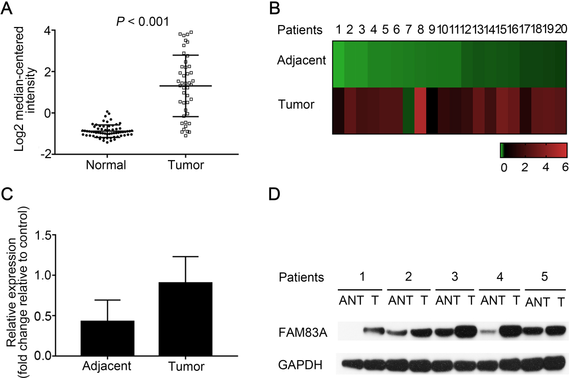
FAM83A overexpression in LUAD correlates with poor overall survival of the patients. A. The tissue microarray including adjacent lung and LUAD tissues, representative IHC images of FAM83A expression in adjacent normal tissues (ANT) and LUAD with low (LOW) and high (HIGH) FAM83A expression. The scale equals to 50 micron. B. The IHC staining index of FAM83A in LUAD tissues (Tumor) and adjacent normal lung tissues (Adjacent). C. The IHC staining index of FAM83A in LUAD tissues with Stage I-II and Stage III-IV. D. Kaplan-Meier survival curves of 86 LUAD patients grouped by low and high expression of FAM83A.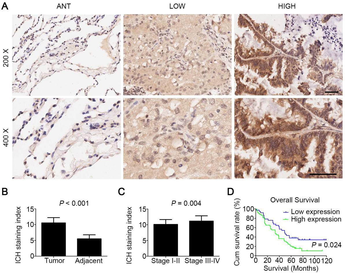
Clinicopathological characteristics and FAM83A expression of the patients
Correlation between clinicopathological characteristics and FAM83A expressions
Univariate and multivariate cox-regression analysis of diverse prognostic factors in patients with lung adenocarcinoma
The clinicopathological features of the patients used in this study were summarized in Table 1. We then examined FAM83A protein in LUAD tissue sections by immunohistochemistry staining. The results showed that FAM83A was mainly located in cytoplasm (Fig. 2A). The staining index of immunohistochemistry for FAM83A was significantly higher in LUAD tissues than that in the adjacent normal tissues (
FAM83A overexpression predicts poorer survival and prognosis
Univariate Cox regression analysis showed that high FAM83A expression increased the risk of death in comparison to low FAM83A expression (Hazard Ratio: 1.765, 95% Confidence Interval: 1.065–2.926,
Discussion
FAM83A is a novel signaling protein that found to be overexpressed in some tumors and confer chemo-resistance to breast cancer [11]. Previous study indicated FAM83A expression was elevated in lung cancer at mRNA levels. In this study, we further demonstrated that FAM83A was also overexpressed in LUAD at protein in addition to mRNA levels, and the heightened FAM83A expression correlated with advanced clinicopathological features and poor prognosis.
FAM83A overexpression was firstly discovered in breast cancer. It may promote proliferation, invasion, and EGFR-TKI resistance of breast cancer cell [5]. FAM83A overexpression was also found later in some other tumors including pancreatic cancer, and overexpressed FAM83A may promote cancer stem cells-like traits and chemo-resistance [16]. However, there have been only a few studies on FAM83A expression in lung cancer. In present study, by analyzing the data from Oncomine database, we confirmed that FAM83A was overexpressed in LUAD at mRNA levels. To exclude the impact of individual differences, we compared twenty paired data and confirmed the earlier findings by others [12, 14].
It is well known that the expression of mRNA and protein are not always in agreement [17], meaning an elevated mRNA expression is not necessarily accompany with an increased protein expression. To ascertain the expression of FAM83A protein in LUAD, we detected FAM83A levels in fresh LUAD tissue samples using Western blotting. Our results demonstrated that FAM83A protein was indeed overexpressed in LUAD as suspected. Therefore, we are the first to report FAM83A protein is overexpressed in LUAD in addition to mRNA overexpression.
Previous studies have demonstrated that FAM83A may be important in tumor cell proliferation, invasion, metastasis and drug resistance [5, 18]. It has been suggested that FAM83A achieves this by serving as the downstream of EGFR, and significantly activate the downstream signaling pathway, such as PI3K/AKT and RAS/MAPK [18]. In the study of breast cancer, FAM83A expression was found to be invertly associate with patient’s survival. Given that EGFR signaling cascade is essential to the proliferation and survival of LUAD [19, 20], it is conceivable to hypothesize that FAM83A may also play an important role in LUAD and affect the clinical course of the disease as demonstrated in breast cancer. In this study, the results of immunohistochemistry analysis performed on the paraffin-embedded LUAD specimens and adjacent lung tissues showed FAM83A is indeed overexpressed in LUAD tissues. Furthermore, patients with higher FAM83A expression were associated with early lymph node metastasis and advanced tumor stages, poorer survivalship as presented by M-H survival analysis. Log-rank test showed a significant difference between patients with high and low FAM83A expressions (
In conclusion, our findings demonstrated aberrant overexpression of FAM83A in LUAD and it predicts more advanced clinicopathological features and poor prognosis. The results of our study suggested a possible oncogenic role of FAM83A in LUAD. It may also potentially serve as a biomarker for LUAD, which requires additional investigation and testing.
Footnotes
Acknowledgments
We sincerely thank Dr. Wu Xiaosheng (Mayo Clinic, Minnesota, US) for his help in language editing this research paper.