
Abstract
BACKGROUND:
Hepatocellular carcinoma (HCC) is a common cancer and exhibits high morbidity and mortality in the world. We recently identified LHX3 as a preferentially expressed gene with a possible involvement in HCC.
OBJECTIVE:
To determine the expression, clinical relevance, prognostic significance and functions of LHX3 in HCC.
MATERIALS AND METHODS:
LHX3 expression was assessed in 190 cancerous and 40 adjacent non-cancerous tissues by PCR, western blot and immunohistochemistry. Associations between LHX3 expression and clinicopathological characteristics of patients were investigated. Correlations between LHX3 expression and overall survival of patients were analyzed by Kaplan-Meier and Cox-regression methods. Functional roles of LHX3 were evaluated by transwell assays.
RESULTS:
LHX3 expression is significantly increased in carcinoma tissues, and associated with clinical stage and metastasis of patients. LHX3 expression is much higher in the advanced-stage patients than the early-stage patients, and is sharply increased in metastasic patients. High LHX3 expression is associated with unfavorable overall survival, and is an independent prognostic factor of patients. Moreover, LHX3 is an unfavorable and independent prognostic factor unique to advanced-stage patients. Knockdown expression of LHX3 obviously inhibits tumor cell migration and invasion.
CONCLUSION:
LHX3 is an advanced-stage prognostic biomarker, and acts as a new potential metastatic oncogene in HCC.
Introduction
Hepatocellular carcinoma (HCC) is one of the most common cancers with high morbidity and mortality worldwide [1, 2, 3]. Although the most effective treatment is surgery for HCC patients at early stage, the patients at advanced HCC is un-resectable. Moreover, there is still deficiency of effective methods for patients diagnosed at the advanced stages [4]. In general, despite advances in treatment strategies, the rate of 5-year survival remains very poor due to limit treatments and lack of effective prognostic markers [5, 6]. Therefore, identification of new targets for treatment and practical prognostic markers for prognostic evaluation are very important in HCC.
LIM homeobox (LHX) genes are important LIM-homeodomain factors. Previous studies have revealed that LHX members play key roles in development of various tissues [7, 8, 9, 10, 11, 12]. Researches have identified a few LHX genes involved in various type cancers, such as Lhx2, Lhx4 and Lhx1 [13, 14, 15, 16, 17, 18]. LHX3, as an important LHX member, is critical in the development of spinal cord motor neurons and pituitary [19, 20, 21, 22]. Recently, a study has revealed a clear correlation between LHX3 methylation and breast cancer [23]. However, the clinical, prognostic significance and potential role of LHX3 in cancer, especially in HCC, remain unknown.
In our present study, we identified LHX3 as a new differentially expression gene in tumor and normal liver tissues. The clinical, prognostic significance and potential roles on metastasis of LHX3 were explored in human HCC. Our study indicates that LHX3 is an important advanced-stage prognostic factor and functions as a potential metastatic promoter in HCC.
Materials and methods
Patient samples and cell lines
Total 190 HCC patients undergone surgical resection between 2004 and 2010 were obtained from the Southwest Hospital in Chongqing, China. The clinico-pathologic information includes gender, age, tumor size, histological grade, lymph node status, clinical stage, radiotherapy, tumor location and overall survival (OS). Patients were followed up for a maximum period of 80 months, with a median of 32.9 months. OS was defined as the period from surgery to death or the last observation. The clinico-pathologic information also includes AFP level and the expressions of Ki67, P53, C-ERBB-2, CK19, HEP1, CK8 and CD34. The level or expressions of these markers were measured by the Hospital. When the tumor cell staining of cancer samples was completely no positive signal, it was identified as negative. The HCC cell lines (Huh7 and 7721) were obtained from the Cell Bank of the Chinese Academy of Science (Shanghai, China), and cultured in RPMI-1640 (HyClone, Logan, USA) supplemented with 10% fetal bovine serum (HyClone). This study was approved by the ethics committee of Southwest Hospital Affiliated to the Third Military Medical University. All experiments were carried out in accordance with approved guidelines of Third Military Medical University. Informed consent was signed by all the patients.
Total RNA isolation
Total RNA was extracted from frozen HCC or normal liver tissues. About 3.5
RT-PCR
Series of PCR with different cycles were performed and the appropriate cycles were chosen. Human
RT-qPCR
RT-qPCR was performed using GoTaq
Tissue microarray
To construct the tissue microarray (TMA), two cores were taken from cancerous and matched adjacent noncancerous liver tissues within a distance of 35 mm. The adjacent noncancerous tissues were stained with hematoxylin & eosin and determined by comparing with normal tissues. The core size of TMA is 1.5 mm. The TMA was constructed as previously described [24].
LHX3 expression is sharply increased in HCC. (A) RT-PCR and qRT-PCR analysis of LHX3 mRNA expression in normal and cancerous tissues. N represents normal tissue; M represents marker; T represent cancerous tissue. The 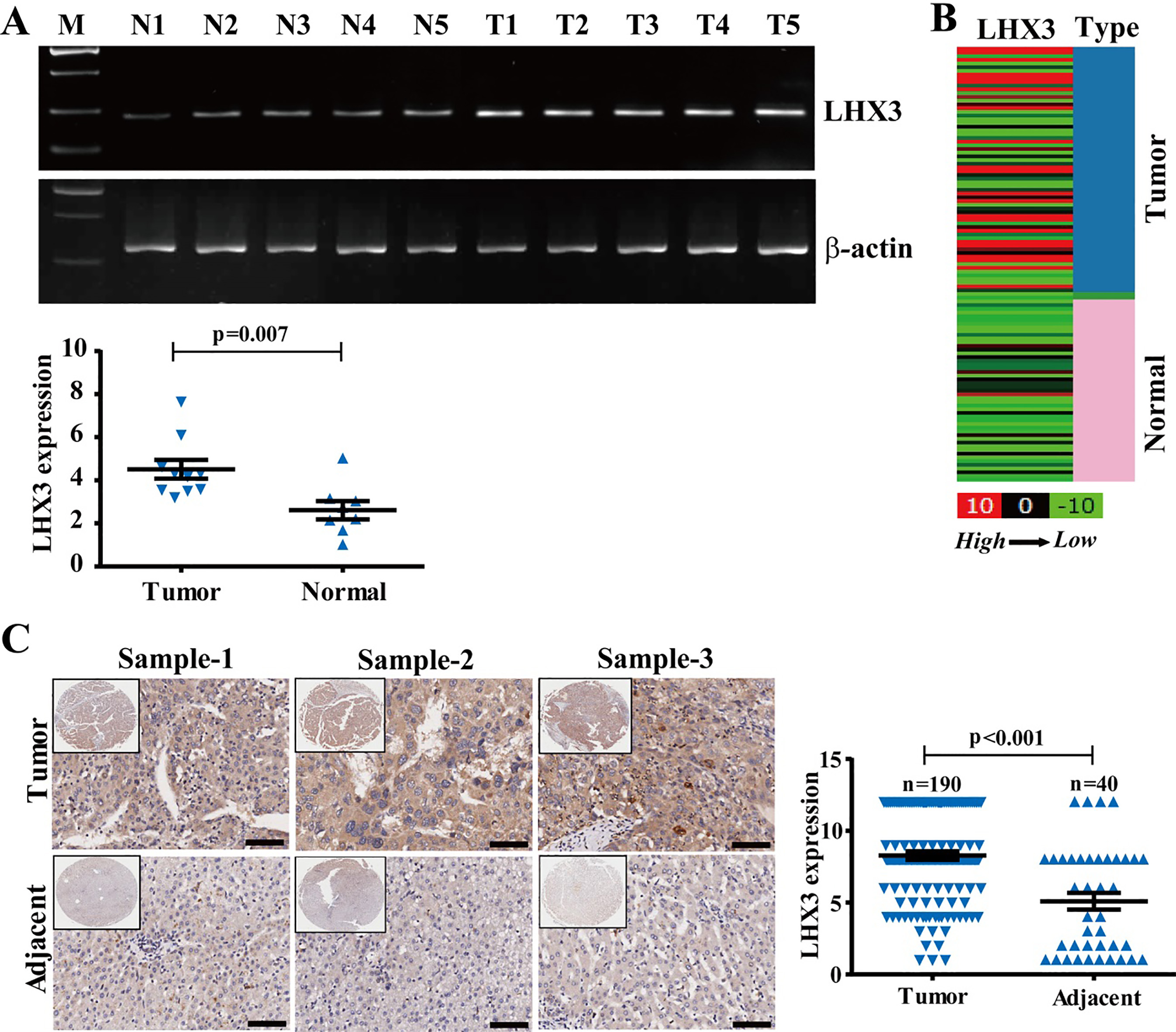
IHC was used to determine the expression of LHX3 in HCC tissues with LHX3 antibody (1:150, Abcam) as previously described [25]. The staining percentage of tumor cells was quantified into 5 grades:
LHX3 siRNA
The LHX3 siRNA used in the present study was purchased from Santa Cruz Biotechnology (sc-38712).
Western blotting analysis
Western blotting (WB) was performed as previous study [28]. Briefly, the protein was run on 10% SDS-PAGE, transferred onto PVDF membrane (Millipore, USA), and incubated with LHX3 antibody (1:1500, Abcam) or
Migration and invasion assays
Transwell assays were performed to determine migration or invasion activities of cancer cells in 24-well plates without or with matrigel (8
The expression of LHX3 is increased in the HCC patients at advanced stage or with metastasis. (A) LHX3 protein expression is shown in the patients at different clinical stages. Scale bars represent 50 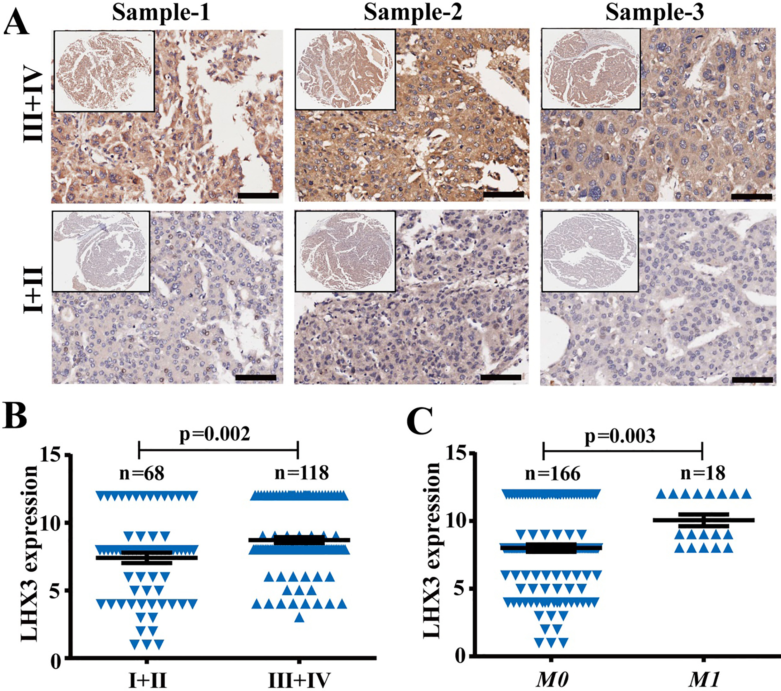
To get the expression of LHX3 in cancer and normal samples in an independent cohort, we used the UCSC Cancer Genomics Browser (
Statistical analysis
SPSS 18.0 software (SPSS, Inc., Chicago, IL, USA) was used for statistical analyses. The difference in categorical variables was analyzed by Chi-square or Linear-by-Linear test. Kaplan-Meier method and log-rank test were used to evaluate the OS. Cox regression was performed for multivariate analysis of prognostic predictors. The high or low expression was categorized by the median.
Results
Increased LHX3 expression is observed in HCC tissues
To further investigate the expression pattern of LHX3 in HCC, LHX3 expression level was checked in normal and tumor tissues by RT-PCR and RT-qPCR. LHX3 expression was increased in tumor tissues compared to normal liver tissues (Fig. 1A). The mean level of LHX3 expression was 4.511
LHX3 expression is associated with clinical stage and metastasis of HCC patients
Based on quantified expression, LHX3 expression was classified into high (greater than and equal to the mean expression,
Correlations of LHX3 expression with clinicopathologic features in human HCC patients (
190)
Correlations of LHX3 expression with clinicopathologic features in human HCC patients (
High LHX3 expression is associated with poor OS of HCC patients. (A) High and low expression level of LHX3 as seen in representative HCC tissues. Low represents staining weak (the expression score 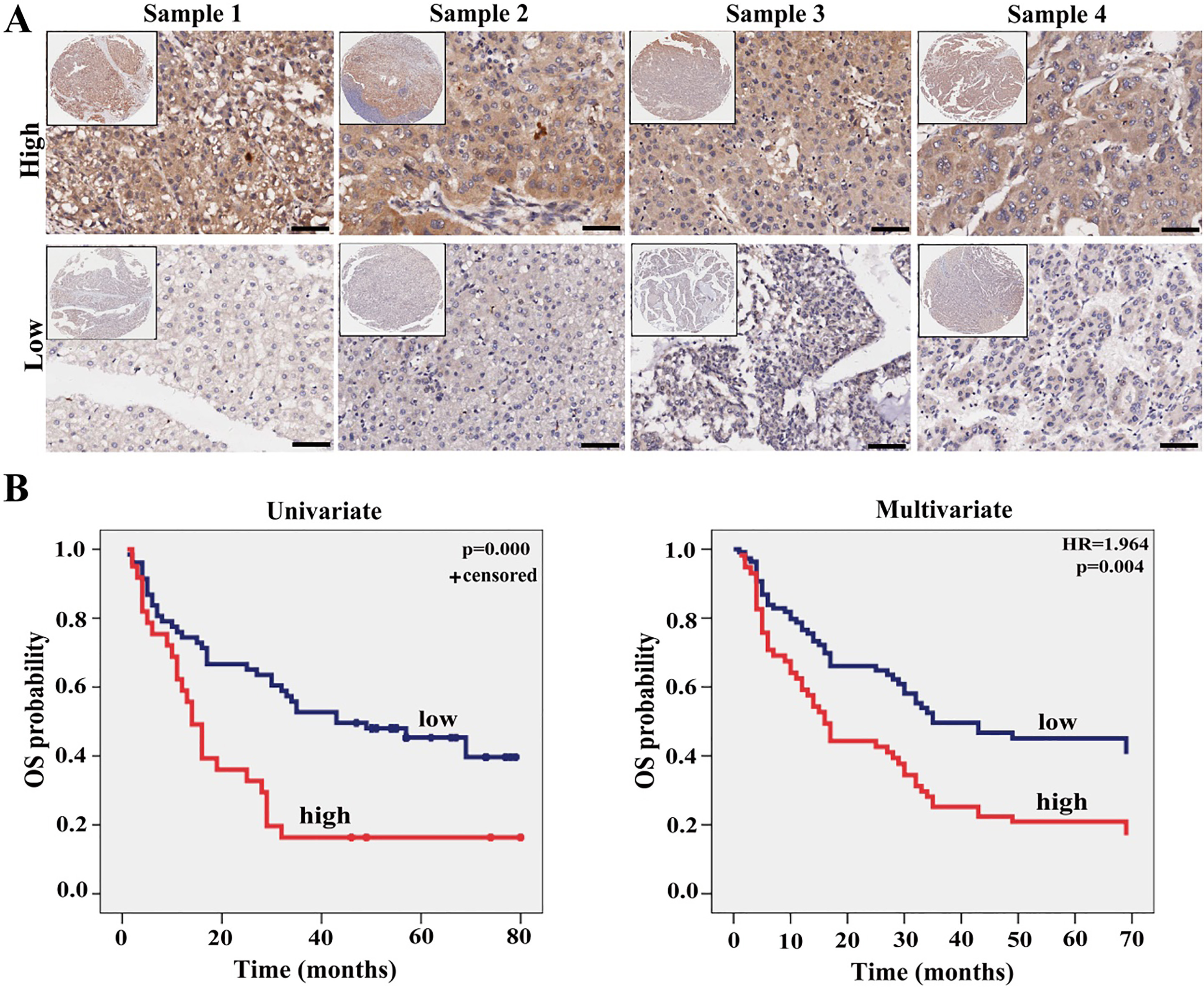
We then analyzed the correlations of LHX3 expression with other maker expression or virus infection in HCC patients. LHX3 expression is clearly associated with hepatitis C virus (HCV,
Correlations of LHX3 expression with other maker expression in human patients (
144)
Correlations of LHX3 expression with other maker expression in human patients (
LHX3 expression was then analyzed with respect to survival data of the HCC patients. Kaplan-Meier analysis revealed a poor OS in the patients with high LHX3 expression compared to the patients with low LHX3 expression (
Multivariate analysis of different prognostic factors in human HCC patients (
190)
Multivariate analysis of different prognostic factors in human HCC patients (
Abbreviations: HR, hazard ratio; CI, confidence interval.
The correlations between LHX3 expression and survival of the patients at early stages (I
LHX3 is an advanced stage prognostic factor of the HCC patients. Kaplan-Meier and Cox regression survival analysis of LHX3 expression in 68 early stage (stage I 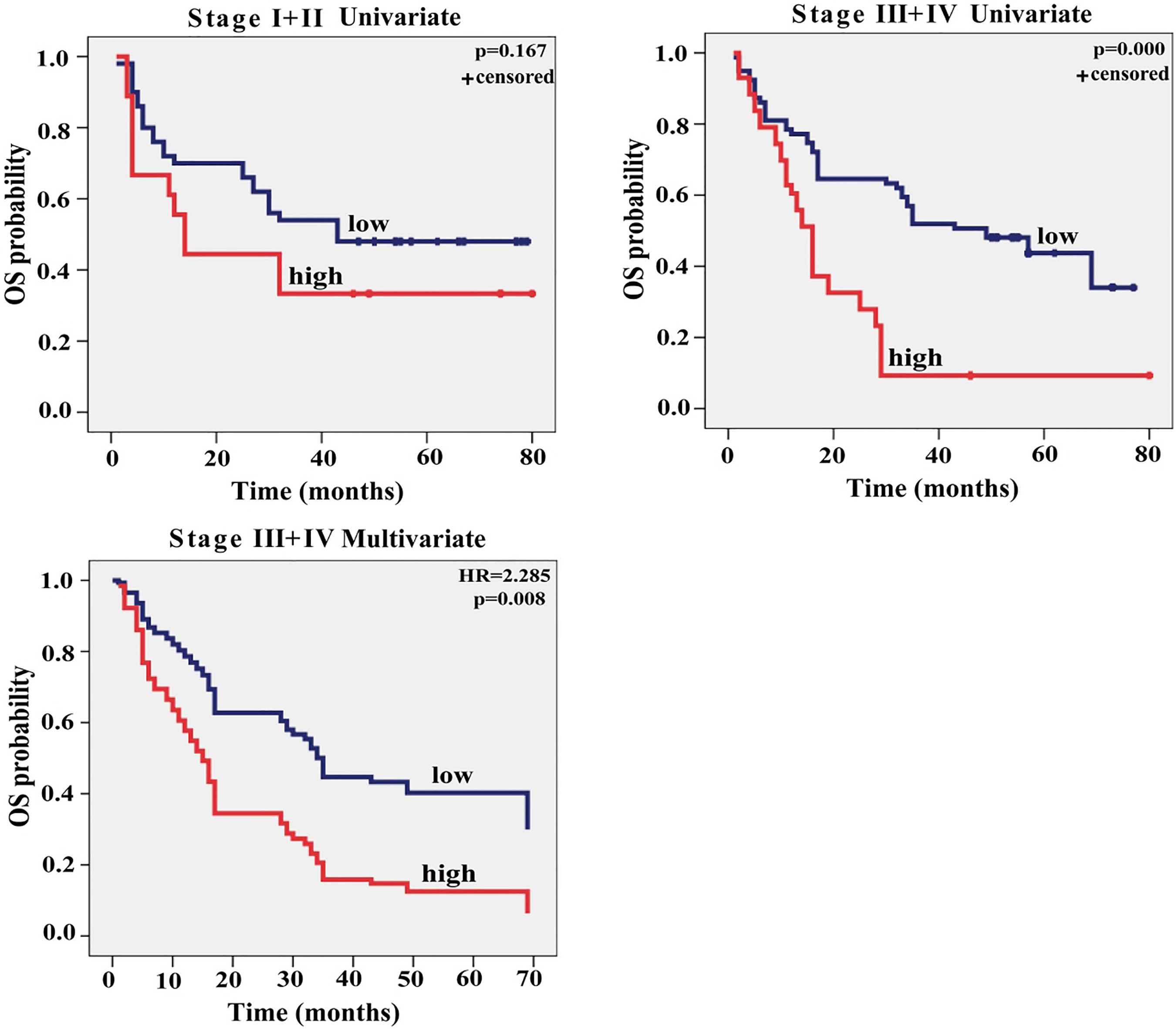
LHX3 expression is associated with clinical stages and metastasis of HCC patients, indicating a role in tumor metastasis. To investigate the effects of LHX3 on HCC cell migration and invasion, we transfected LHX3 siRNA into HCC Huh7 cells with high LHX3 expression and performed the transwell assays with or without matrigel (Fig. 5A and B). The data showed that a significant decrease of cancer cell migration and invasion was found in LHX3 siRNA cells compared with negative cells (Fig. 5C). These data indicate that LHX3 strongly stimulates cancer cell migration and invasion.
Multivariate analysis of different prognostic factors in advanced HCC patients (
118)
Multivariate analysis of different prognostic factors in advanced HCC patients (
Abbreviations: HR, hazard ratio; CI, confidence interval.
LHX3 knockdown inhibits cancer cell migration and invasion. (A) RT-PCR analysis of LHX3 mRNA expression in HCC cell lines and tissues. N represents normal tissue; T represents cancerous tissue. The 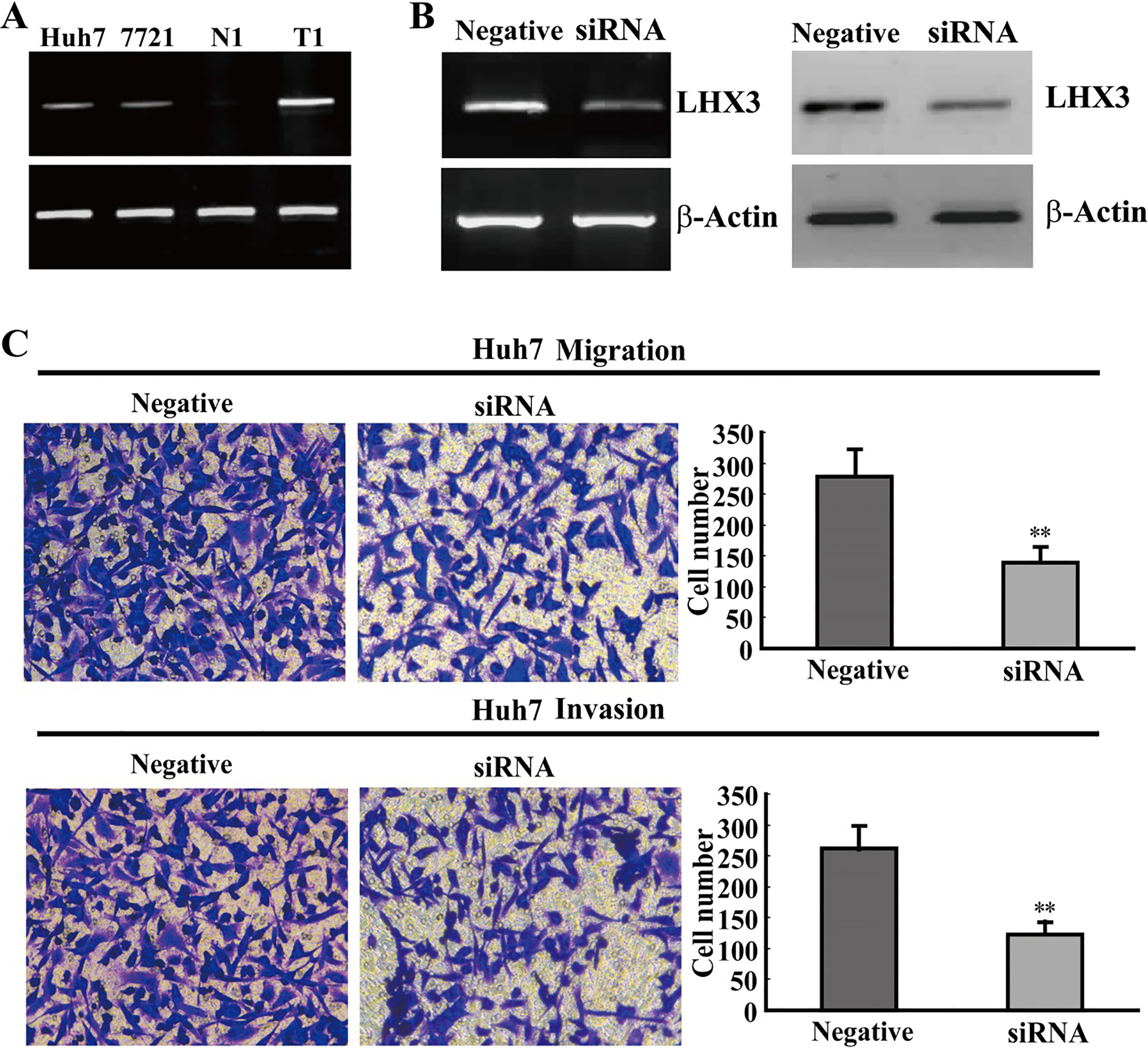
In our present study, the clinical relevance and functional roles of LHX3 for the first time were investigated in HCC. LHX3 expression is generally increased in HCC tissues, and is associated with clinical stages and metastasis. LHX3 expression is an unfavorable and independent prognostic factor of patients in HCC. Moreover, LHX3 expression is an advanced-stage prognostic factor of HCC patients. Functionally, LHX3 strongly promotes HCC cell migration and invasion.
Previous studies have demonstrated that LHX genes are involved in tumorigenesis of different cancers [13, 14, 15, 16, 17, 18]. However, little is known about their clinical and prognostic relevance in these cancers. In this study, LHX3 expression is found to be closely associated with clinical stages and metastasis of HCC patients. To evaluate the prognostic significance of LHX3 expression on HCC patients, Survival analyses were performed by Kaplan-Meier and Cox-regression. The patients with low LHX3 expression have correspondingly prolonged OS compared to the patients with high LHX3 expression. Furthermore, LHX3 expression is an independent prognostic factor for OS in HCC patients. Noticeably, LHX3 is an advanced-stage prognostic factor in HCC patients. These data demonstrates that LHX3 expression is an important prognostic biomarker in HCC patients at advanced stages. It is worth noting that the number of cases in some tables is not consistent in the text, which is due to missing the clinico-pathologic features of some patients. In addition, this study here mainly focuses on the clinical and prognostic relevance of LHX3. So, we included the clinico-pathologic features, and excluded the factors of AFP level and the expressions of Ki67, P53, C-ERBB-2, CK19, HEP1, CK8, and CD34 in multivariate analysis.
Previous evidences have revealed that LHX3 plays key roles in the development of spinal cord motor neurons and pituitary [19, 20, 21, 22]. Recently, it is reported that the DNA methylation of LHX3 is closely related to breast cancer [23]. However, there is few functional study of LHX3 on tumorigenesis. In the present study, LHX3 expression is significantly elevated in cancer tissues. LHX3 expression is much higher in the advanced-stage patients than in the early-stage patients. Moreover, LHX3 expression is sharply increased in the patients with metastasis. These data suggest that LHX3 may be involved in HCC metastasis. So, gain/loss-of-function assays were investigated to determine the roles of LHX3 on tumor metastasis. LHX3 indeed significantly promotes cancer cell migration and invasion, revealing that LHX3 functions as a metastatic promoter in HCC. However, the effects of LHX3 on cancer cell proliferation, apoptosis, and cycle still remain unclear, and further studies are needed to solve this problem in HCC, as well as confirm its function in vivo.
It seems that LHX3 is expressed in the non-neoplastic stromal cells between HCC cells of stage III
In conclusion, LHX3 expression is a potential advanced-stage biomarker for prognosis evaluation of patient, and LHX3 acts as a candidate metastatic promoter in HCC. However, further studies are needed be performed to explore the molecular mechanism of LHX3 in HCC.
Footnotes
Acknowledgments
This work was supported by the Natural Science Foundation of Chongqing (cstc2018jcyjAX0473), and the Natural Science Foundation of Chongqing (cstc 2015jcyjBX0060). The authors thank all the patients in this study.
Conflict of interest
None declared.