
Abstract
Heat-induced antigen retrieval (HIAR) is routinely employed on aldehyde-fixed tissue sections to enhance the reactivity of antibodies that exhibit weak or no specific interactions with tissue antigens when applied in conventional immunohistochemical protocols. A major drawback of HIAR protocols is, however, the heat-induced detachment of sections from the microscope slide with resultant impaired tissue morphology or loss of the section. We developed a method in which tissue sections mounted on glass slides are temporally coverslipped, and a clamp is used to compress the sections on the microscope slide during HIAR treatment. This “pressurized coverslipping” during HIAR was tested on various formalin-fixed tissues (murine kidneys and temporal bones, human tonsils and temporal bones) that were embedded in paraffin or celloidin. The method reliably kept the sections adherent to the slide, preserved the tissue morphology, and effectively retrieved tissue antigens for improved results in immunohistochemical labeling, even for exceptionally delicate, large, and poorly adhering sections, that is, decalcified human temporal bone sections. In summary, we present a simple method for improved slide adherence and morphological preservation of tissue sections during HIAR treatment that can be combined with all HIAR protocols and that requires only basic lab equipment.
Introduction
Heat-induced antigen retrieval (HIAR) as an enhancement method for immunohistochemical staining1,2 is routinely employed for formalin-fixed, paraffin-embedded (FFPE) tissue sections. This method enables the use of antibodies which otherwise fail to react with FFPE tissue sections, allows the use of antibodies at more dilute concentrations, and can obtain higher signal-to-noise ratios in immunolabeling experiments. 2 However, HIAR techniques all have a common major drawback, which is the risk of heat-induced detachment of mounted tissue sections from the surface of the glass slide. Partial section detachment impairs the tissue morphology and results in artifactual immunolabeling gradients, whereas complete detachment leads to the loss of the section and precludes further processing. Tissue sections are especially prone to detachment during HIAR treatment when particularly high temperatures and long heat exposure times are applied, when sections from tissues with low adhesive properties—such as skin, cartilage, or (decalcified) bone—are exposed to heat, or when the tissue exhibits a delicate morphology, for example, tissues that harbor extensive hollow spaces.3,4 In general, the factors that determine whether sections tend to detach during HIAR treatment are manifold and include the conditions of tissue processing, such as fixation, mounting, sectioning, and storage of the sections.
Previous approaches have attempted to overcome the problem of detaching tissue sections during HIAR treatment by using so-called “gentle” HIAR protocols5,6 in which lower temperatures (approximately 70C), but considerably extended heating times (several hours), are applied. Standard HIAR protocols apply temperatures of approximately 90C to 100C and heating times <30 min, 1 with the aim to avoid the high temperature peaks that are a major cause of section detachment. Other more popular approaches use slide coatings with charged compounds to improve the adhesive properties of the glass slide on which the tissue sections are mounted for HIAR treatment. 6 However, all of these methodological refinements have limitations. The “gentle” HIAR protocols are ineffective for the retrieval of certain tissue antigens that require exposure to a critically high temperature, and adhesive slide coatings become degraded and lose their binding properties when HIAR treatment exceeds certain temperatures and/or exposure times.
Here, we present a simple method that effectively prevents HIAR-induced section detachment by using a pressurized coverslipping technique that exerts mechanical force and presses the section onto the coated slide surface during heat application. We tested this method on different formalin-fixed tissues that were embedded in paraffin or celloidin, including exceptionally delicate and fragile sections of decalcified human temporal bones that are not accessible with conventional HIAR protocols. After HIAR treatment, we performed immunohistochemical staining with various antibodies that were nonreactive with the tissue sections when HIAR treatment was omitted. We compared the morphological preservation of the tissue and the intensity and the quality of the immunostaining on sections that were either exposed to (1) no HIAR treatment, (2) conventional HIAR treatment, or (3) HIAR treatment with pressurized coverslipping of the tissue sections. Our results show that the pressurized coverslipping technique dependably prevents section detachment during HIAR treatment, preserves the tissue morphology, including that of exceptionally delicate tissues (decalcified human temporal bone), effectively retrieves tissue antigens and produces improved immunolabeling results, can be combined with almost all HIAR protocols, does not add further substances/chemicals to the protocol, and requires only basic lab equipment.
Materials and Methods
Ethics
This study was performed in accordance with the Policy on Human Care and Use of Laboratory Animals of the Public Health Service, the National Institutes of Health (NIH) Guide for the Care and Use of Laboratory Animals, and the Animal Welfare Act (7 U.S.C. et seq.). The animal use protocol was approved by the Institutional Animal Care and Use Committee of the Massachusetts Eye and Ear Infirmary.
Human temporal bones and tonsil tissues were extracted at autopsy, as approved by the Human Research Protections Program of the Massachusetts Eye and Ear Infirmary.
Animal Tissue Processing
After induction of deep anesthesia with sodium pentobarbital (100 mg/kg, i.p.), 6- to 8-week-old CBA/CaJ mice underwent intracardiac perfusion with phosphate-buffered saline (PBS), followed by perfusion with 10% buffered formalin. The entire heads and 0.5 mm slices of the kidneys were postfixed overnight in 10% buffered formalin. The skulls were then cut open in the horizontal plane, the brains (except of the lateral parts of the cerebellum) were removed, and adherent soft tissues were cleaned off using a scalpel. Decalcification was performed in 120 mM ethylenediaminetetraacetic acid (EDTA) for 7 days. All tissue specimens were then processed for embedding in paraffin according to standard methods. In brief, the tissues were dehydrated in a graded series of ethyl alcohols, cleared in xylene, and embedded in paraffin (Paraplast X-TRA; McCormick Scientific, St. Louis, MO). Paraffin sections were cut at 10 μm thickness using a rotary microtome (Reichert 2030 Microtome, Bensheim, Germany) and mounted on precoated glass slides (Superfrost Plus, Thermo Fisher, Pittsburgh, PA). The slides were placed on a slide warmer at 50C overnight and stored at room temperature. For further use, the sections were deparaffinized in xylene and rehydrated in a graded series of ethyl alcohols and distilled water (dH2O). For one experimental series, unmounted FFPE murine kidney sections were deparaffinized and rehydrated as described above. Those sections were then placed on glass slides (Superfrost Plus) and HIAR with pressurized coverslipping was performed as described in the respective paragraph below.
Human Tissue Processing
The human temporal bones and tonsil tissues were removed after death and fixed in 10% buffered formalin. Temporal bones were decalcified in EDTA. All tissue samples were embedded in celloidin (parlodion strips; Mallinckrodt Chemicals, Phillipsburg, NJ) according to previously described techniques. 7 All celloidin-embedded (CE) specimens were serially sectioned at a thickness of 20 μm (temporal bones were sectioned in the horizontal plane) and stored in 80% ethyl alcohol at room temperature.
The CE tissue sections were mounted on albumin-subbed glass slides, and the celloidin was removed according to previously described methods. 8 In brief, glass slides were smeared with albumin, and the sections were fixed in place with 10% buffered formalin-soaked bibulous paper. A wooden block was placed on top of the bibulous paper, and a 500-g weight was placed on the block. The sections were dried for 1 hr in a flat horizontal position under the weights. Celloidin was removed using washes in freshly prepared methanol saturated with sodium hydroxide and diluted 1:2 with methanol. Finally, the sections were rehydrated in 70% methanol and dH2O.
Pressurized Coverslipping of Mounted Tissue Sections
Before HIAR treatment, the mounted tissue sections were mechanically pressed to the glass slide using the materials listed in Table 1 and then underwent the pressure coverslipping method that is illustrated in Fig. 1 and described in the following protocol:
Place the microscope slide with the rehydrated tissue section on a paper towel. Use a transfer pipette to cover the section generously with antigen retrieval buffer solution (Fig. 1A).
Cover the section with a piece of Teflon sheet. Avoid trapping air bubbles between the section and the Teflon sheet (Fig. 1B).
Position a second glass slide on top of the Teflon sheet (Fig. 1C).
Use a plastic tubing clamp to press together the two sandwiched glass slides, the tissue section and the Teflon sheet. Center the clamp above the area of the tissue section and forcefully tighten the ratcheted closure of the clamp (approximately six to seven ratchets; Fig. 1D).
Figure 1E shows a coverslipped and clamped slide.
Place the pressure-coverslipped slide in a plastic beaker. Add a sufficient volume of antigen retrieval buffer solution to immerse the microscope slide completely and to account for evaporative volume loss during heating (Fig. 1F). (For the heating devices and HIAR protocols used in the present study, please see the respective paragraphs below.)
After heat exposure, remove the (hot) plastic beaker from the heat source. After a cool-down period (protocol specific), use forceps to transfer the clamped slides to a glass jar filled with dH2O and wait for another 5 min (Fig. 1G).
Remove the clamped slides from the dH2O and carefully remove the clamp and the microscope slide that resides on top of the Teflon sheet (Fig. 1H).
Place the microscope slide with the loosely attached Teflon sheet vertical (in a slide holder) in a glass jar filled with dH2O and wait until the Teflon sheet floats off the slide (Fig. 1I).
Materials Used for the Pressurized Coverslipping and HIAR.
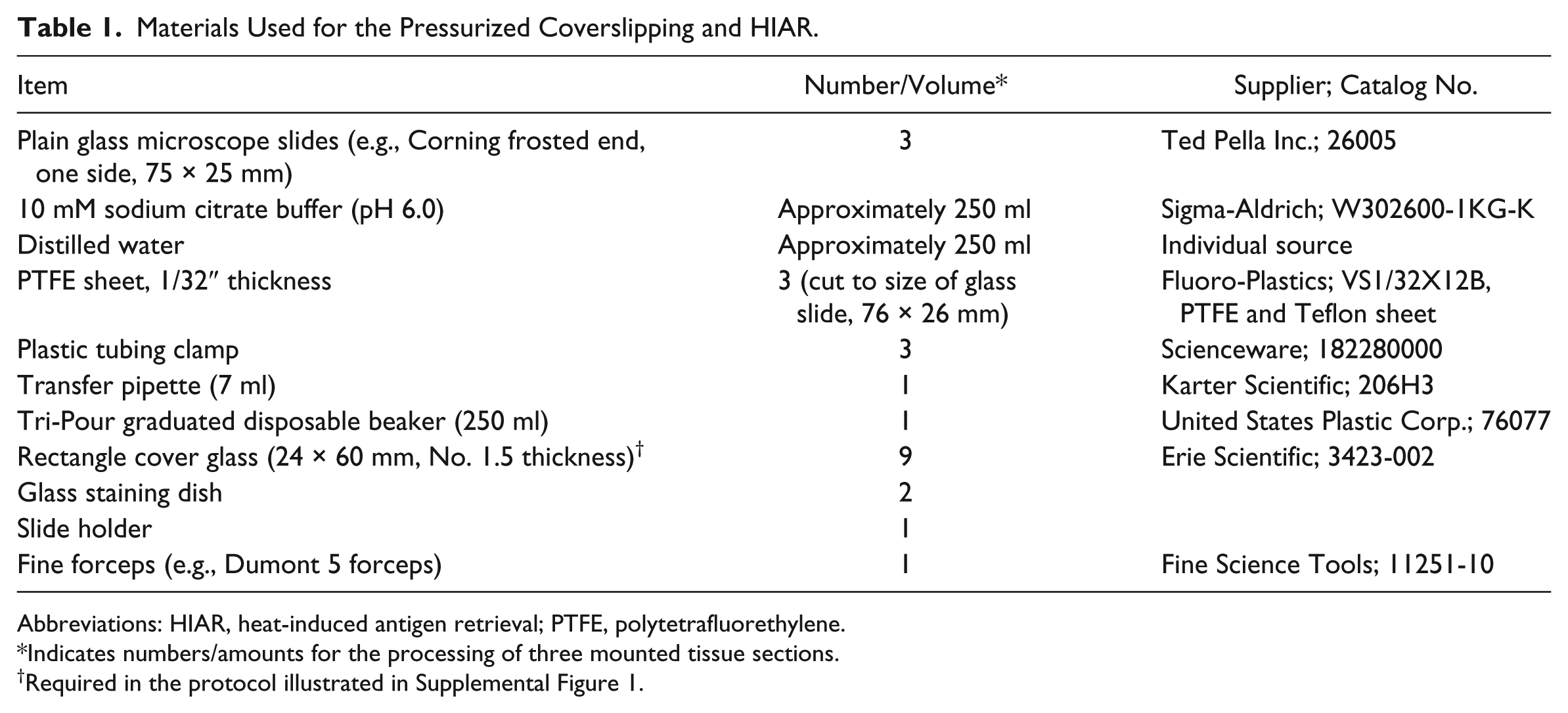
Abbreviations: HIAR, heat-induced antigen retrieval; PTFE, polytetrafluorethylene.
Indicates numbers/amounts for the processing of three mounted tissue sections.
Required in the protocol illustrated in Supplemental Figure 1.
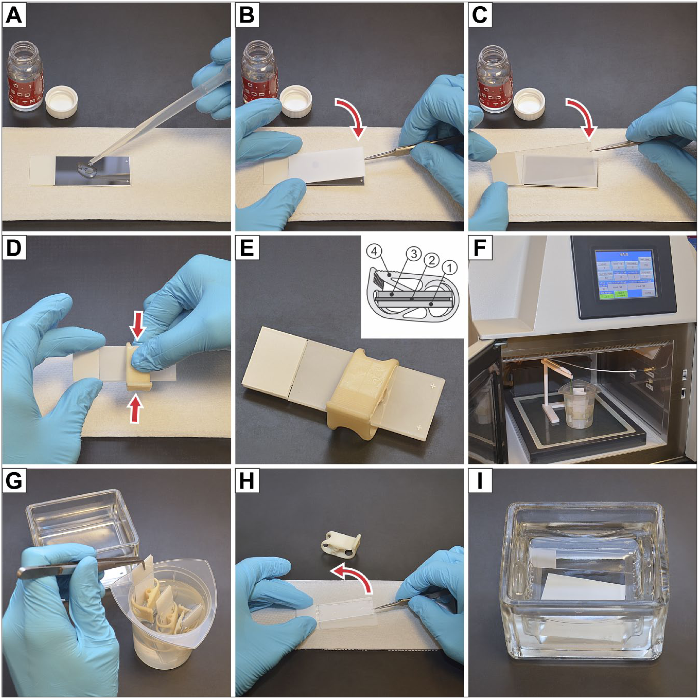
Step-by-step instructions for pressure coverslipping of mounted tissue sections for heat-induced antigen retrieval treatment. The detailed protocol, (A) to (I), is given in the “Materials and Methods” section. Inset in (E), schematic illustration of a pressure-coverslipped slide: (1) microscope slide with mounted tissue section, (2) Teflon sheet, (3) second microscope slide, and (4) plastic tubing clamp.
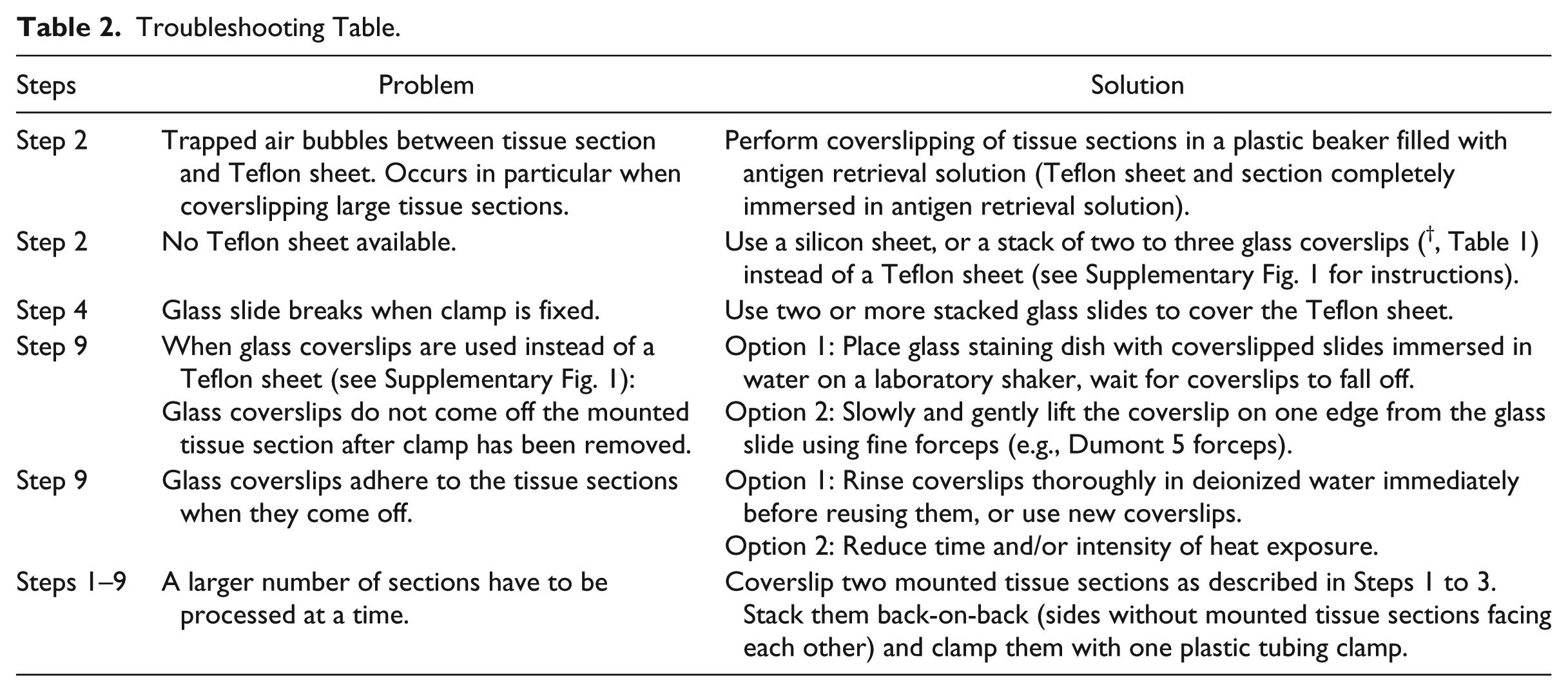
Potential problems that have occurred during the development of this protocol and possible solutions are given in Table 2.
Troubleshooting Table.
Heat-induced Antigen Retrieval
HIAR was performed in a temperature-controlled microwave processing system (PELCO BioWave Pro; Ted Pella Inc., Redding, CA) at 98C for 20 min. After heating, the clamped slides were immediately transferred to a glass jar with dH2O (Fig. 1G) and allowed to cool down to room temperature for 10 min. Alternatively, the plastic beaker that contained the clamped slides immersed in antigen retrieval buffer solution was placed in a microwave pressure cooker (Tender Cooker, Nordic Ware, Minneapolis, MN) that was heated in a domestic microwave oven (80% power for 15 min; Model AT738A, rotary plate, 980 Watts, Emerson Radio Corp., Hackensack, NJ). After the heating period, the Tender Cooker was allowed to depressurize at room temperature, and the clamped slides were then transferred to a glass staining dish containing dH2O (Fig. 1G) and allowed to cool to room temperature for 10 min.
Immunohistochemistry
Sections were blocked in 5% normal horse serum (NHS) diluted in PBS for 30 min and incubated overnight at room temperature with either a monoclonal mouse anti-mineralocorticoid receptor (MR) antibody (dilution 1:5000; Developmental Studies Hybridoma Bank, University of Iowa, Iowa City, IA), a polyclonal rabbit anti-proliferating cell nuclear antigen (PCNA) antibody (dilution 1:2000; Abcam, Cambridge, MA), or a polyclonal rabbit anti-serum and glucocorticoid-regulated kinase 1 (SGK1) antibody (dilution 1:1000; Abcam). The sections were then respectively incubated with biotinylated secondary antibodies raised in donkey against rabbit or mouse IgG (dilutions 1:400; Jackson, West Grove, PA) for 1 hr at room temperature, followed by avidin-biotin-horseradish peroxidase (Standard ABC Kit, Vector Laboratories, Burlingame, CA) for 1 hr at room temperature. All primary and secondary antibodies were diluted in 1% NHS in PBS. The sections were washed in PBS between each incubation step. Finally, the sections underwent color development with 0.01% diaminobenzidine and 0.01% hydrogen peroxide diluted in 100 mM phosphate buffer (pH 7.3) for 5 to 10 min. In some sections, the nuclei were counterstained with Harris’s hematoxylin for a few seconds. All sections were dehydrated in a graded series of ethyl alcohols, cleared in xylene, and coverslipped in permount.
Microscopic Analysis and Image Processing
Microscopic analysis was performed using a dissection microscope (Carl Zeiss, Germany) equipped with an InfinityX-32 digital microscope camera (Lumenera Corp., Ottawa, ON) or an Olympus BX51 microscope (Olympus Corp., Tokyo, Japan) equipped with an Olympus DP70 digital camera (Olympus Corp.).
Digital images were sharpened by unsharp masking and were gamma corrected using Photoshop CS6 (V 13.0.1, Adobe Systems, San Jose, CA).
Results
Pressurized Coverslipping Preserves Delicate Tissue Morphology and Enables Efficient Heat-induced Antigen Retrieval in Formalin-fixed, Paraffin-embedded Tissue Sections
FFPE sections from a decalcified mouse skull were used in these experiments. The sections were either exposed to (1) no HIAR treatment, (2) conventional HIAR treatment, or (3) HIAR with pressurized coverslipping. All sections were then immunolabeled for MR, and nuclei were counterstained with hematoxylin. At least 10 sections were exposed to each of the three experimental conditions. Representative results for each group are shown in Fig. 2. In sections that were not exposed to HIAR treatment, the anatomical structures in the posterior cranial fossa, including the superior semicircular canal (SSC), the cerebellum (Cer) and the extraosseus portion of the endolymphatic sac (ES), showed excellent morphological preservation (Fig. 2A and A’). No tissue reactivity was detected for the anti-MR antibody in sections from this experimental group. Immunolabeling signals for MR were expected to be localized in the ES epithelium, according to previous reports. 9 On slides that were exposed to conventional HIAR treatment, large parts of the sections were detached after heating, and the integrity of the sections was largely disrupted (Fig. 2B). Immunolabeling for MR resulted predominantly in the artifactual deposition of DAB in areas of the sections that became nonadherent; the ES epithelium could not be identified in its expected location due to the severely impaired tissue morphology (Fig. 2B′). In contrast, sections that were pressure-coverslipped before HIAR treatment showed an excellent morphological preservation that was almost indistinguishable from sections that were not exposed to HIAR treatment (Fig. 2C). Moreover, pressure coverslipping for HIAR allowed an effective retrieval of tissue antigens and considerably improved the tissue reactivity of anti-MR antibodies. Immunolabeling for MR on these sections resulted in an intense nuclear signal in the ES epithelial cells (Fig. 2C′).
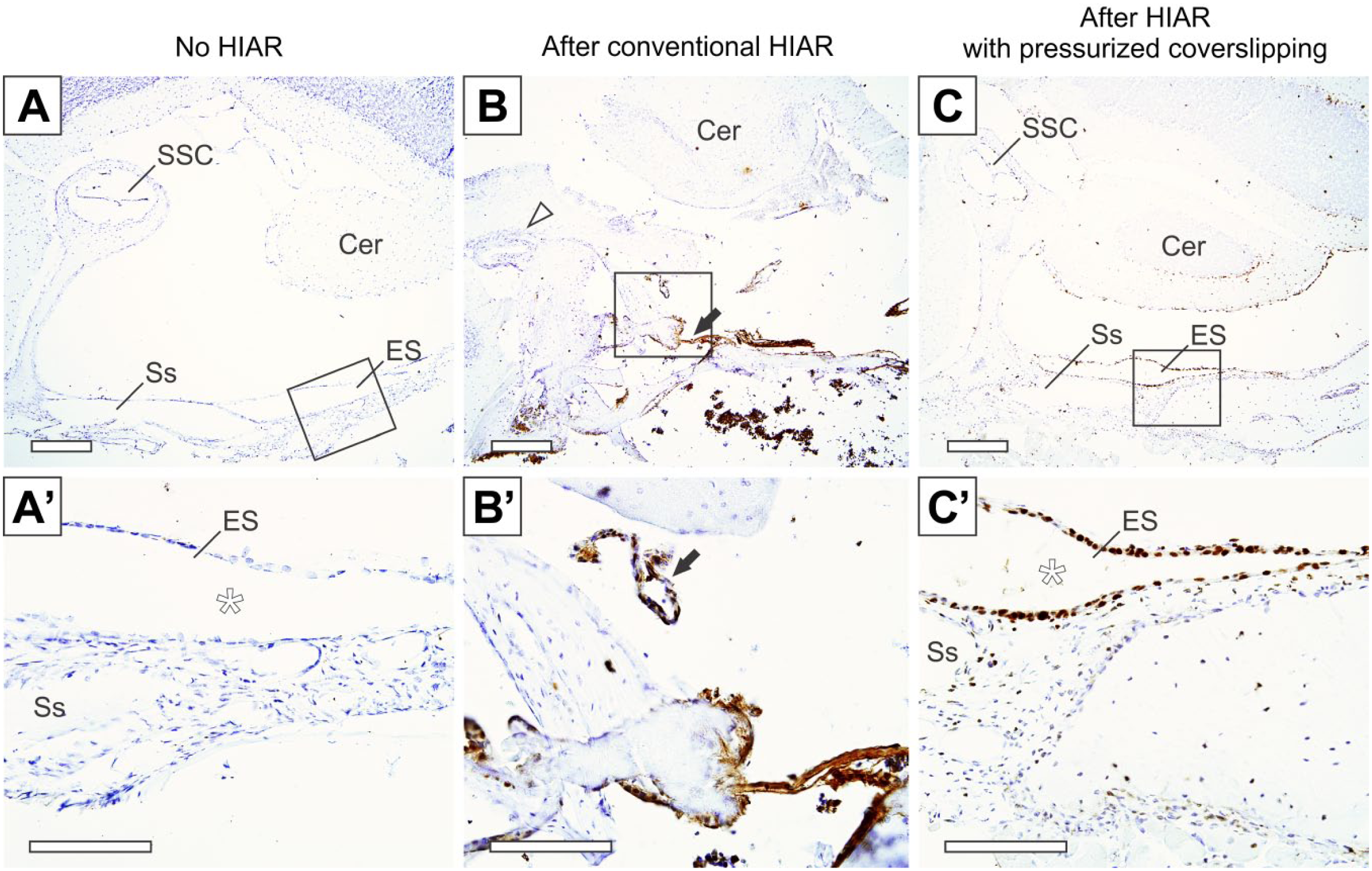
Comparison of tissue morphology and effect of HIAR treatment on paraffin sections of the decalcified mouse skull (area of the posterior cranial fossa; horizontal section plane). Corresponding areas from three different sections are shown in (A), (B), and (C). (A–A′) Control section that was not exposed to HIAR treatment (arrowhead, superior semicircular canal [SSC]; arrow, endolymphatic sac [ES]; Cer, cerebellum). Higher magnification (A′) shows the delicate morphology of the ES with its mono-layered epithelium (arrowhead) that encloses the ES lumen (*). Immunolabeling with an antibody against the mineralocorticoid receptor (MR) was performed on this section; however, without HIAR pretreatment, no immunolabeling signals were detected. (B–B′) After HIAR without pressure coverslipping, large parts of the mounted section were detached from the slide (B), resulting in a vast disruption of the delicate anatomical structures of the SSC (arrowhead), the Cer, and the ES (arrow). A dislocated piece of the ES epithelium shows immunolabeling for MR—arrow in (B′); artifactual DAB reaction products are observed at detached tissue parts. (C–C′) HIAR treatment with pressure coverslipping of the tissue section results in an excellent morphological preservation (C), as well as a distinct and intense nuclear immunolabeling signal for MR in the ES epithelium (C′). Scale bars: 100 μm. Abbreviations: HIAR, heat-induced antigen retrieval; Ss, sigmoid sinus.
Heating of Pressure Coverslipped Tissue Sections Can Re-adhere Detached Parts of Sections and Free-floating Sections
When testing the pressurized coverslipping technique on FFPE sections, we observed that sections that had partially detached during the steps of deparaffinization or rehydration could be permanently re-adhered to the glass slide after subsequent pressurized coverslipping and HIAR treatment. This finding was then systematically reproduced using FFPE sections of murine kidney that were deparaffinized, rehydrated, placed free-floating on microscope slides, pressure-coverslipped, heated using the above-described HIAR protocol, and finally immunolabeled for MR. Figure 3 compares the effect of pressurized coverslipping and heating on section adherence, tissue morphology, and MR-immunolabeling between two sections that remained adherent during all experimental steps (“1” and “2” in Fig. 3A – B′) and one section that randomly detached during the rehydration step (“3” in Fig. 3A and A′). After pressurized coverslipping and HIAR treatment, the previously detached section was able to be re-adhered to the glass slide (“3” in Fig. 3B and B′), and it remained adherent during all following experimental steps. In all three sections, intense immunolabeling signals for MR were detected in the epithelium (nuclear localization) of the so-called aldosterone-sensitive distal nephron (ASDN, 10 ). No apparent differences were noted regarding the morphological preservation or the quality of the MR-immunolabeling (no intensity gradients, high signal-to-noise ratio) between the two sections that remained adherent throughout (Fig. 3C– D′ ) and the section that was re-adhered after pressurized coverslipping and heating (Fig. 3E and E′), expect for a narrow band of artifactual DAB deposition at the margin of the re-adhered section (Fig. 3E). These results were consistently reproduced in the series with 10 FFPE sections of murine kidney tissue.
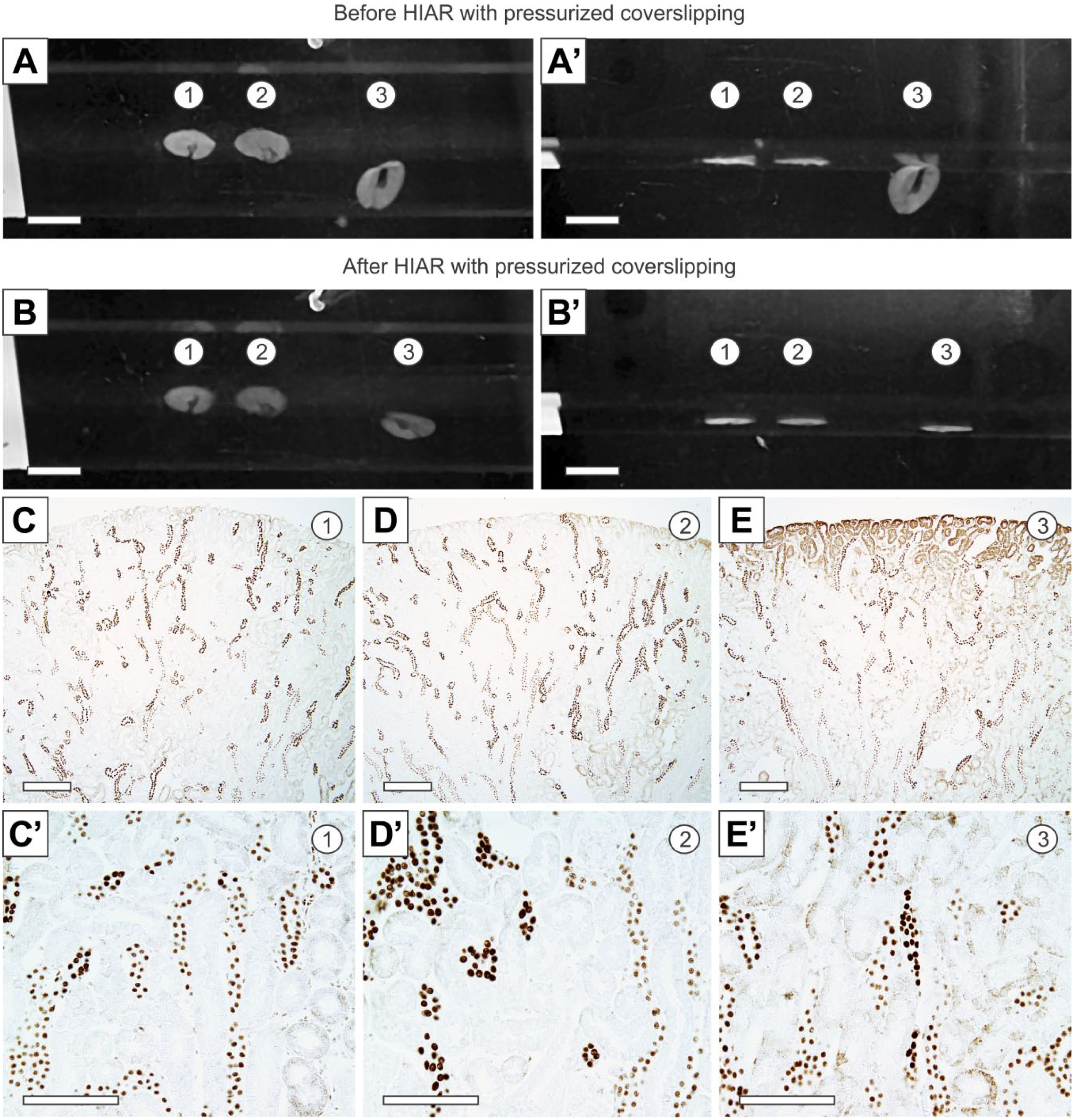
Reattaching partially detached tissue sections using pressure coverslipping and HIAR. (A and A′) Three paraffin-embedded mouse kidney sections were mounted on a microscope slide. After paraffin removal and rehydration, two sections were still properly attached to the slide, whereas the third section was almost completely detached. (A′) shows the same slide as in (A) at a different angle. (B and B′) After HIAR treatment using pressure coverslipping, all three sections were properly attached to the slide. (B′) and (B) are at different angles. (C–E) The three sections from (A–B′) were immunolabeled with an antibody against the mineralocorticoid receptor (MR). In all three sections, specific nuclear labeling in distinct segments of the tubular epithelium that belong to the aldosterone-sensitive distal nephron was observed. In the reattached section (E and E′), artifactual DAB reaction product was observed at the margin of the section. Scale bars: (A–B′), 400 μm; (C–E′), 100 μm. Abbreviation: HIAR, heat-induced antigen retrieval.
Although the procedure of re-adhering (partially) detached tissue sections is not the primary intended application of the method described here, nor a common issue in routine immunohistochemical tissue processing, these results underscore the method’s power in maintaining and creating a permanent adhesion of tissue sections on the slide surface using a combination of mechanical pressure and thermal energy.
Pressurized Coverslipping Preserves Delicate Tissue Morphology and Enables Efficient Heat-induced Antigen Retrieval of Celloidin-embedded Tissue Sections With Delicate Morphology
The use of CE tissue sections for immunohistochemistry is challenging for several reasons. Mounting and adhering CE sections on slides must be performed under “wet-conditions” (permanent immersion of the CE sections in alcohol or water), which creates only a limited adhesiveness between the sections and the slide surface compared with FFPE sections that are air-dried and “baked” (slide heater) onto the glass slide after mounting. Moreover, the removal of celloidin from the mounted CE sections is a prerequisite for the subsequent immunolabeling of most tissue antigens, and this involves repeated washes that expose the mounted sections to considerable mechanical stress, increasing the risk of section detachment.
The CE sections from human tonsil tissue were either exposed to (1) no HIAR treatment (Fig. 4A – B″), (2) conventional HIAR treatment (Fig. 4C – D″), or (3) HIAR with pressurized coverslipping (Fig. 4E – F″) before immunolabeling for PCNA and hematoxylin staining. Sections that were not exposed to HIAR treatment remained properly adhered to the slide; occasionally small nonadherent spots were observed within the sections (arrowheads in Fig. 4B). Sections from this experimental group could be stained homogeneously with hematoxylin; however, the anti-PCNA antibody showed almost no tissue reactivity on these sections (Fig. 4B – B″). After conventional HIAR treatment, the tissue margins (arrows in Fig. 4D), as well as numerous large spots within the sections (arrowhead in Fig. 4D), were detached from the slide surface, which caused tears within the sections (arrowhead in Fig. 4D′). HIAR considerably improved the reactivity of the anti-PCNA antibody (specific nuclear staining predominantly in lymph follicles); however, PCNA-immunolabeling and hematoxylin staining exhibited artifactual gradients throughout the partially detached sections (Fig. 4D′ and D″). In contrast, after HIAR with pressurized coverslipping, the sections were properly adhered to the slide surface; occasionally small spots within the sections remained nonadherent (arrowhead in Fig. 4F). HIAR treatment efficiently improved the tissue reactivity of the anti-PCNA antibody, which resulted in specific and intense immunolabeling signals. No artifactual gradients were observed for PCNA-immunolabeling and hematoxylin staining (Fig. 4F′ and F″). The described differences between conventional HIAR and HIAR with pressurized coverslipping were consistently observed over a larger series of consecutively processed sections (>50 sections per condition).
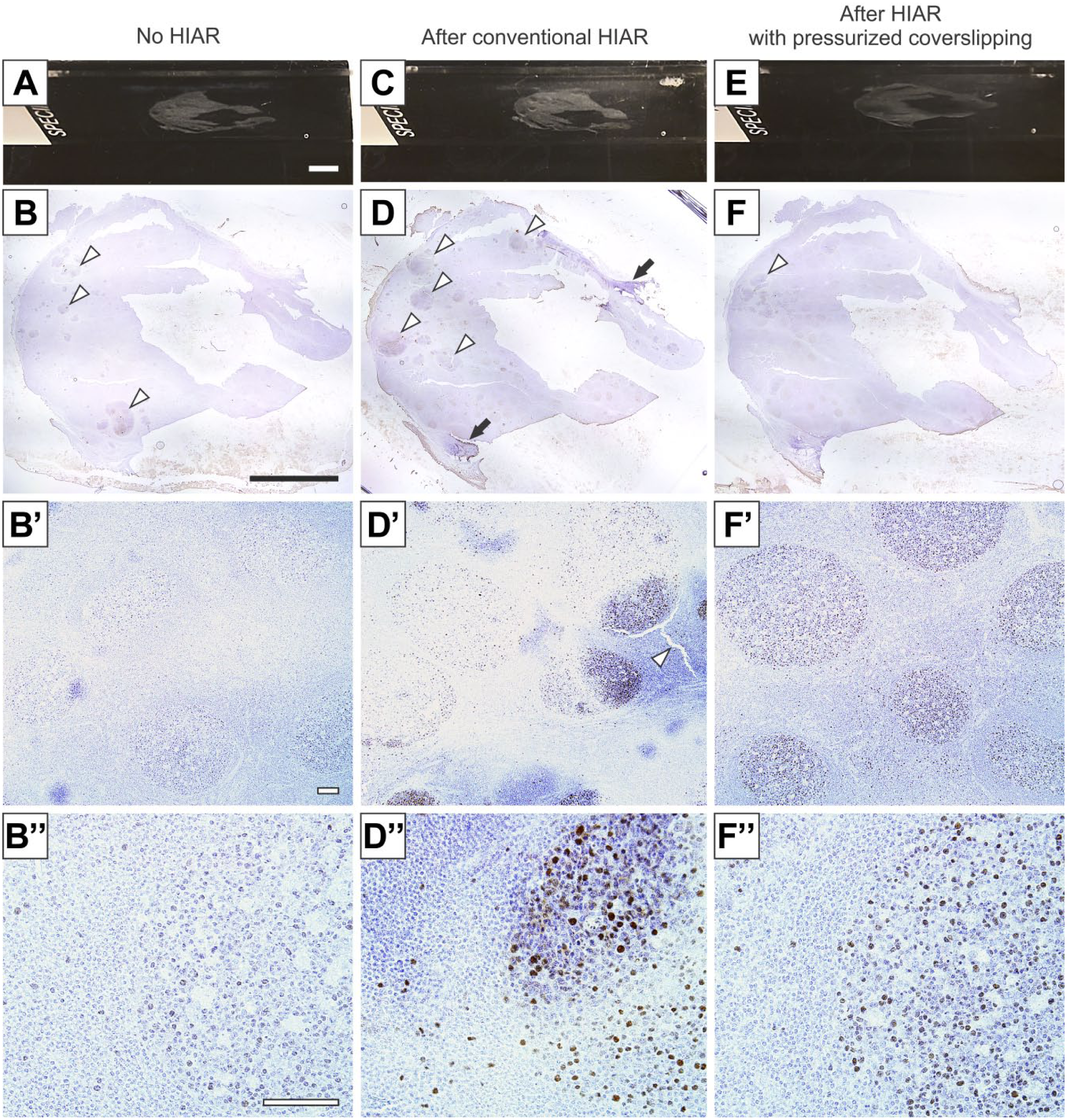
Pressure coverslipping during HIAR enables a distinctly more homogeneous immunolabeling of large tissue sections. (A–B″) A celloidin-embedded section of human tonsil tissue was mounted on a microscope slide (A). Overview (B) and higher magnifications (B′ and B″) of the same section after immunolabeling for PCNA and hematoxylin staining. Only a few weakly PCNA-labeled cells were observed in the lymphoid follicles. (C–D″) After HIAR without pressure coverslipping, disseminated focal detachment of the tissue section occurred—arrowheads and arrows in (D). These areas exhibited a more intense PCNA-immunolabeling and hematoxylin staining compared with adherent regions of the section—arrowhead in (D′), which resulted in nonhomogeneous labeling of the entire tissue section. (E–F″) Using HIAR with pressure coverslipping, the entire tissue section remained adherent to the slide (F). An intense and homogeneous immunolabeling for PCNA and hematoxylin staining was achieved (F′ and F″). Scale bars: (A, B, C, D, E, and F), 5 mm; (B′, B″, D′, D″, F′, and F″), 100 μm. Abbreviations: HIAR, heat-induced antigen retrieval; PCNA, proliferating cell nuclear antigen.
In another experimental series, the same three treatment conditions (no HIAR, conventional HIAR, HIAR with pressurized coverslipping) were applied to archival CE sections from formalin-fixed and decalcified human temporal bones. These sections exhibit an exceptionally delicate morphology; they consist of dense bone tissue that forms a pneumatized trabecular meshwork in the mastoid bone and harbors numerous bony channels and hollow spaces that are filled with soft tissue structures, including the interconnected spaces of the inner ear labyrinth. The latter are lined by the exceptionally sophisticated neuroepithelial structures of the auditory (cochlea) and vestibular sense organs. Sections that were stained with hematoxylin or immunolabeled with an anti-SGK1 antibody without previous HIAR treatment (Fig. 5A – B″) demonstrated an excellent preservation of all anatomical structures within the temporal bone, including the neuroepithelial membranes in the cochlea and the saccule of the inner ear (Fig. 5B′). No immunolabeling for SGK1 was detected in these sections (Fig. 5B″). HIAR treatment without pressurized coverslipping (Fig. 5C – D′) had a deleterious effect on these tissue sections. All sections became almost completely detached from the slide and could not be used for further immunolabeling. In contrast, the morphological preservation of sections that were exposed to HIAR with pressurized coverslipping was nearly comparable to that observed without HIAR treatment. Heat-induced focal gradients of hematoxylin staining and small tears were observed only in areas with dense bone tissue, for example, the otic capsule (Fig. 5F′). HIAR treatment efficiently improved the reactivity of the anti-SGK1 antibody, which resulted in nuclear and cytoplasmic labeling in nonsensory supporting cells of the organ of Corti (Fig. 5F″).
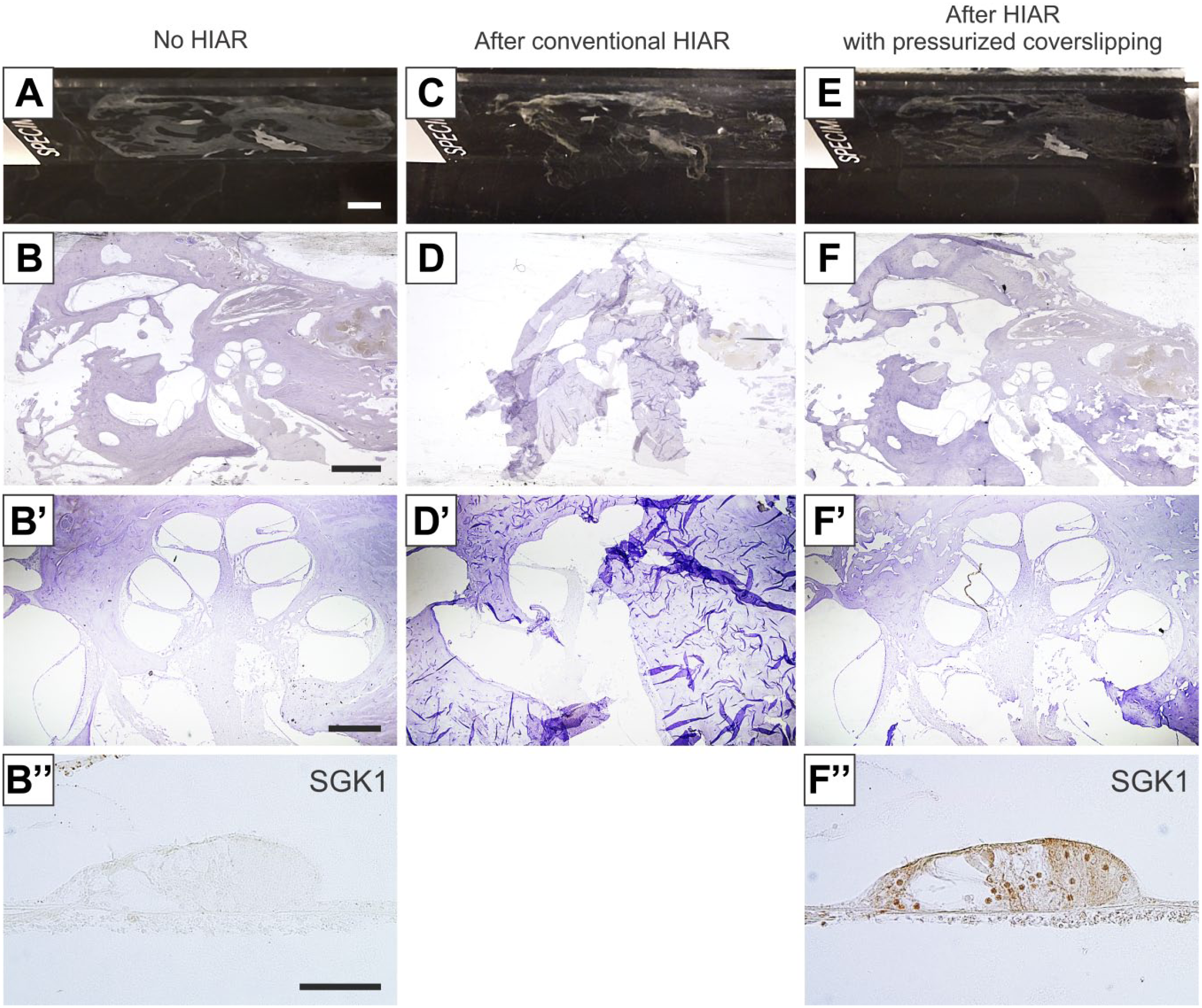
Pressure coverslipping during HIAR preserves the delicate morphology of decalcified, celloidin-embedded human temporal bone (TB) sections. (A–B″) A celloidin-embedded human TB section after mounting on a microscope slide (A). Hematoxylin staining reveals the trabecular bony structure of the mastoid (left part of the section), cross-sections through the cavities of the middle and inner ear (middle part of the section), and the compact bone of the petrous apex (right part of the section). In particular, the delicate membranous structures of the cochlea—auditory portion of the inner ear (B′)—are vulnerable to the mechanical stress that occurs during histological processing and immunolabeling of the tissue. Without HIAR, no immunolabeling for the serum and glucocorticoid-regulated kinase 1 (SGK1) is observed in the organ of Corti (B″). (C–D′) After HIAR without pressure coverslipping, the tissue section almost completely detached from the slide (C and D). The delicate structures of the cochlea were almost completely disrupted (D′). The section could no longer be used for immunohistochemical labeling. (E–F″) After HIAR with pressure coverslipping, the section remained properly attached to the microscope slide (E). The morphology of all structures within the TB section was preserved (F), including the delicate architecture of the cochlea (F′). Immunolabeling for SGK1 showed nuclear and cytoplasmic signals in most supporting cells of the organ of Corti (F″). Scale bars: (A, B, C D, E, and F), 5 mm; (B′, D′, and F′), 1 mm; (B″ and F″), 100 μm. Abbreviation: HIAR, heat-induced antigen retrieval.
Discussion
The present study demonstrates that pressure coverslipping of tissue sections during HIAR treatment reliably prevents heat-induced morphological damage and detachment of the tissue sections from microscope slides, while at the same time the method efficiently retrieves tissue antigens for improved results of immunohistochemical labeling. The method proved to be advantageous compared with conventional HIAR protocols, in particular for sections with delicate tissue morphology and for tissues with generally weak adherence properties.
Section detachment and impaired tissue morphology caused by conventional HIAR treatment are technical drawbacks that were previously described in the first studies that introduced HIAR to the field of immunohistochemistry.1,11 Despite the current widespread use of HIAR techniques in research laboratories and clinical histopathology laboratories, the technical problem of heat-induced section detachment has not been completely solved. 12 Former technical attempts that addressed this issue mainly aimed to strengthen the adhesive forces between the tissue section and the slide surface by coating the slide with charged molecules. However, this approach turned out to have only limited success because the slide coating techniques for HIAR all harbor a critical weakness. Heat application retrieves antigens in formalin-fixed tissues presumably by hydrolytic cleavage of formalin-induced cross-links, thermal unfolding of protein structures, and other proposed mechanisms that break and/or reorganize chemical bonds within the tissues. 13 In addition to these desired effects, the application of heat to mounted tissue sections also clearly affects the chemical bonds that link the tissue with the coated surface of the microscope slide—an undesired side effect that ultimately leads to section detachment.
The approach described in the present study successfully overcomes this problem by using a pressurized coverslipping technique to maintain the physical contact between the tissue section and the coated slide surface. During the “vulnerable” period of heat application, when the adhesive chemical bonds become thermally destabilized, this technique prevents the section from floating off the slide, and during the subsequent cool-down period, the mechanical depression of the section on the coated slide surface promotes reestablishment of the adhesive chemical bonds between the tissue and the slide surface coating. In fact, the assumption that chemical bonds between the slide surface and the tissue section reestablish after heating is supported by our observation that initially detached or free-floating tissue sections are successfully (re)adhered after pressurized coverslipping and heat application. In addition to the application to FFPE and CE sections, the pressurized coverslipping technique might also be useful for performing HIAR treatment on cryosections and flat-mount preparations (method successfully tested, data not shown), or on cell smears and cell cultures that are mounted or grown on glass surfaces.
In summary, we describe a simple method that uses mechanical pressure during heat application for antigen retrieval to keep the section on the glass slide and prevent heat-induced section detachment and morphological damage. The method requires only basic lab equipment, can easily be incorporated in all HIAR protocols, and is compatible with all commonly used heating sources.
Supplemental Material
DS_10.1369_0022155419826940 – Supplemental material for Mechanical Compression of Coverslipped Tissue Sections During Heat-induced Antigen Retrieval Prevents Section Detachment and Preserves Tissue Morphology
Supplemental material, DS_10.1369_0022155419826940 for Mechanical Compression of Coverslipped Tissue Sections During Heat-induced Antigen Retrieval Prevents Section Detachment and Preserves Tissue Morphology by Andreas H. Eckhard, Jennifer T. O’Malley, Joseph B. Nadol and Joe C. Adams in Journal of Histochemistry & Cytochemistry
Footnotes
Acknowledgements
We are grateful for the technical expertise of Barbara Burgess and Diane Jones in preparing the human temporal bone specimens. We thank Leslie Liberman and David Bächinger for technical help. We thank Garyfallia Pagonis for her expert assistance with graphics.
Competing Interests
The author(s) declared no potential conflicts of interest with respect to the research, authorship, and/or publication of this article.
Author Contributions
AHE and JTO performed the experiments. AHE and JCA conceived and designed the study and wrote the manuscript. JBN provided critical review of the manuscript.
Funding
The author(s) disclosed receipt of the following financial support for the research, authorship, and/or publication of this article: This study was funded by the American Hearing Research Foundation, the GEERS Foundation (S 030–10.051), and National Institutes of Health (NIH) grant U24 DC011943. The author A.H.E. was supported by a research fellow grant from the German Research Council (Deutsche Forschungsgemeinschaft; EC 472/1-1).
References
Supplementary Material
Please find the following supplemental material available below.
For Open Access articles published under a Creative Commons License, all supplemental material carries the same license as the article it is associated with.
For non-Open Access articles published, all supplemental material carries a non-exclusive license, and permission requests for re-use of supplemental material or any part of supplemental material shall be sent directly to the copyright owner as specified in the copyright notice associated with the article.