
Abstract
In this study, an acquired pigmentation in Nero Siciliano pigs is reported and evaluated by a multidisciplinary approach to support the hypothesis it is caused by an ingested material. A total of 18 pigs were studied. Fourteen conventionally slaughtered animals showed black discoloration of lymph nodes. The lymph nodes were normal in size and shape but showed diffuse black discoloration of the cortex and medulla. Melanosis of fat was observed in 2 animals and was limited to the back. Histochemical tests performed on tissues enabled identification and differentiation of the pigment. Immunohistochemical staining for macrophage markers showed macrophages containing a variable amount of melanin-like granules. Stains for human melanoma, as well as S-100 protein, did not show any reaction. Histochemical methods for tyrosinase showed colorimetric patterns that confirmed the presence of the enzyme in acorns. The activity was mostly latent. A high tannin content was demonstrated, reaching about 76% of the total phenolic compounds. Our data, and the well-known steps on melanin formation, permit us to hypothesize that swine tyrosinase could act on phenolic substances found in acorns. Tyrosinase activation could take place in genetically predisposed swine after acorns are eaten, and this event could increase the biosynthesis and the anomalous storage of melanin.
Introduction
Melanosis is a disorder in which anomalous storage of melanin occurs. It has been reported in domestic animals, but rarely described in swine as a generalized pigmentation 23 or as a localized disorder of the mammary gland, 5 thymus, and retro pharyngeal lymph nodes. 32 Granules of melanin are produced by melanocytes, which can release them to melanophages, and these cells can store the pigment and carry it to distant sites. Abnormal storage of melanin can be caused by modification of the processes of production or transport. The term “melanosis” is used for a group of conditions characterized by an abnormal black or brownish-black pigmentation of an organ or tissue. It does not confirm that the pigment is melanin. For example, in melanosis coli the pigment granules contain lipofuscin, in melanosis ilei the pigment granules may contain hemosiderin, and in melanosis esophagi the pigment is melanin. 26 The term “pseudomelanosis” is used for the blue-green discoloration of tissue by iron sulfide formed by the reaction of hydrogen sulfide generated by putrefactive bacteria on iron from hemoglobin released from lysed erythrocytes; it usually takes a day or more to develop after the death. Pseudomelanosis, associated with dietary compounds, has rarely been reported in swine 6 and guinea pigs. 16 These pigmentations could have economic importance affecting carcass quality, decreasing consumer appeal and the marketability of the pigs, but they do not pose a health risk for the consumer. “True” melanosis in swine is usually congenital, and in some breeds, such as Sinclair, Duroc-Jersey, Hormel, Vietnamese pot-bellied and their cross-breeds, it has been be linked to tumors. 7, 11, 21, 27 Awareness of the pathogenesis of melanosis will aid its differentiation from melanoma, lipofuscinosis, hemosiderosis, and ceroid pigmentation.
The Nero Siciliano pig, native to rural areas in north Sicily, Italy, lives freely in the woods of the Nebrodi mountains (maximum altitude 1,800 meters above sea level). This pig is found in fossil remains and in writings from the Greek period (VIII–VI BCE). The breed shows good adaptation to the often unfavorable environmental conditions and lives on undergrowth products (roots, tubers, acorns, uncultivated fruits, etc.). Exploitation of free living native breeds not only provides high-quality meat products and limits imports. The addition of these production could allow wider and more rational utilization and recognition of the value of marginal land. 8
As yet, little is known of the prevalence and incidence of diseases in such breeds. 15, 25 In this study, an abnormal pigmentation occurring occasionally in Nero Siciliano pigs is reported and evaluated by a multidisciplinary approach to support the hypothesis that the cause is related to ingested materials.
Materials and Methods
Over a 3-year period, a total of 18 Nero Siciliano pigs of heterogeneous genetic background were studied. Thirteen animals (13), conventionally slaughtered in winter 2005, after containment for 3 months in movable shelters while fed on acorns, showed black discoloration of all the lymph nodes. In 2 of these pigs, pigmentation also involved the subcutaneous fat in the lumbar region. Five pigs comprising a second group were slaughtered at the same age (12 months) as the first group, in late spring 2007, when acorns are less available or no longer available at all. These pigs, belonging to the same breed, coming from the same farm, and fattened in the same place, were examined postmortem at the slaughterhouse. Only 1 subject of the second group had black pigmentation, localized in the retro-pharyngeal lymph nodes.
Lymph nodes and fat tissue samples were collected both unfixed and fixed in 10% buffered formalin. Serial 5-µm cryostat sections were stained with hematoxylin and eosin (HE), and deparaffinized sections were stained using HE, Nile blue, Millon and Lillie stains, and decolorized with hydrogen peroxide and melanin bleach. Retro-pharyngeal lymph node samples collected from 5 subjects, belonging to the first group, with melanosis, were also fixed in a 10% buffered formalin solution and routinely processed for paraffin embedding and sectioned at 5 µm. The following immunohistochemical stains were applied using the Avidin-biotin complex technique: macrophage marker (Novocastra, Newcastle Upon Tyne, UK; dilution 1 : 100), mouse monoclonal antibody with specificity toward macrophages, monocytes and histiocytes, monoclonal mouse anti-human melanoma antibody (Dako, Glostrup, Denmark; dilution 1 : 25, 1 : 50, 1 : 100, 1 : 200), 9 S-100 (Dako; dilution 1 : 1,000), and a calcium-binding protein detected in almost all benign nevi and malignant melanocytic tumors of the skin. Additionally, the Millon reaction 28 was carried out on lymph node sections to show tyrosine. Biochemical and histochemical evaluations were carried out on acorns (Fig. 1) collected in the forest area where the pigs were fattened. The presence of tyrosinase was shown by qualitative and quantitative assays. For the qualitative methods, histochemistry was performed on cryostat sections of acorns, and the Okun method 20 was used in an assay on extracted acorn liquid. Quantitative determination of tyrosinase extracted from acorns was performed by spectrophotometric analysis using catechol as a substrate. 31 The activity of tyrosinase in the crude extract was determined in triplicate by melanin-like pigments formed in the polymerization of quinone. The reaction mixture, in a final volume of 1.0 ml, contained 10 mM catechol and 20 µl enzyme preparation in 50 mM phosphate buffer (pH 7.0). One activity unit (U) is defined as the amount of enzyme that causes an increase of absorbance at 410 nm of 0.001 per minute at pH 7.0 at 25°C. A total phenolic compounds analysis was carried out on a methanolic acorn extract by the Folin-Ciocalteau reagent 30 ; the data were expressed as gallic acid equivalents. Tannin content was monitored using binding properties of polyvinyl polypyrrolidone. 24 All values were expressed as a mean of three replications ± SD.
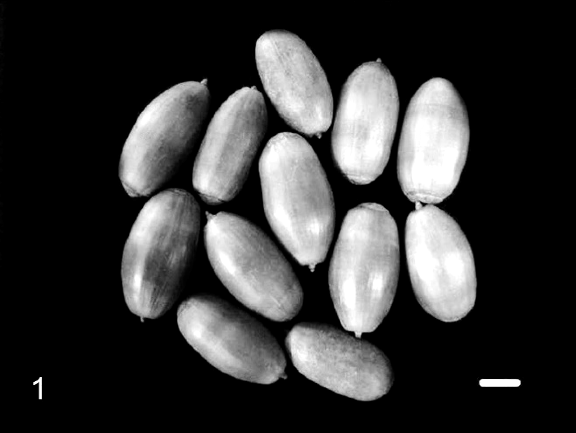
Acorns collected in the Nebrodi area used to feed Nero Siciliano pigs during fattening. Bar = 1 cm.
Results
At macroscopic evaluation, the lymph nodes were normal in size and shape, and showed a diffuse black color consistently involving the cortex and sometimes the medulla (Fig. 2). The melanotic fat tissue observed in 2 animals was limited to the back region, and the cut surface of the fat tissue was mottled (Fig. 3).
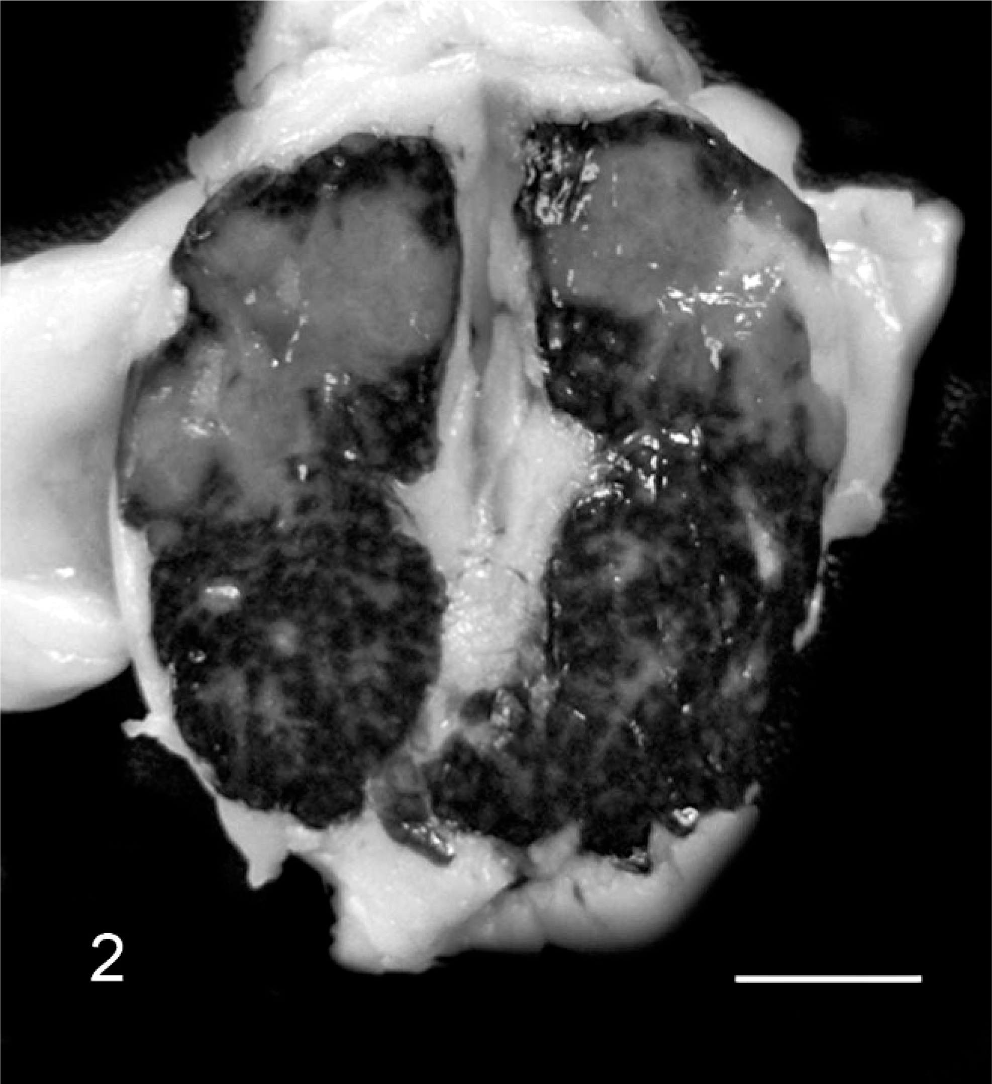
Nero Siciliano pig. Cut surface of a retropharyngeal lymph node showing melanosis. Bar = 1 cm.
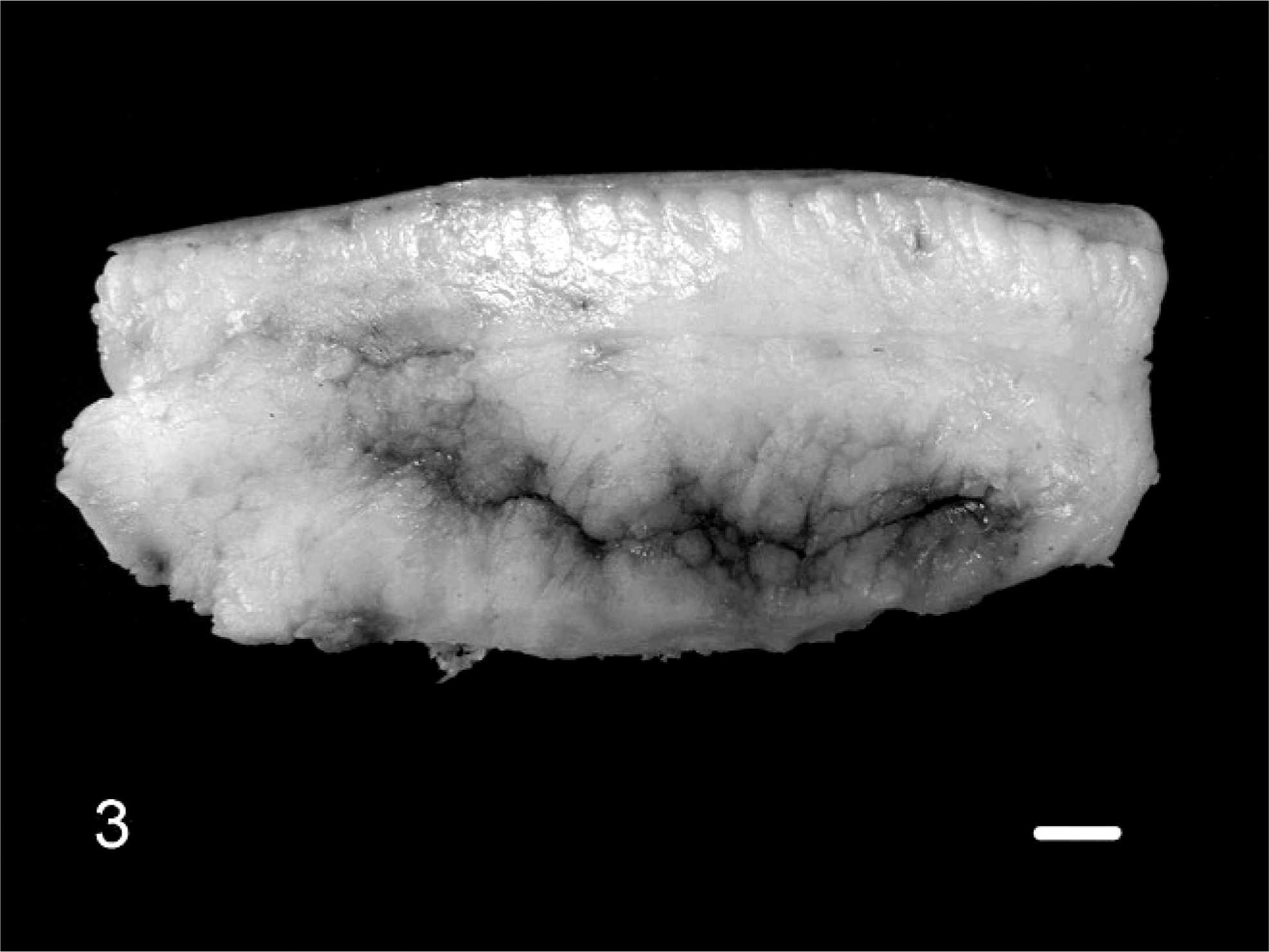
Nero Siciliano pig. Melanotic fat tissue from the region of the back. Bar = 1 cm.
Lymph node sections stained with HE showed infiltrating macrophages with brown/black granular pigment (Fig. 4). Normal melanocytes were present in the skin. To identify and differentiate the pigment further, histochemical tests were carried out. Staining with Nile blue (hydrogen sulphate) showed a dark green pigment, typical of melanin, differentiating it from lipofuscin, which would have stained blue. Additionally, 2 decolorizing agents, melanin bleach and hydrogen peroxide, applied for 24 hours made the pigment totally disappear, as would be expected only for melanin. Millon's reaction to tyrosine, which is a precursor of melanin, was positive too, giving a red-yellowish staining of the lymph nodes. Finally, Lillie's method, 3 a highly specific reaction, showed dark green granules within the macrophages (Fig. 5), giving further confirmation of the presence of melanin. The positive immunohistochemical reaction for macrophage marker, as well as the negative staining for S-100 and human melanoma, combined with the histologic appearance support the hypothesis that this is melanosis rather than melanoma.
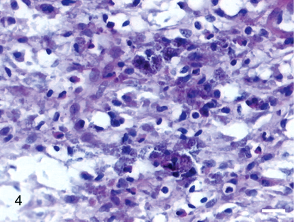
Lymph node. Histologic section showing melanin granules within macrophages. HE 40×. Bar = 30 µm.
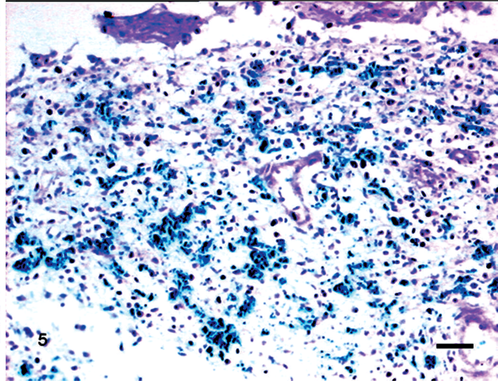
Lymph node. Lillie's method showed dark green granules within macrophages (30×). Bar = 50 µm.
The histochemical and biochemical methods for tyrosinase showed colorimetric patterns that confirmed the presence of this enzyme in acorns. The results obtained by the quantitative determination of the tyrosinase activity in acorn extracts from the samples analyzed are shown in Table 1; the activity was present mainly in latent form, as reported for other vegetable species. 10, 19 The amount of phenolic compounds was 0.137 ± 0.012 mg gallic acid equivalents/mg of acorn tissue. Results also showed a strong tannin content, reaching about 76% of the total phenolic compounds.
Acorn tyrosinase activity.*
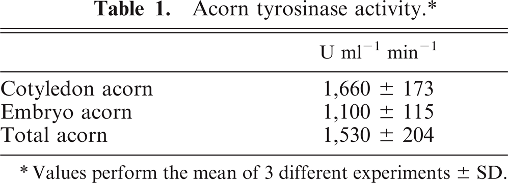
Values perform the mean of 3 different experiments ± SD.
Discussion
Melanosis is considered an unusual congenital pigmentation in swine, only occasionally observed in the slaughter house. The aim of the present paper was to identify an acquired pigmentation detected in a local swine breed and to relate it to the ingestion of acorns.
Our results showed a high content of phenols in acorns. These substances have various physiologic functions, and some authors have recently reported their effect on melanogenesis. 4, 14, 17, 18, 29 For this reason, we hypothesize an involvement of phenols in this lesion, probably acting as a substrate instead of tyrosine. Our data and current understanding of the process of melanin formation permit us to hypothesize that swine tyrosinase, being a polyphenol oxidase, could accelerate the use of phenol-rich substrates in acorns. After acorns are eaten, tyrosinase activation could take place in genetically predisposed pig breeds, leading to increased biosynthesis of melanin-like pigment, followed by anomalous storage. This hypothesis is supported by the different prevalence of melanosis in the 2 groups studied: 100% (13/13) in pigs fattened in acorn season versus 20% (1/5) in those ones fattened in late spring, when acorns are less available or no longer available at all. Moreover, melanosis has never been found in Nero Siciliano pigs reared in intensive systems. In conclusion, the data reported defend the hypothesis that ingestion of acorns can cause acquired melanosis. Kitt 13 published a picture showing dark lymph nodes of a pig fed on acorns, without any explanation with regard to the nature of the pigment. Carta 6 reported a prevalence of 100% in half-wild pigs fed on acorns in Sardinia, Italy. He called this pigmentation “pseudomelanosis” and recognized the cause in tannins. It is interesting that acorn bread is a typical local food for people in the same Italian region. More recently, there have been reports of melanosis in slaughtered swine. 1, 32 Modern husbandry and intensive farming do not include use of acorns as feed. Basic commercial pig feed has a very low concentration of phenolic compounds. This could explain why this pigmentation is rare in farm animals. Using the movable shelter farming system, this swine breed could be proposed as a model for the study of unusual lesions, such as melanosis, which today have disappeared in other breeds intensively reared. Acorns in ground cover are frequently damaged by parasite attack. 12, 22 Further investigations are needed to understand the parasite's role, if any, in eliciting melanosis in the tissues of this black swine breed. Acquired melanosis has been described in man's intestines, associated with the use of vegetable-derived laxatives, 2 supporting the possibility of the cause being ingested material. In our study, a melanin-like pigment was found, and other possible pigmentations have been excluded histochemically. Immunohistochemical and histological findings permit us to exclude melanoma, which is a likely cause of abnormal melanin deposition. As a final confirmation, we are now carrying out a trial to evoke acquired melanosis by feeding Nero Siciliano pigs on acorns and using white pigs as controls.
Footnotes
Acknowledgements
The authors are particularly grateful to Prof. Vincenzo Chiofalo, Coordinating Sect. Animal Production University of Messina, Italy, for providing data on this local pig and for the contact with the farmers.