
Abstract
Bovine viral diarrhea virus (BVDV) infection in goats can result in severe reproductive losses, with abortion rates reaching 80%. Infection with BVDV in aborted goat fetuses and stillborn kids can result in placentitis, encephalitis, myocarditis, and thymic depletion. This study investigates the distribution of viral antigen within the organ systems of aborted goat fetuses, stillborn kids, and nonviable kids infected with BVDV at various stages of gestation using immunohistochemistry (IHC). Virus antigen was detected within the placenta (8/13), thymus (4/9), heart (4/11), and brain (4/15) of affected goats. Uncommonly, BVDV antigen was detected within the skin (1/14), liver (1/13), kidney (1/12), lung (1/11), and trachea (1/3). BVDV antigen was not detected within the spleen (0/9), nasal turbinate (0/2), or thyroid (0/3). The results of this study indicate that placenta, heart, thymus, and brain are the most reliable tissues for BVDV antigen detection using IHC in aborted goat fetuses.
Keywords
Bovine viral diarrhea virus (BVDV) is an enveloped single-stranded RNA virus that can infect ruminants and camelids. 8, 16 Infection with BVDV can result in severe economic and reproductive losses, especially in cattle. Goats directly inoculated with BVDV and those exposed to cattle persistently infected with BVDV can experience abortion rates from 50 to 80%. 2, 3, 5, 12, 15, 16
The histologic lesions associated with reproductive losses due to BVDV infection in ruminants have been well described. 8 BVDV infection in a goat fetus can result in lesions within multiple organ systems. Encephalitis, cerebellar hypoplasia, gliosis, and choroid plexitis have been described in aborted fetuses. 3, 12 Nonsuppurative myocarditis, necrotizing placentitis, and thymic depletion have also been reported in cases of BVDV infection in goat fetuses, but other fetal organ systems appear to be relatively unaffected. 3
BVDV can be detected in a wide variety of aborted bovine and ovine tissues with immunohistochemistry (IHC). 1, 6, 7, 9, 17– 19 Though the gross and histologic changes associated with BVDV infection in goat fetuses have been described, the viral distribution within the fetal tissues remains unknown. 3 In previous studies, BVDV antigen has been detected with immunohistochemistry (IHC) in the brain of aborted goat fetuses but was not demonstrated in other organs. 1, 20 The present study will demonstrate the distribution of BVDV antigen in infected goat fetuses using IHC.
The tissues examined in this study originated in a larger study detailed elsewhere. 2, 3 To summarize the parent study, 24 pregnant female meat goats that were serologically and ear-notch negative for BVDV were obtained. These goats were introduced at various stages of gestation to 3 black Angus heifers persistently infected with BVDV, and the pregnant goats remained with the heifers for the duration of their pregnancy. Approximately 60% of the does aborted, had stillborn kids, or had nonviable kids that were euthanatized. The fetuses and kids from these does were subjected to complete necropsy at the Oklahoma Animal Disease Diagnostic Laboratory (OADDL) between February 2007 and April 2007. All goats were handled in accordance with the Oklahoma State University Institutional Animal Care and Use Committee.
Tissues from 18 aborted fetuses, stillborn kids, and nonviable kids were included in this study. Representative tissues were immersion fixed in 10% buffered formalin. Tissues were routinely processed, paraffin embedded, and 4–5 µm thickness sections were produced. The gross and histologic lesions in these goats are described in detail elsewhere. 3
Streptavidin-horseradish peroxidase methods with a mouse monoclonal antibody for BVDV (OADDL) were used on all available tissues from affected goats as previously described. 4 Novared (Vector Laboratories, Burlingame, CA) was used as the immunolabel and the sections were counterstained with Mayer's hematoxylin. IHC was performed on sections of brain, heart, kidney, liver, lung, nasal turbinate, placenta, spleen, thymus, thyroid, and trachea when available. Skin and kidney from a calf persistently infected with BVDV (confirmed with virus isolation and polymerase chain reaction) was used as a positive control. Duplicate slides from each affected animal without addition of the primary antibody to BVDV were used as negative controls.
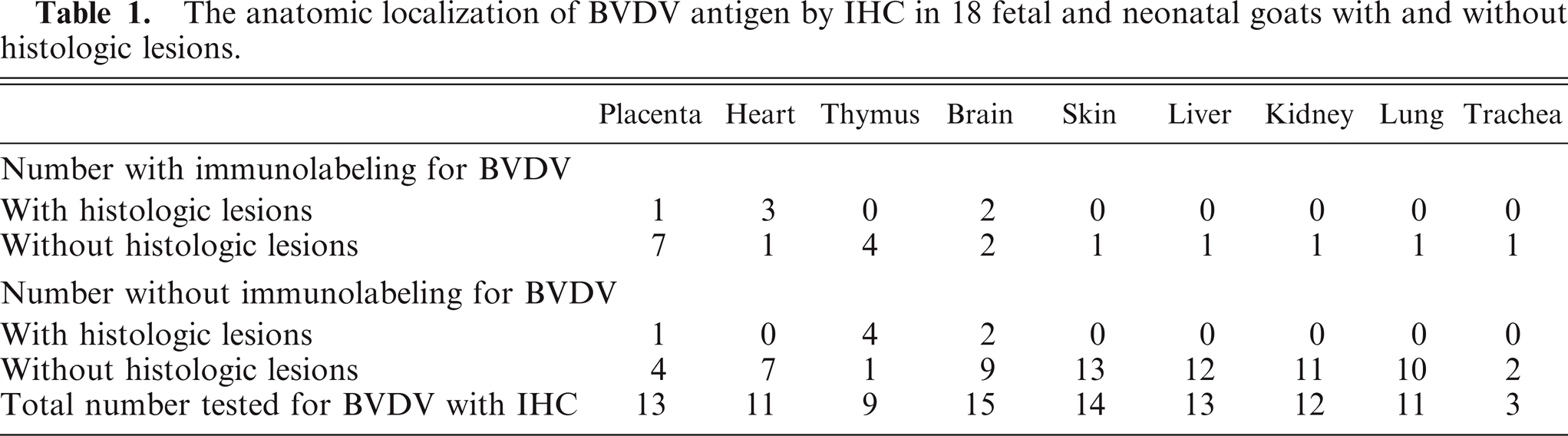
BVDV antigen was detected in numerous tissues from all of the goats with IHC. Immunolabeling was observed in organs with and without lesions (Table 1). BVDV antigen was frequently detected in the placenta (62%, 8/13 goats) and less often within the thymus (44%, 4/9 goats) with IHC (Figs. 1, 2). BVDV antigen was detected within the trophoblasts and mononuclear inflammatory infiltrate within the placenta. In the thymus, BVDV antigen was detected within the lymphocytes and reticular cells of Hassall's corpuscles. BVDV antigen was detected within 27% (4/15 goats) of the brains and 36% (4/11 goats) of the hearts of affected goats (Figs. 3, 4). In the brain, BVDV antigen was detected within the glial cells, neurons, endothelial cells, and mononuclear cells. BVDV antigen was detected within the myofibers and mononuclear cells of the heart. Rarely, BVDV antigen was detected within the skin (1/14 goats), liver (1/13 goats), kidney (1/12 goats), lung (1/11 goats), and trachea (1/3 goats) (Figs. 5, 6). In the skin, BVDV antigen was detected within the epidermis, follicular epithelium, and glandular epithelium. BVDV antigen was detected within circulating mononuclear cells within the lung and liver. In the kidney, BVDV antigen was detected within the medullary tubular epithelium. In the trachea, BVDV antigen was detected within the glandular epithelial cells and smooth muscle cells. BVDV antigen was not detected within the spleen (0/9 goats), nasal turbinate (0/2 goats), or thyroid (0/3 goats).
The anatomic localization of BVDV antigen by IHC in 18 fetal and neonatal goats with and without histologic lesions.
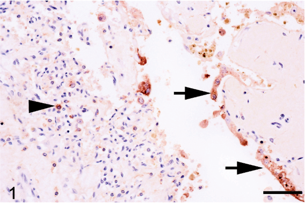
Placenta; goat. BVDV antigen is detected within the trophoblastic epithelium (arrows) as well as the mononuclear inflammatory cells (arrowhead). Streptavidin-horseradish peroxidase for BVDV counterstained with Mayer's hematoxylin. Bar = 100 μm.
Thymus; goat. Strong antigen signal is present within mononuclear cells (arrow) and reticular cells (arrowhead). Streptavidin-horseradish peroxidase for BVDV counterstained with Mayer's hematoxylin. Bar = 100 μm.
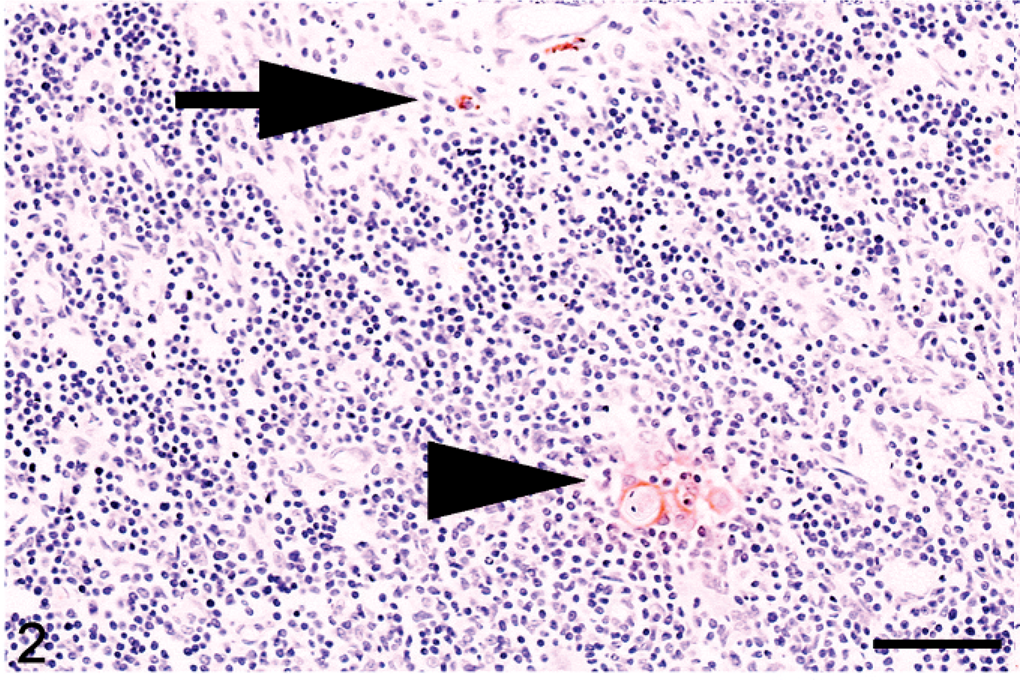
Brain; goat. BVDV antigen is detected within glial cells (arrowheads) and endothelial cells (arrow). Streptavidin-horseradish peroxidase for BVDV counterstained with Mayer's hematoxylin. Bar = 100 μm.
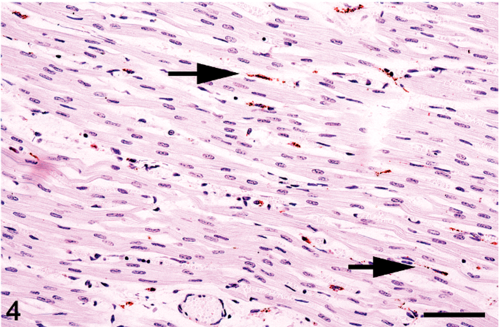
Heart; goat. BVDV antigen is detected within rare myofibers (arrows). Streptavidin-horseradish peroxidase for BVDV counterstained with Mayer's hematoxylin. Bar = 100 μm.
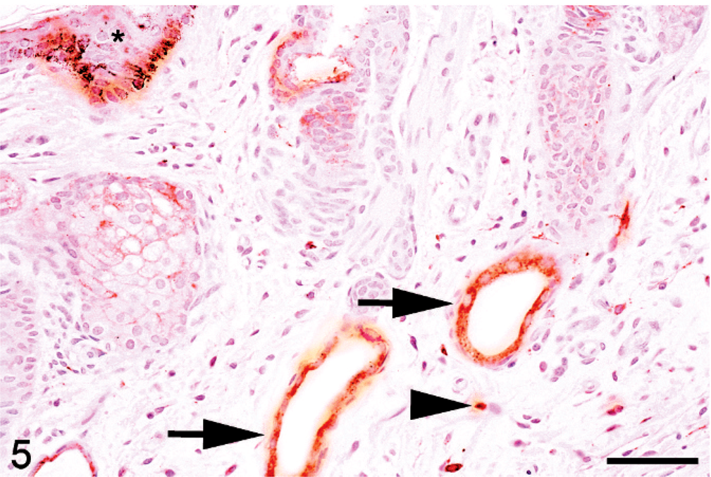
Skin; goat. The epidermis (asterisk), glandular epithelium (arrows), and mononuclear cells (arrowhead) have strong antigen signal. Streptavidin-horseradish peroxidase for BVDV counterstained with Mayer's hematoxylin. Bar = 100 μm.
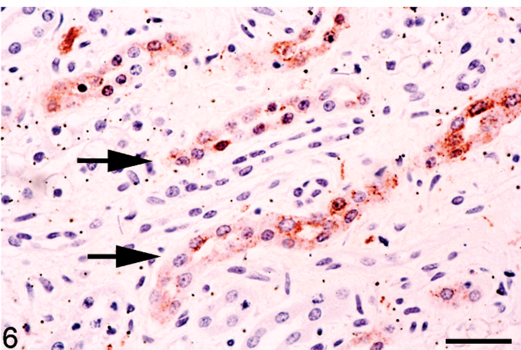
Kidney; goat. BVDV antigen is detected within the medullary tubular epithelial cells (arrows). Strepavidin-horseradish peroxidase for BVDV counterstained with Mayer's hematoxylin. Bar = 100 μm.
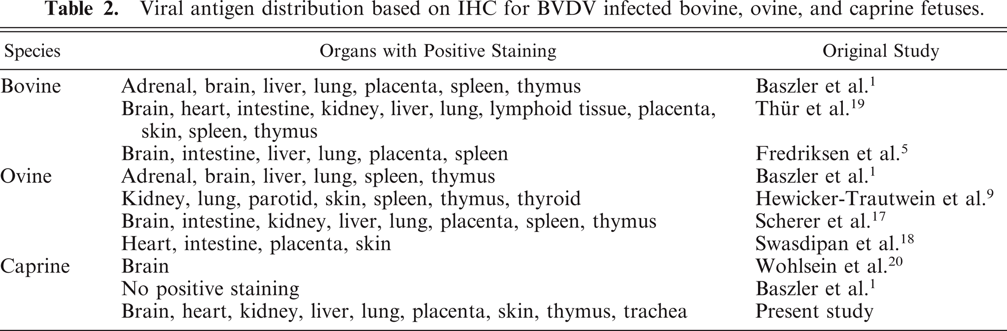
The distribution of BVDV antigen based on IHC in aborted goat fetuses is similar to, though not as widespread as that in aborted bovine and ovine fetuses (Table 2). Based on the results of this study, BVDV antigen was most commonly detected within the placenta, thymus, heart, and brain of aborted goat fetuses and stillborn or nonviable goat kids infected with BVDV. BVDV antigen was also typically detected within the brain, heart, placenta, and thymus of ovine and bovine fetuses with IHC. 1, 17– 19 Though BVDV antigen is commonly detected within the lung, liver, skin, and kidney of ovine and bovine fetuses, antigen was rarely detected within these tissues in the goat fetuses included in this study. Furthermore, other bovine and ovine fetal tissues, such as endocrine organs and spleen, consistently contain BVDV antigen. These tissues were not immunolabeled in the goat fetuses.
Viral antigen distribution based on IHC for BVDV infected bovine, ovine, and caprine fetuses.
The reason for the more limited distribution of viral antigen in fetal goats compared with sheep and cattle is not known. Limited distribution may be related to limited viral replication in goat fetuses, with replication being restricted to small clusters of cells in certain organ systems. The limited distribution of BVDV antigen in goat tissues may also be related to differences in the virulence and/or tissue tropism between the strain of virus used in this study compared with those used in previous studies in sheep and cattle. Or the immunolabeling differences may be related to the level of virus within the tissues; the levels in some organs in the goats in this study were below the detection limit of the IHC used.
BVDV antigen can be detected within a variety of cell types in infected ruminant fetuses. In cattle, BVDV infection is widespread and can be detected within the placental trophoblasts; endothelial cells; mononuclear white blood cells; mucosal and glandular epithelial cells in multiple organ systems; and multiple endocrine organs, including follicular cells of the thyroid. 1, 3, 4, 10, 11, 13, 19 Viral antigen has also been demonstrated within cerebral neurons, glial cells, vascular smooth muscle cells, and mononuclear cells. 14, 19 BVDV antigen distribution is somewhat more limited in sheep. Viral antigen can be detected in the trophoblast cells of the placenta, tubular epithelial cells of the kidney, mononuclear white blood cells, endothelial cells in multiple organs, and pneumocytes. 17, 18
In a previous study, BVDV antigen were found in the glial cells and neurons of aborted goat fetuses. 19 The immunolabeling pattern within the brain of the fetuses in the present study was extensive, involving more cell types than previously reported. Furthermore, immunolabeling was noted in several additional organ systems not reported in previous studies. The immunolabeling pattern within the placenta, thymus, brain, kidney, trachea, skin, and liver were similar to that described in cattle where a wide variety of cell types contain demonstrable antigen. The BVDV antigen distribution in the lung of the goat fetuses was restricted to the circulating mononuclear cells, which was a striking difference from the extensive epithelial and glandular staining reported in cattle and sheep.
The mechanism of BVDV infection within ruminant fetuses is still not clearly understood. Several studies suggest that the placenta is infected first through the dam's vascular supply. 17, 18 The infection then crosses the placenta to the fetus and spreads through the fetal blood supply to the fetal organs. The uterus of the dam is infected only after fetal infection is well established. We can only speculate on the mechanism of fetal infection in the goat fetus based on the findings of this study. In this study and the larger parent study, BVDV antigen was most commonly detected within the placenta: fetal infection was not documented in goats in which the placenta was negative for BVDV antigen. 3 This would suggest that placental infection precedes fetal infection. Additional time-course studies are needed to further understand the pathogenesis of fetal infection and abortion due to BVDV in goats.
Based on the results of this study, placenta, heart, thymus, and brain appear to be the most reliable tissues for detecting BVDV antigen using IHC in aborted goat fetuses. BVDV antigen can be detected in organs with and without histologic lesions.
Footnotes
Acknowledgements
We thank Mr. Curtis Andrew and Mrs. Darlene Giracello for their excellent technical assistance in preparing the immunohistochemistry slides. We also thank our collaborators on the larger study, Dr. Lionel Dawson and Dr. Sanjay Kapil.