
Abstract
A 20–year old male cotton-top tamarin (Saguinus oedipus) was presented with unilateral enlargement of an intrascrotal testicle. Fine-needle aspiration cytology demonstrated a neoplastic population with Call-Exner-like bodies and features of malignancy. The animal was castrated, and histologic examination revealed a biphasic sex cord-stromal tumor, with one region resembling Sertoli-cell tumor and one region resembling granulosa-cell tumor, with extensive microfollicular pattern and many Call-Exner bodies. Eight months after castration, the animal was euthanized on discovery of a caudal abdominal mass that displaced organs, was highly infiltrative, and extended into the paravertebral musculature with lysis of vertebral bone. Metastases to lymph node and lung were also present. Histologic examination of the abdominal tumor showed multifocal formation of Call-Exner bodies in an otherwise highly dedifferentiated population. Positive immunolabeling for alpha inhibin confirmed the sex cord-stromal origin of the abdominal and paravertebral tumor masses. This case has similarities to malignant testicular granulosa-cell tumor of humans.
Sertoli-cell tumors and Leydig-cell tumors comprise the common testicular sex cord–stromal tumors in domestic animals, with prevalence varying according to species. 22 Mixed testicular sex cord–stromal tumors in animals have rarely been reported and have been limited to tumors of combined Sertoli-Leydig–cell composition. 27, 36 Mixed germ cell–sex cord–stromal tumors are more commonly recognized and consist of neoplastic Sertoli cell and neoplastic germ cell components admixed in a single tumor. 8, 19, 25, 26, 28 To the authors' knowledge, only a single case of testicular granulosa-cell tumor in a non-human species has been reported. 1 However, there are multiple published reports of testicular tumors containing Call-Exner-like bodies suggestive of granulosa cell differentiation in animals. 5, 8, 28, 32, 36 In humans, granulosa-cell tumors of the testes are rare but constitute the most common testicular tumor in patients under 6 months of age, in which they consistently display benign behavior. 3, 4, 34 Of the 25 reported testicular granulosa-cell tumors in adult men however, 5 developed metastases, either within the abdominal cavity or distantly to bone. 10, 14, 21, 30, 34 Testicular tumors in general and testicular sex cord–stromal tumors in particular have been very infrequently reported in non-human primates. 13, 18 Testicular tumors in the cotton-top tamarin (Saguinus oedipus), a small, endangered species of monkey native to Colombia, have not previously been documented to the authors' knowledge.
During a routine, quarterly, preventative health check, a 20-year-old male cotton-top tamarin, that had lived in the New England Primate Research Center tamarin colony since birth, was noted to have an enlarged, firm left testicle. Both testes were intrascrotal, and the contralateral testicle was clinically unremarkable. The remainder of the physical examination and the results of a complete blood cell count were within normal limits. A fine-needle aspirate of the enlarged testicle was obtained and stained with Wright's-Giemsa for cytologic examination, yielding a sample of moderate cellularity on a variegated pink-to-gray background, intermixed with profuse, 1–20-µm-diameter, nonstaining accretions of granular mineral (Fig. 1). Cells present predominantly occurred clustered around streaming bands and pools of smooth, amorphous, pink, extracellular material (Fig. 1), with a marked tendency for cells to encircle accumulations of this material in a manner reminiscent of Call-Exner bodies (Fig. 2). The cells present had wispy, moderately basophilic, finely granular cytoplasm, occasional moderate punctate cytoplasmic vacuolation, and variably well-defined cytoplasmic margins (Fig. 3). Nuclei were round to ovoid, often eccentrically positioned, with delicate lacy to coarse ropy chromatin, and 1–3 indistinct-to-prominent pale-blue nucleoli, which were occasionally angular, elongate, or unusually large. The nuclear-to-cytoplasmic ratio was variable but often high, and there was up to threefold variation in cellular and nuclear size. Infrequent thick strands of fibrillar collagen, occasional well-differentiated mast cells, and low numbers of large macrophages, with copious cytoplasmic vacuolation and phagocytized mineral material, were also present (not shown). Given the suspicion of a malignant neoplasm based on results of the cytologic examination, the animal was castrated, and both testicles were submitted for histopathologic analysis.
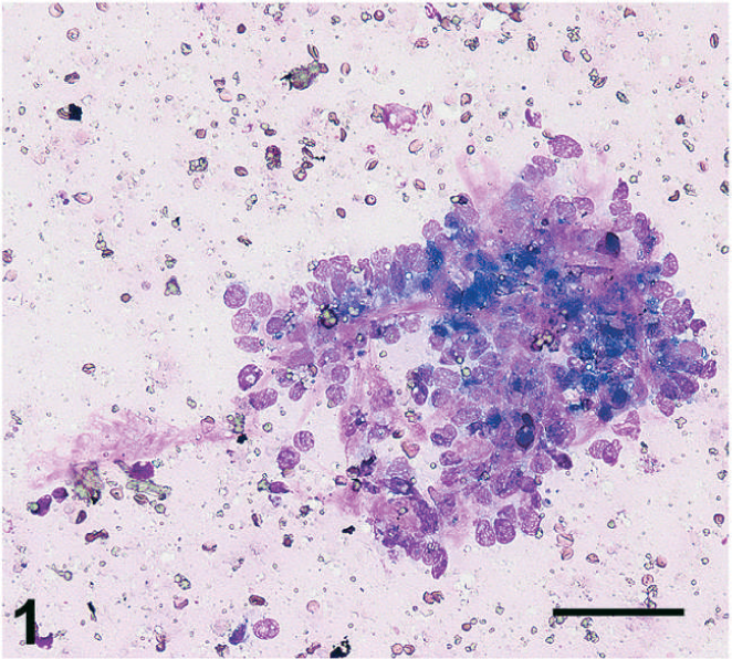
Left testis, fine-needle aspirate. Dense aggregates of cells clustered around streaming, smooth, pink extracellular material, consistent with aspiration of granulosa cell tumor microfollicles. Wright's-Giemsa. Bar = 100 µm.

Left testis, fine-needle aspirate. Ruptured cells partially encircle a pool of smooth, pink, extracellular material, suggestive of Call-Exner body. Wright's-Giemsa. Bar = 20 µm.
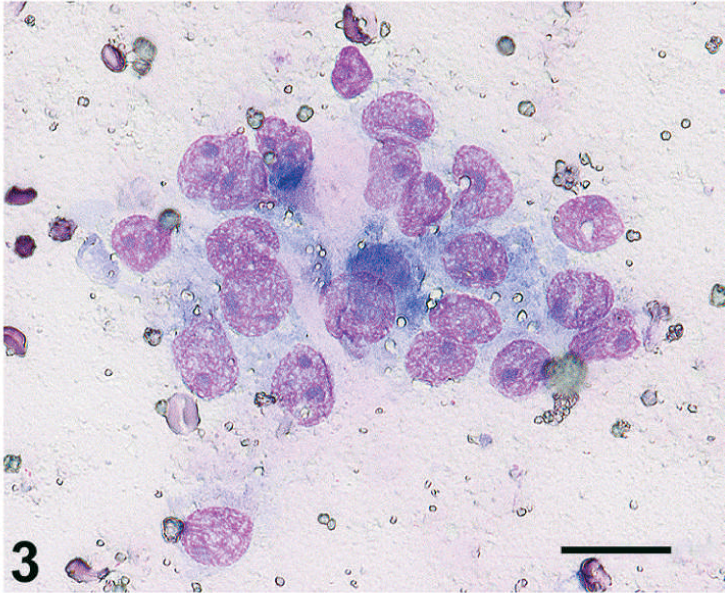
Left testis, fine-needle aspirate. Cluster of disorganized, atypical cells with small amount of associated pink extracellular material. Wright's-Giemsa. Bar = 20 µm.
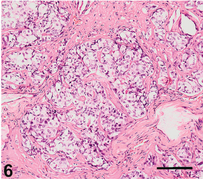
On gross examination, the left testicle was greatly enlarged, measuring 1.3 × 0.8 × 0.8 cm compared with 0.5 × 0.3 × 0.3 cm for the contralateral gonad. The tissue was soft, with a variegated pink, gray, and tan surface on cut section. On histologic examination, the normal tissue architecture was effaced and expanded by a densely cellular neoplasm composed of 2 distinct populations of cells. Each of these populations constituted roughly half of the total tumor mass, a feature retained throughout 18 sections examined by light microscopy. Cells of the first population were arranged in ribbons, cords, and cellular rosettes. Prominent pools of hyalinized, eosinophilic, extracellular material were present within the cellular rosettes in a microfollicular Call-Exner body pattern (Fig. 4). Cells of this population were cuboidal to columnar, with scant-to-moderate quantities of finely vacuolated eosinophilic cytoplasm, basally positioned nuclei with coarsely stippled nuclear chromatin, and 1–3 dark basophilic nucleoli of variable size and shape. As in the cytologic specimens, there was approximately threefold anisocytosis and anisokaryosis. Cellular atypia increased in densely cellular, solid areas, away from organized follicular or cord structures. Mitotic figures numbered 2 per 400× field. There were moderate numbers of individual apoptotic cells throughout, along with multifocal large areas of necrosis and mineralization, and multiple regions of invasion into the tunica albuginea. The extracellular material present in the Call-Exner-like bodies showed intense histochemical staining with the Periodic Acid-Schiff (PAS) (Fig. 5).
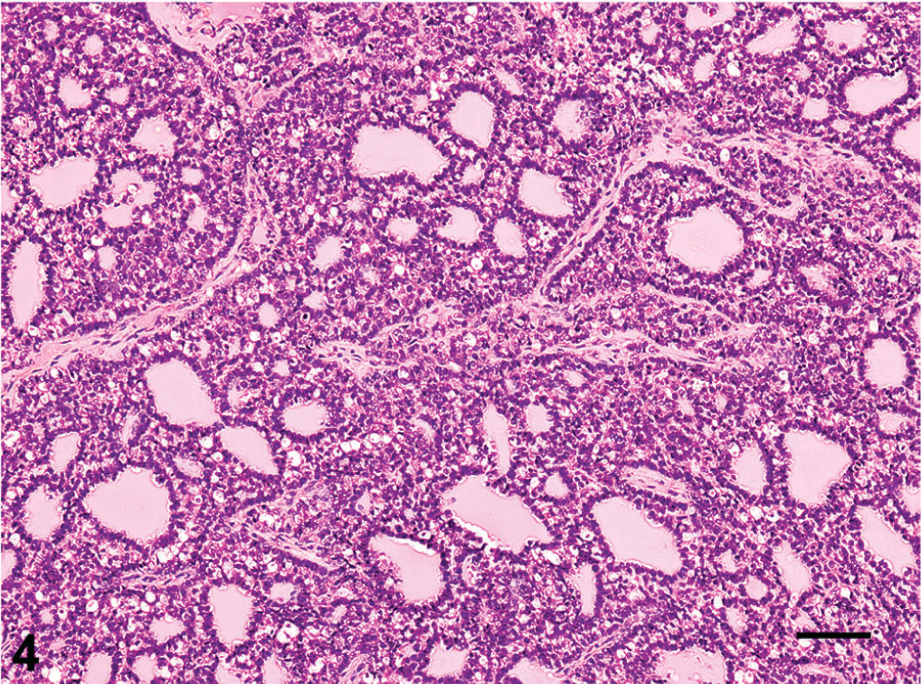
Left testis. Neoplastic granulosa cells forming cords and follicular structures, with many prominent Call-Exner bodies. HE. Bar = 70 µm.
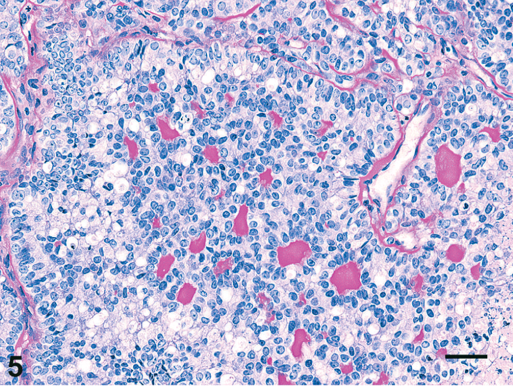
Left testis. The intrafollicular material comprising the Call-Exner bodies is strongly PAS positive. PAS reaction. Bar = 35 µm.
The second population, which occupied the remaining half of the tissue, consisted of clusters of polygonal cells subdivided by thick collagenous bands into irregular nodules and tubules (Fig. 6). The collagenous nature of the bands was confirmed with trichrome staining (Fig. 7). Cells in this region differed from those in the previous population in being considerably larger, having abundant, highly vacuolated, pale eosinophilic cytoplasm; larger nuclei, with more open, finely stippled nuclear chromatin; only rare mitotic figures; and a higher frequency of apoptotic cells. Like the first population, well-defined regions of necrosis and mineralization were present throughout, along with rare Call-Exner-like bodies bordered by cells indistinguishable from the remainder of cells in this section of the tumor.
Left testis. Atypical proliferative Sertoli cells in a tubular to diffuse distribution with extensive fibroplasia. HE. Bar = 200 µm.
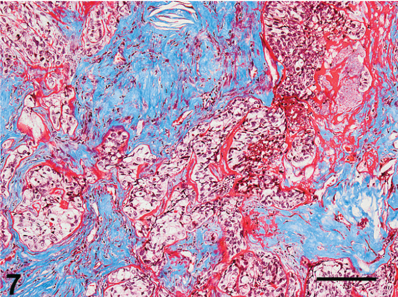
Left testis. Extensive intratumoral collagen deposition is confirmed by histochemical staining for collagen, which appears blue by this method. Masson's trichrome. Bar = 200 µm.
In regions where the 2 populations abutted one another to form a transitional zone, cellular morphology and tumor architecture became highly ambiguous, with cells no longer discretely classifiable as belonging to either one population or the other (not shown). Cells within a focal region at the intersection of the 2 populations showed intense cytoplasmic staining for cytokeratin by immunohistochemistry (rabbit polyclonal Z0622, Dako, Carpinteria, CA), with intermittent cytokeratin positivity along the periphery of this region (Fig. 8). Strong cytoplasmic immunoreactivity for cytokeratin in adjacent epididymal epithelial cells (not shown) provided an internal control for specificity of staining. Histologic examination of 2 HE stained sections of the contralateral gonad revealed moderate tubular degenerative changes and an absence of spermatogenesis but no evidence of neoplasia.
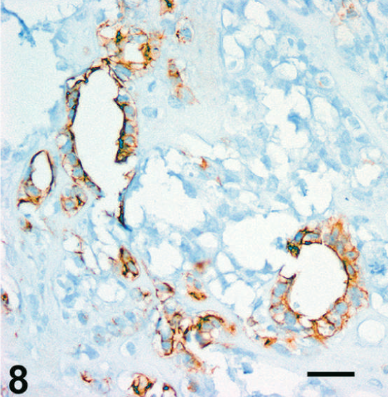
Left testis. A subset of the follicular structures and surrounding cells showed strong cytoplasmic reactivity for cytokeratin. Wide-spectrum cytokeratin immunohistochemistry, avidin-biotin complex method, Mayer's hematoxylin counterstain, DAB chromogen. Bar = 100 µm.
At 8.5 months after castration, a large mass was palpated in the animal's caudal abdomen during a routine physical examination. Because of a high suspicion for colonic adenocarcinoma, which is common in cotton-top tamarins, 17 the animal was euthanatized. At necropsy, an approximately 4-cm-diameter, firm, irregularly nodular, pale tan-yellow mass, which greatly distorted the lower left quadrant of the abdomen, was identified (Fig. 9). The mass was not associated with any segment of the gastrointestinal tract and was adhered to the left dorsolateral abdominal wall, with an extension that projected dorsocranially to the level of the kidneys. There was extensive infiltration, effacement, and expansion of the left seminal vesicle, as well as infiltration of the bladder wall, and deep infiltration of paravertebral musculature with lysis of vertebral bone. On histologic examination, the abdominal mass consisted of a densely cellular, partially encapsulated, multinodular neoplasm, with nodules defined by thick bands of collagen (Fig. 10). Nodules were subdivided into packets and cords by fine fibrovascular stroma. Cells of the mass had indistinct cytoplasmic margins, with scant-to-moderate quantities of eosinophilic cytoplasm; a high N : C ratio; round-to-ovoid nuclei with open, vesicular chromatin; and 1–3 small, dark, basophilic nucleoli. Up to fourfold anisokaryosis was present, along with multiple large discrete areas of necrosis, frequent individual cell apoptosis, and multifocal tumor emboli within lymphatics. At several sites, cells within the tumor nodules formed individuated microfollicular structures that resembled Call-Exner bodies, similar to those identified in the testicular mass (Fig. 10, inset). Rare aggregates of disorganized follicular structures lined by pleomorphic cells and filled with smooth eosinophilic extracellular material also suggested more widespread attempts at Call-Exner body formation (Fig. 11). Immunohistochemical staining for alpha inhibin (clone R1, Thermo Scientific, Fremont, CA) was negative on sections of the testicular tumor itself but showed multifocal, often lobular positivity in sections of the abdominal tumor (Fig. 12) and paravertebral infiltrates, which confirmed sex cord–stromal origin. Randomly distributed, often very strong granular cytoplasmic positivity for alpha inhibin occurred within some areas of the tumor (Fig. 13) but was found only rarely in better differentiated regions that formed Call-Exner-like bodies (Fig. 13, inset). Metastases histologically resembling the main tumor were found effacing the inguinal lymph node and forming an embolus of cells within an intrapulmonary branch of the pulmonary artery. No other metastatic lesions or evidence of additional primary neoplastic disease was detected on either gross or microscopic examination.
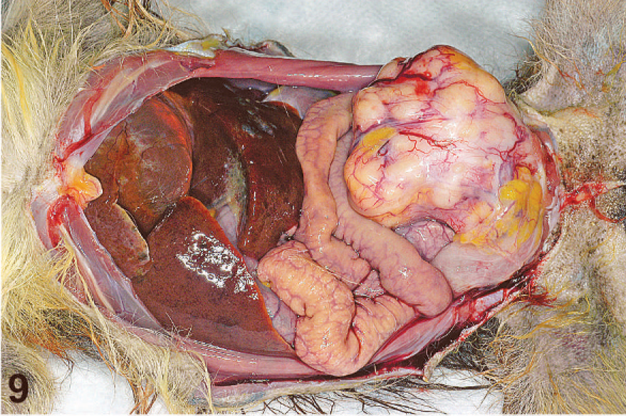
Abdominal cavity. An irregular, multilobulated, pale tan-yellow mass expands and nearly fills the caudal left quadrant of the abdomen.
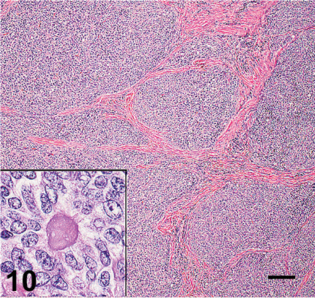
Abdominal mass. Neoplastic cells form densely cellular nodules subdivided by thick bands of collagen. Occasional isolated follicular structures resembling Call-Exner bodies (inset) are present within the sheets of tumor cells. HE. Bar = 400 µm.
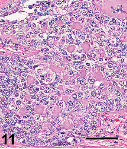
Abdominal mass. Foci of atypical cells forming large numbers of poorly organized follicular structures resembling Call-Exner bodies are found at low frequency. HE. Bar = 100 µm.
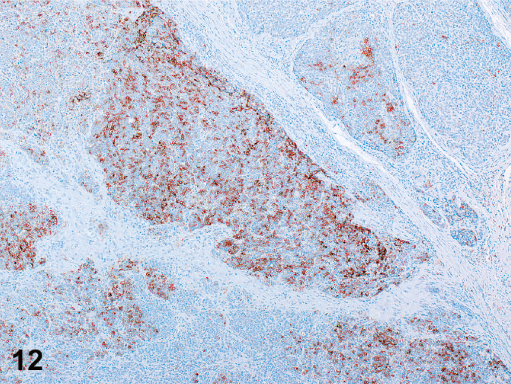
Abdominal mass. Tumor cells immunolabeled with anti-alpha inhibin monoclonal antibody in a multifocal to lobular pattern. Avidin-biotin complex method, Mayer's hematoxylin counterstain, DAB chromogen.
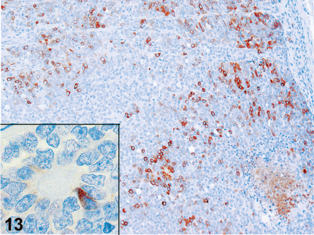
Abdominal mass. Tumor cells show variable intensity, multifocal, granular cytoplasmic immunohistochemical signal with anti-alpha inhibin antibody. Tumor cells forming Call-Exner-like bodies (inset) only rarely demonstrate positive alpha inhibin immunolabeling. Avidin-biotin complex method, Mayer's hematoxylin counterstain, DAB chromogen.
Testicular tumors are rare in nonhuman primates and, to the authors' knowledge, have never been reported in a cotton-top tamarin. The biphasic character of the tumor in this particular animal is of special interest, given the juxtaposition of one component that most closely resembled Sertoli-cell tumor and another component that most closely resembled granulosa-cell tumor, a tumor type rare in the testis regardless of species. Cytologic features of the tumor were consistent with those described in reports of ovarian granulosa-cell tumors, although with a higher degree of atypia than is often found, in keeping with the aggressive behavior of this particular tumor. 6, 20, 23, 31
Metastatic rates for sex cord–stromal tumors vary significantly by species. Sertoli-cell tumors in dogs are common, but the vast majority are benign. 22 In contrast, testicular Sertoli-cell tumors are rare in humans, but, based on long-term follow-up, close to half may be malignant. 35 In domestic animals, the propensity for ovarian granulosa-cell tumors to metastasize varies by species. 22 Ovarian granulosa-cell tumor malignancy rates in humans range from 5 to 25%, with all granulosa-cell tumors being potentially malignant, regardless of histology. 7 Based on current reports, approximately 20% of testicular granulosa-cell tumors in adult men appear to demonstrate malignant behavior. 2, 10, 16, 30 The tumor in this animal was highly malignant, which resulted in distant metastases and extensive tissue invasion, including lysis of vertebral bone. Although the metastases lost much of the defining morphologic character found in the 2 tumor subpopulations within the primary site, they retained a tendency to form isolated-to-regional, sometimes highly disorganized microfollicular structures that resembled Call-Exner bodies, which, along with positive immunolabeling for alpha inhibin, convincingly ties their origin to the primary testicular mass.
The antigenic signature of sex cord–stromal tumors may vary. In many cases, these tumors show a positive immunohistochemical reaction for inhibin, a peptide hormone which suppresses follicle stimulating hormone secretion and serves as a marker of sex cord–stromal differentiation. 9, 12, 34, 37 However, a subset of Sertoli-cell tumors and granulosa-cell tumors may be negative for inhibin, 9, 12, 35, 37 as appears to have been the case with the primary tumor in this animal, despite the fact that metastatic lesions were characterized by abundant inhibin expression. Reported expression of cytokeratins in sex cord–stromal tumors varies extensively. 9, 11, 25, 35 Call-Exner bodies are characteristically PAS positive and Alcian blue positive, and have been demonstrated in normal ovarian follicles to be composed of aggregates of accumulated basal lamina or basal lamina-like material, with positivity for type IV collagen in preantral follicles switching to predominant laminin positivity in antral follicles. 29, 33 Immunohistochemistry for type IV collagen (clone CIV22, Dako, Carpinteria, CA) was attempted on the testicular sections of this case. While Call-Exner-like bodies uniformly stained positive, interpretation of the result was confounded by very weak basement membrane staining in the section (data not shown).
The origin of granulosa-like cells in testicular tissue, where they are not normally resident, parallels the finding of Sertoli cells in human ovarian Sertoli-cell tumors. Although the exact pathogenesis of the development of granulosa-cell tumors in testes or Sertoli-cell tumors in ovaries is not clear, granulosa cells constitute the ovarian cell population whose gene expression profiles most closely match testicular Sertoli cells, and in humans appear to be linked by a similar mesonephric embryologic origin, although this origin may vary across species. 15, 24 The short evolutionary distance between humans and nonhuman primates would make similar embryologic characteristics of gonadogenesis relatively likely. It seems plausible, therefore, that testicular granulosa-like cells such as seen in this case, as well as those that have been described forming Call-Exner-like bodies in numerous other testicular tumors in animals, 5, 8, 28, 32, 36 arise from dysregulated resident Sertoli cells, although this has never been proven. While this tumor differed from the pure testicular granulosa-cell tumors documented in humans because of its biphasic character, it demonstrated pronounced granulosa-cell differentiation at the primary site, which was retained at sites of distant metastases.
Footnotes
Acknowledgements
We thank Douglas Pauley and Dennis Walsh for their technical support, along with Kristen Toohey for photographic assistance. Funding was provided by NIH grants RR00168, T32 RR007000, and K01RR24120.