
Abstract
The enzyme cyclooxygenase-2 (COX-2) is expressed in some tumor and stromal tissues, and catalyzes production of prostaglandins with growth stimulatory, antiapoptotic, proangiogenic, and immunosuppressive properties. Pharmacologic inhibition of COX-2 is associated with antitumor activity in various human and canine malignancies. The purpose of this study was to assess COX-2 expression in a series of equine sarcoids, melanomas, and squamous-cell carcinomas (SCC). COX-2 expression was assessed in formalin-fixed paraffin-embedded tissues from 14 sarcoids, 11 melanomas, and 37 SCC that represent various anatomic sites by using standard immunohistochemical methods. COX-2 was expressed in 2 of 14 sarcoids, 7 of 11 melanomas, and 32 of 37 SCC, 56% of which demonstrated moderate-to-strong immunoreactivity. There were no differences in expression between anatomic sites. In conclusion, most equine SCC and many melanomas appear to express COX-2 and thus could respond to COX-2 inhibitor therapy.
The cyclooxygenase (COX) enzymes, which catalyze the production of multiple prostaglandins from arachidonic acid, play a key regulatory role in numerous physiologic functions, including regulation of renal perfusion, platelet aggregation, and maintenance of gastrointestinal mucosal integrity. The COX-1 isoform is expressed constitutively in many cells, whereas the COX-2 isoform is inducible and is expressed in normal cells, such as macrophages and multiple types of human neoplasia, where it may mediate diverse oncogenic functions, including stimulation of proliferation and angiogenesis, inhibition of apoptosis, and immune suppression. 10, 11
Recent studies demonstrated COX-2 expression in a variety of canine carcinomas, including urinary bladder and mammary gland, as well as squamous-cell carcinoma (SCC), melanoma, and osteosarcoma. 3, 5, 7 COX-2 expression has been demonstrated in feline mammary carcinoma and in a subset of feline transitional-cell carcinomas and SCC. 1, 3 Furthermore, strong COX-2 expression may confer a worse prognosis in canine and feline mammary tumors and canine osteosarcoma. 3, 7
There is accumulating epidemiologic and clinical evidence that inhibition of COX-2 with nonsteroidal anti-inflammatory drugs (NSAID) may reduce the occurrence of cancer in humans. 10, 11 Furthermore, the NSAID piroxicam has been shown to induce meaningful antitumor responses in dogs with certain malignancies, such as transitional-cell carcinoma and oral SCC. 4, 9 A single case report exists of long-term control of a metastatic SCC in a horse after the administration of piroxicam. This tumor was demonstrated to abundantly express COX-2 by immunohistochemistry (IHC). 6 The purpose of this study was to assess the expression of COX-2 by IHC in a series of archived equine sarcoids, melanomas, and SCC.
COX-2 expression was assessed in formalin-fixed paraffin-embedded tissues from 14 sarcoids from 14 individuals; 11 melanomas from 6 individuals, which represent various anatomic sites, including subcutis, lymph node, bone, pituitary, and abdominal viscera; and 37 SCC from 32 individuals, which represent various anatomic sites, including eye/periocular area (n = 9), prepuce/penis (n = 7), vulva (n = 5), gastrointestinal/oral/nasal (n = 7), and metastatic sites (n = 9). In 5 SCC cases, paired primary tumors and lymph node or distant metastases were evaluated from the same individual.
The IHC staining was performed by using standard techniques on an automated stainer (Discovery, Ventana Medical Systems, Tucson, AZ). Briefly, 4-µm sections were cut and mounted on positively charged slides. The sections were deparaffinized and then rehydrated with descending alcohol concentrations to tris-buffered saline solution with 0.05% Tween 20. The melanomas were bleached with 10% H2O2 for 24 hours at room temperature before epitope retrieval. Heat-induced epitope retrieval with a proprietary EDTA-containing antigen retrieval buffer, pH 8.0 (Ventana Medical Systems) at 95°C for 30 minutes was followed by blocking endogenous peroxidase with 3% hydrogen peroxide and incubation with the primary antibody at room temperature for 10 hours.
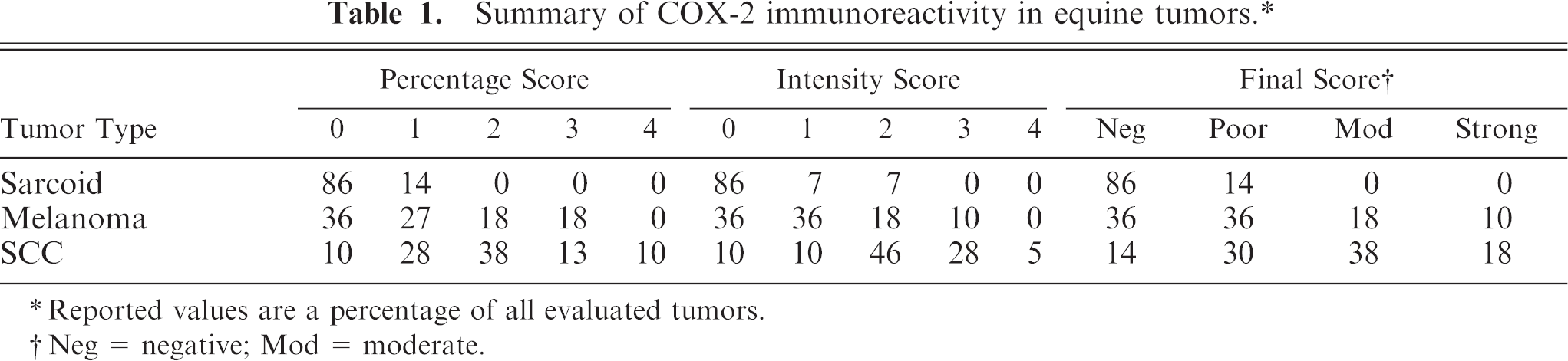
The primary antibody used was a polyclonal rabbit anti-human prostaglandin H synthetase-2 at a dilution of 1 : 250 (Cat. no. PG27b, Oxford Biomedical Research, Oxford, MI). A prediluted, universal biotinylated secondary antibody and the Ventana DAB detection kit (Ventana Medical Systems) were used to detect the immunoreactive complexes. The slides were then counterstained with Mayer's hematoxylin. Normal equine kidney served as a positive control, and substitution of normal rabbit serum for the primary antibody served as a negative control. This control was used, because positive COX-2 immunoreactivity is present in the juxtaglomerular apparatus of other species.
Immunoreactivity scoring was performed in a blinded fashion by a single pathologist (EJE). The percentage of COX-2 positive tumor cells was scored as either 0 = 0%, 1 = 1–5%, 2 = 6–20%, 3 = 21–50%, or 4 = >50% of cells. In addition, the intensity of the stain uptake was graded as 0 = negative, 1 = weak, 2 = moderate, 3 = strong, and 4 = intense. Intensity was based on a comparison with control tissue staining. The percentage score and the intensity grade were then multiplied to give a final immunoreactivity score, which ranged from 0 to 16. The categories for the final immunoreactivity score were as follows: negative (0), weak (1–3), moderate (4–7), and strong expression (8–16). Differences in final immunoreactivity scores between matched primary and metastatic tumors were compared by using a paired, 2-tailed t-test.
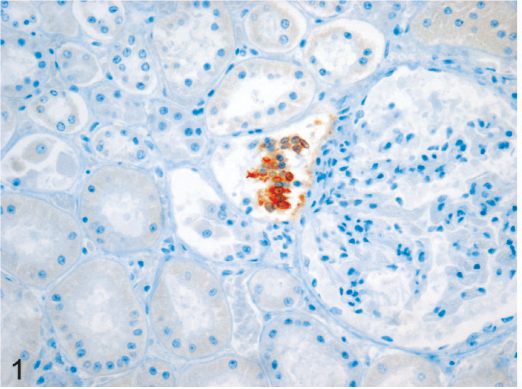
Positive control tissue had intense staining confined to the juxtaglomerular apparatus of the kidney (Fig. 1); there was no immunoreactivity in negative control sections. Staining in all tumor cells was cytoplasmic, with increased intensity in perinuclear zones. Staining was heterogeneous throughout tumor sections. A summary of tumor COX-2 immunoreactivity is provided in Table 1. COX-2 was expressed in 2 of 14 sarcoids (14%) and 7 of 11 melanomas (63%). In the 2 sarcoids that express COX-2, final immunoreactivity scores were weak in both. Three of 6 horses with melanoma had at least 1 COX-2–positive lesion. Final COX-2 immunoreactivity scores in the melanomas were negative in 36%, weak in 36%, moderate in 18%, and strong in 10%. A photomicrograph of equine melanoma tissue that demonstrated COX-2 immunoreactivity is provided in Fig. 2.
Summary of COX-2 immunoreactivity in equine tumors.∗
Reported values are a percentage of all evaluated tumors.
Neg = negative; Mod = moderate.
Normal equine kidney. Rabbit polyclonal anti-COX-2. DAB substrate and hematoxylin counterstain. Intense staining is localized to the juxtaglomerular apparatus.
Equine melanoma. Rabbit polyclonal anti-COX2. DAB substrate and hematoxylin counterstain.
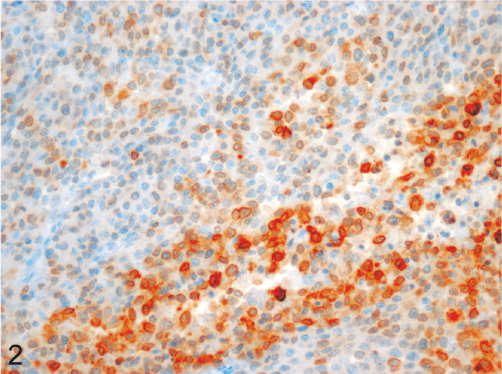
Thirty two of 37 SCC (86%) expressed COX-2, and 28 of 32 patients with SCC had at least 1 COX-2–positive lesion. Final immunoreactivity scores were negative in 14%, poor in 30%, moderate in 38%, and strong in 18%. Photomicrographs of SCC tissue that demonstrated absent and strong COX-2 immunoreactivity are provided in Figs. 3, 4, respectively. There were no observable differences in final immunoreactivity scores or percentage positives between different anatomic sites, but there was a trend (P = .12) toward reduced scores in metastases versus primary tumors from the same individual (Fig. 5). This is in contrast to what is reported in the veterinary literature, in which COX-2 expression is more often upregulated in metastases and/or associated with a worse outcome after therapy. 3, 7
Equine SCCs. Examples of negative (
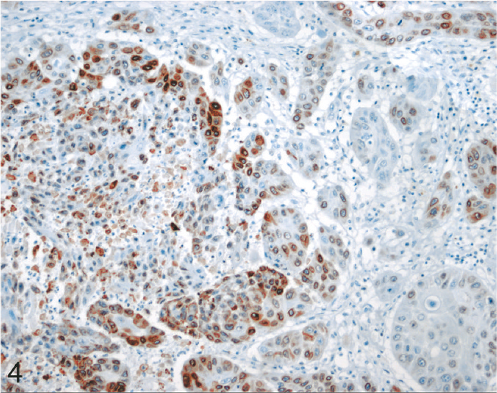
(
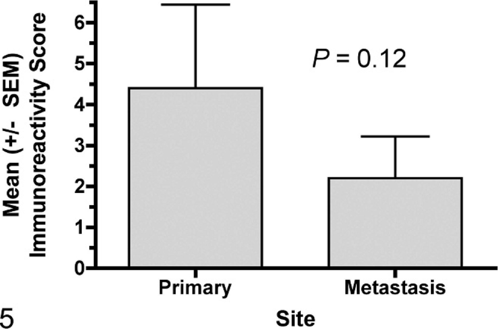
Mean (±SEM) COX-2 immunoreactivity scores between paired equine SCC primary tumors and metastases from the same individual.
Based on the data provided in this study, COX-2 appears to be expressed in very few equine sarcoids, a substantial number of melanomas, and the large majority of SCC. Most SCC demonstrated poor-to-moderate immunoreactivity, and immunoreactivity was independent of anatomic site. These results are similar to what has been reported in dogs, in which approximately 65% of SCC and 60% of melanomas are positive for COX-2 by IHC 5 but in contrast to what is observed in cats, in which only a minority of SCC express COX-2. 1
A recent publication that used Western analysis rather than IHC techniques demonstrated ubiquitous expression of both COX-1 and COX-2 in a 100% of equine SCC and in all normal tissues evaluated. 2 Although the study reported here did not seek to comprehensively evaluate normal equine tissues, most anatomic structures within the equine kidney were IHC negative, as were a significant number of equine tumor samples. It is not obvious why the results of Elce et al 2 and the results reported here differ. The Western methodology and IHC may have different sensitivities. The potentially higher threshold for a positive signal obtained with IHC might allow better discrimination between malignant and normal tissues than that achieved with Western analysis. In addition, Elce et al 2 used a different antibody for their investigations. Western and IHC approaches to assess protein expression have different benefits. IHC allows the precise cellular and subcellular localization of protein expression, whereas Western analysis allows the molecular weight of the immunoreactive protein to be determined as an added measure of specificity. Although Western analysis was not performed by using this antibody in equine tissues, the cellular, normal tissue, and intratumoral distribution of COX-2 immunoreactivity observed in our study was similar to that reported in feline and canine tumors. 1, 5
Although NSAIDs may exert antitumor effects by COX-independent mechanisms, and a recent study of dogs with canine transitional-cell carcinoma failed to demonstrate a relationship between COX-2 expression or prostaglandin production and responsiveness to COX-2 inhibition with the NSAID piroxicam, 8 the bulk of information suggests that NSAID therapy is most effective in veterinary tumor types that commonly express COX-2. 4, 5, 9 Indeed, the single case report that documented an antitumor response to piroxicam in a horse with SCC concurrently demonstrated abundant COX-2 expression in the lesions. 6
With the exception of the aforementioned case report and the recent report by Elce et al., 2 which used a different methodology, this is the first study undertaken to systematically evaluate the expression of COX-2 in multiple equine-tumor types. Taken together, these data and the available veterinary literature suggest that equine SCC and melanoma might be logical targets for the clinical investigation of therapy with COX-2 inhibitors, e.g., piroxicam. Given the range of COX-2 immunoreactivity detected in these tumors, it will be interesting to determine if a pretreatment level of COX-2 expression can be correlated with NSAID sensitivity.
Footnotes
Acknowledgements
This study was supported with funds from the Colorado State University Animal Cancer Center and the Department of Clinical Sciences, College of Veterinary Medicine and Biomedical Sciences, Colorado State University. Dr. Thamm is supported by American Cancer Society grant RSG-04-219-01-CCE.