
Abstract
Amputation is commonly performed to both treat and diagnose conditions affecting the digits of dogs. Although histopathologic evaluation of these digits is routinely done, data on the prevalence and prognosis of neoplasms of the digit are scarce. The records of multiple veterinary diagnostic laboratories were searched to identify submissions of amputated digits from dogs. Four hundred twenty-eight separate submissions were reviewed for diagnosis, age, sex, limb of origin, and digits affected, and the original submitting clinics were surveyed to determine clinical outcome of the animal. No diagnosis could be agreed upon in 24 animals, and these were excluded from the study. Kaplan-Meier product-limit method was used to determine the disease-free interval and survival time. Neoplastic disease was identified in 296 of 404 submissions, with exclusively inflammatory lesions composing 108 cases. A total of 30 different neoplastic processes were identified. In 233 (77.7%) of the neoplastic cases, a malignant tumor was identified. Squamous cell carcinoma was the most commonly identified tumor (n = 109, 36.3%), and 11 of 42 dogs for which clinical follow-up information was available developed metastatic disease. Squamous cell carcinoma of the digit appears to have a greater metastatic potential than that occurring elsewhere in the body. Other common diagnoses included melanoma (n = 52, 17.3%), soft-tissue sarcoma (n = 29, 9.7%), and mast cell tumor (n = 20, 6.7%). Melanomas were associated with poor prognoses, with a median survival time of 365 days.
Amputation is commonly performed in veterinary medicine to both treat and diagnose conditions affecting the digit. Clinical manifestations of digital lesions in dogs are similar regardless of the cause, and histopathologic evaluation is commonly used to determine the underlying disease condition and the prognosis. Although neoplasia is reported to be common in the digits of dogs, studies of the prevalence of neoplastic disease in digital conditions are scarce. Most studies have concentrated on squamous cell carcinoma (SCC) and, to a lesser degree, melanoma of the digit. The purpose of this study was to examine the diagnoses in a large number of digits from dogs submitted to multiple veterinary diagnostic laboratories to better define the prevalence of neoplasia and the prognoses for various neoplastic diseases.
Materials and Methods
Specimens were obtained from veterinary diagnostic laboratories in the United States and Canada: Colorado State University Diagnostic Laboratory (CSUVDL), Department of Veterinary Pathology at the University of Saskatchewan, and Prairie Diagnostic Services (PDS) in Regina and Saskatoon, Saskatchewan. Biopsies of amputated digits from dogs were identified by using computer-based record searches.
Biopsies during the 6-year period between January 1, 1996, and December 31, 2001, inclusive, at the CSUVDL; the 9.5-year period from January 1, 1995, to June 30, 2004, inclusive, at the Department of Veterinary Pathology at the University of Saskatchewan and PDS laboratory Saskatoon; and the 7-year period between January 1, 1996, and December 31, 2002, inclusive, at PDS Laboratory Regina were included. For the purposes of this study, amputated digits had to include some portion of bone within the biopsy to be considered.
The HE-stained histologic slides and original diagnostic reports from each case were retrieved. These original diagnoses were confirmed histologically by an experienced pathologist (ALA) in conjunction with a pathology graduate student (BKW) viewing slides together at a multiheaded microscope. This second diagnosis was made without knowledge of the prior diagnosis or any information related to animal signalment or history. A comparison of the second and original diagnoses was then made, and agreement was recorded. Disagreements between this diagnosis and the original diagnosis were resolved by submission to a Diplomate of the American College of Veterinary Pathologists (BAK) for further assessment. This third diagnosis was used in conjunction with the other 2 diagnoses, and the majority diagnosis was recorded. Those cases in which no majority diagnosis existed were not used. Submission records for the digits were used to determine patient signalment, including breed, age, sex, neuter status, and site of amputation. Animals diagnosed with non-neoplastic inflammatory disease were considered as a group.
A survey was sent to all original submitting veterinarians to collect follow-up information regarding each case, including treatment; date of recurrence of disease, if any; metastasis, if any; survival; and cause of death, if applicable; or last date on which animal was examined.
Disease-free interval was defined as the time, in days, between amputation of the digit and recurrence of disease or identification of metastatic sites. Survival time was defined as the interval between surgical amputation of the digit and death or euthanasia of the animal. Animals that died or were euthanized due to unrelated disease or were lost to follow-up were censored at the date of this occurrence. Only animals that died or were euthanized as a result of the disease diagnosed in the amputated digit were classified as “deaths.”
Data analysis
Risk factors considered for associations with the development of neoplasia were as follows: sex and neuter status (male, female, neutered male, spayed female), age, breed, leg of origin, and digit of origin. Diagnoses were evaluated in a pair-wise fashion for these risk factors using a chi-square goodness-of-fit test, and the threshold of significance was set at P ≤ .05. Dunnett t-tests were used to compare the age of dogs with a given type of neoplasm to the age of dogs with inflammatory disease. The Kaplan-Meier product-limit method was used to determine the disease-free interval and survival time. Analysis was performed with the aid of a statistical software package (SPSS Version 13, SPSS Inc., Chicago, IL).
Results
Amputated digits from 428 dogs were evaluated. Submissions for which no diagnosis could be agreed upon (n = 24) were excluded from further evaluation. Multiple digits were affected in 34 dogs (7.9%). A total of 30 different diagnoses were identified. Malignant neoplastic processes were present in 229 (53.5%) submissions; benign neoplastic or noninflammatory non-neoplastic processes, in 67 (15.7%); and inflammation, in 108 (25.5%).
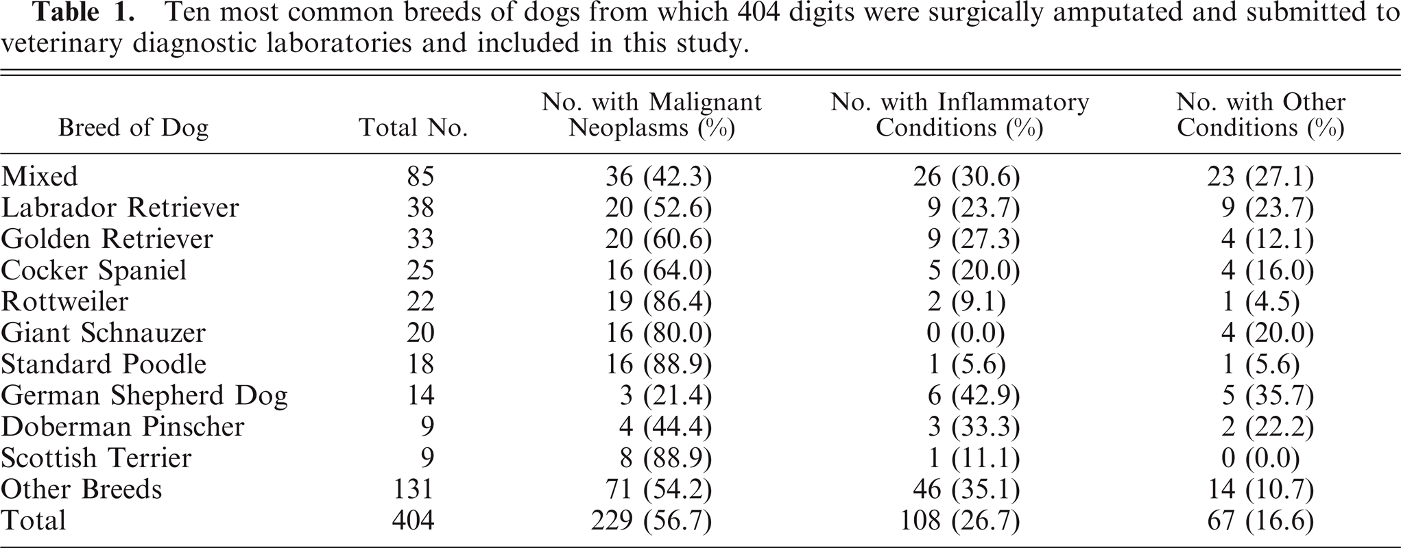
The overall study population consisted of 67 breeds of dogs. The 10 most common breeds are listed in Table 1. Two hundred two dogs were females, of which 172 were spayed; and 184 dogs were males, 132 of which were castrated. In 18 submissions, no sex was specified.
Ten most common breeds of dogs from which 404 digits were surgically amputated and submitted to veterinary diagnostic laboratories and included in this study.
Two hundred fifty-five submissions specified the affected leg in the clinical history. All 4 legs were affected in 16 submissions. In those submissions where only the front or only the hind limbs were affected, the front leg was more often affected (n = 150, 62.8%) than the hind (n = 89, 37.2%) (P < .01). Lesions were less likely to occur on a dew claw than on any other digits (20/203, 9.9%) (P < .01).
A survey was sent to the original submitting veterinarian for each of the 428 cases. Two hundred of these were returned for a response rate of 46.7%.
Inflammatory conditions, primarily pyogranulomatous in nature, were the only abnormalities in 108 submissions. In this group, the mean age at the time of amputation was 7.1 years and the age range was 1–14 years. Fifty were females and 50 were males; no sex was specified on the submission form in 8 cases. Multiple digits were reported to be involved in 19 dogs at the time of amputation. Comparisons to this group of dogs regarding age and sex distribution were made for groups of dogs with other diagnoses. The mean age of animals with inflammatory conditions was younger than that for animals with SCC (P < .01), melanoma (P < .01), soft-tissue sarcoma (P = .02), mast cell tumors (P < .01), and benign neoplasms (P < .01).
Squamous cell carcinoma was the most common diagnosis (n = 109, 26.7%) and therefore the most common malignant neoplasm. Age distribution was 3–17 years at the time of surgical amputation, with a mean age of 9.8 years. Fifty female dogs and 59 male dogs were affected. Multiple digits and feet were involved at the time of amputation in 7 of the submissions (3 standard Poodles, 2 Labrador Retrievers, 1 Giant Schnauzer, and 1 Gordon Setter). The diagnosis of SCC was over-represented in several breeds of dogs. The diagnosis of SCC compared with all diagnoses in these breeds of dogs was as follows: 11 of 22 Rottweilers, 11 of 20 Giant Schnauzers, 13 of 18 standard Poodles, 5 of 6 Dachshunds, and 3 of 3 Flat Coated Retrievers. Forty-two surveys were returned for dogs with SCC. Ten of these dogs developed metastatic disease with a mean time to metastasis of 309 days. One dog was euthanized due to recurrence of its disease at 395 days postamputation. Of the 11 dogs that died or were euthanized due to SCC, most died within the first year after amputation and diagnosis. Less than 50% of dogs died as a result of SCC, so no median survival time can be determined (Fig. 1).
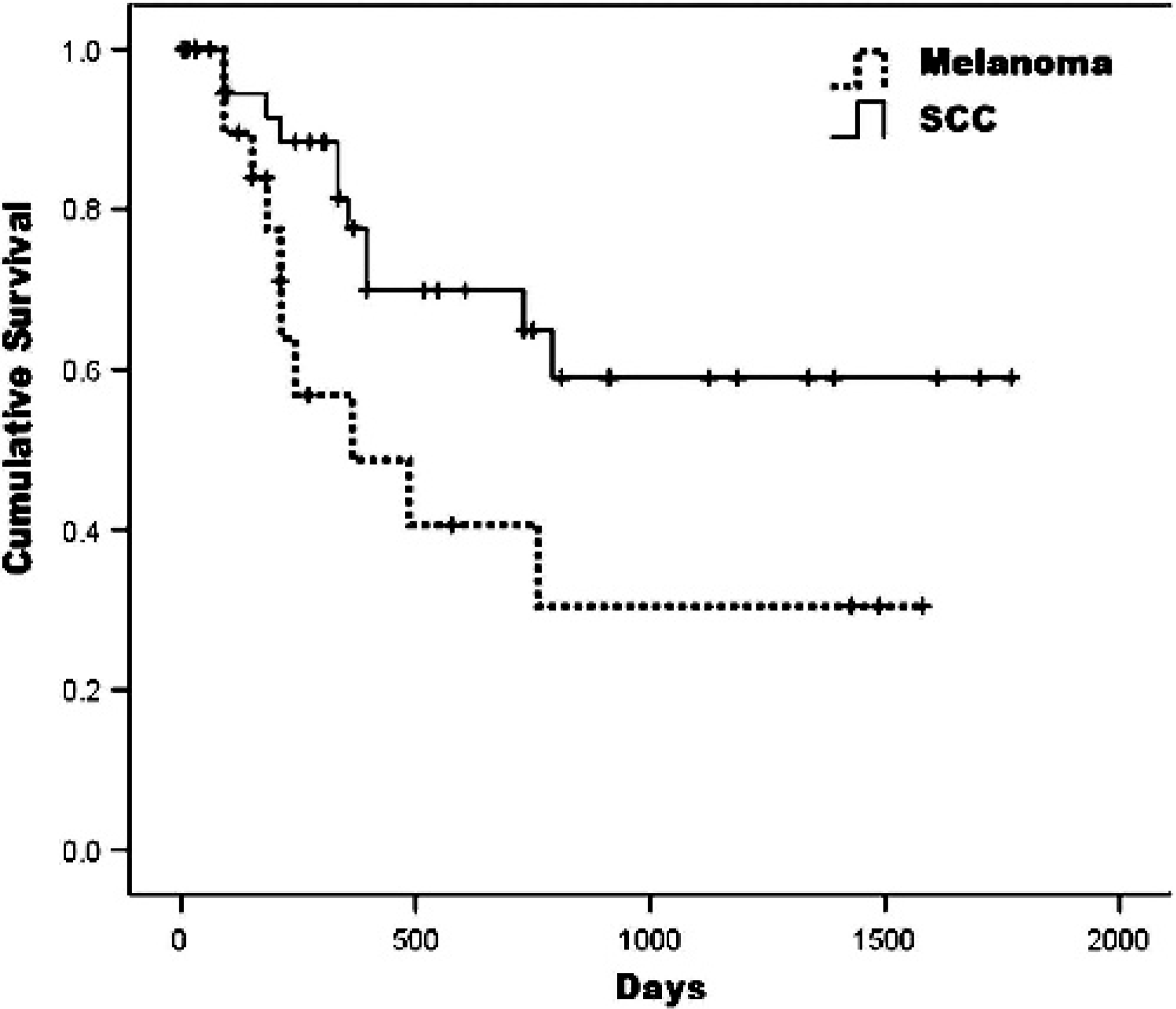
Kaplan-Meier survival curve for dogs with squamous cell carcinoma (SCC, n = 42) and melanoma (n = 22) of the digit.
Melanoma was the second most common malignant neoplasm (n = 52, 12.7%). Age ranged from 4–14 years at the time of surgical amputation, with an average age of 9.9 years. Females (n = 31) were more commonly affected than males (n = 18, P = .06). Multiple digits were involved in 2 submissions at the time of amputation. Scottish Terriers were over-represented, with 6 of 9 dogs of this breed in the entire study diagnosed with melanoma. Surveys were returned on 22 dogs, with 11 dogs having metastasis with a median disease-free interval of 322.1 days. All of these 11 dogs died or were euthanized due to metastatic disease, resulting in an overall median survival time of 365 days (Fig. 1).
Soft-tissue sarcomas were the third most common malignant neoplasm (n = 30, 7.4%). The age range was 3–15 years at the time of surgical amputation, with a mean age of 9.3 years. The sex was identified in 22 submissions, with 11 being female and 11, male. No breeds were over-represented. Surveys were returned on 17 dogs. Two dogs developed metastatic disease, 1 at 274 days and the other at 577 days. One dog was euthanized at the time of diagnosis of the metastasis (577 days), and the other was euthanized at 608 days.
Mast cell tumors were diagnosed in 20 dogs, with a mean age of 9.2 years, with a minimum age of 5 years and with a maximum age of 17 years at the time of surgical amputation. Thirteen females and 7 males were diagnosed with mast cell tumors. A single dog had multiple digits on the same foot involved at the time of amputation. Surveys were returned on 8 dogs, with 3 dogs having metastatic disease at 8, 152, and 486 days postsurgery. All 3 of the dogs with metastatic disease died or were euthanized due to this disease at 182, 152, and 486 days postsurgery, respectively, resulting in a 1-year survival rate of 75% and a 2-year survival rate of 62.5%.
Osteosarcoma (OSC) was diagnosed in 7 dogs. The average age was 11.1 years, with a range from 9–14 years at the time of surgical amputation. Four female and 3 male dogs were diagnosed. Of the 7 dogs, 6 were large breed dogs and 1 was a small breed (Dachshund). Of the 4 surveys that were returned, 2 dogs had metastatic spread of their disease and were euthanized at the time of diagnosis of metastasis at 584 and 714 days postsurgery. The remaining dogs were lost to follow-up 184 and 547 days postamputation.
Other malignant neoplastic processes were diagnosed infrequently. Due to the small numbers of animals involved, no conclusions about their prognoses can be determined (Table 2).
Proportion of the type of malignant neoplasms diagnosed in digits surgically amputated from dogs and submitted to multiple veterinary diagnostic laboratories included in this study.
One dog had both a melanoma and soft-tissue sarcoma within the same digit.
Various benign neoplastic and noninflammatory, non-neoplastic diseases were diagnosed, although in smaller numbers than malignant ones. The average age at the time of surgical amputation for these lesions was 9.4 years. The most common benign changes were epithelial inclusion cysts and keratoacanthomas (n = 20 and n = 15, respectively) (Table 3). A single dog diagnosed with keratoacanthoma had metastatic disease reported 729 days postsurgery and was euthanized due to this disease 912 days postsurgery.
Proportion of various types of benign neoplasms and noninflammatory, non-neoplastic lesions diagnosed in digits surgically amputated from dogs and submitted to veterinary diagnostic laboratories and included in this study.
Discussion
Few studies in the veterinary literature have described the prevalence of tumors in the digits of dogs, apart from SCC. Therefore, the incidence of neoplasms of different histogenesis is unclear. This study examined the frequency of primary neoplasms in the digits of dogs surgically amputated and submitted to multiple veterinary diagnostic laboratories. Previous smaller scale studies have examined digital lesions in dogs7,11,16 or specific diagnoses,9,10,13,14 but a study on the scale of the one described here has not been reported. Four hundred four submissions of surgically amputated digits from dogs from multiple veterinary diagnostic laboratories were examined; neoplastic changes were present in 296 of these digits, with only inflammatory changes in 108 submissions. This is markedly different from a previous study of nail and nail bed lesions in dogs,16 in which only 24 of 196 claw disorders were neoplastic. This may be accounted for by the fact that many lesions affecting the claw are diagnosed clinically and the digit is not amputated. Thirty different neoplastic diagnoses were identified in these digits, and malignant neoplastic disease was much more common than benign changes (229:67).
The clinical presentation of dogs from which these digits were amputated was similar, and determination of the underlying disease process would be difficult without histopathologic diagnosis. Multiple digits on the same dog were involved in malignant, benign, and inflammatory conditions. Although the age of animals with both benign and malignant neoplastic conditions was higher than that in dogs with inflammatory conditions, the age range on all conditions was broad and overlapped considerably. Unlike previous studies, the mean age of benign processes was not lower than that for malignant ones.16 A previous study has shown that destruction of bone, particularly the third phalanx, can occur with multiple underlying lesions,1 and this was the case in this study where bony lysis occurred in malignant processes (SCC, melanoma, OSC, giant cell tumor of bone, plasma cell myeloma, and adenocarcinoma), benign processes (epidermal inclusion cysts, keratoacanthoma), and inflammatory conditions and thus was not specific for any condition.
Variability seems to exist in the literature as to the predilection of digital neoplasms for front-versus-hind limbs. This study concurs with what some other studies have found, that most neoplasms in the digit occur on the front limbs.1,7,11 No particular digit was more likely to be affected, with the exception that the first digit (dew claw) was less likely to be involved, which is consistent with other studies.7,16 Dew claw removal in puppies may account for the lower proportion of disease in the dew claw, but this was not examined in this study. Possible reasons for the predilection for neoplasia for the front limbs and the possible sparing of the dew claw has been previously speculated. The front limbs bear more weight than do the hind limbs and are possibly exposed to carcinogens during digging activity. Similarly, as the first digit does not come in contact with the ground, there maybe less opportunity for trauma or carcinogen exposure than in other digits.7
SCC was the most common diagnosis and most common malignant diagnosis (n = 109) in this study, which is in agreement with most other studies.7,8,9,11,13,14,16 Multiple digits were involved at the time of amputation in 7 of these submissions. This has been reported on numerous occasions.1,7,10,11,14,15,16 A breed predilection for large breed, black-coated dogs has been reported,7,10,11 and affected dogs fitting these criteria in this study included the following breeds: Giant Schnauzers, Rottweilers, Flat Coated Retrievers, and standard Poodles. These 4 breeds were all over-represented in dogs diagnosed with SCC compared with dogs with inflammatory disease. Of dogs with multiple digits affected, all were from black-coated breeds (including 1 Gordon Setter). Interestingly, Dachshunds were also over-represented with 5 of the 6 in this study being diagnosed with SCC. Although a report of SCC in the digit of a Dachshund has been published,4 this breed's possible predisposition has not been identified in any previous study. Dachshunds also have a black-hair coat but are of small stature. The reason for the relationship of hair coat color to the incidence of SCC in the digit is unclear, but masses in multiple digits in a black-coated breed should raise suspicion of SCC.
The reported metastatic rate of digital SCC is variable in the veterinary literature, with reported metastatic rates ranging from 4.7%6 to 24.1%.16 Of 42 dogs in this study for which clinical follow-up was available, metastasis occurred in 10 dogs (23.8%) and 1 dog had locally recurrent disease. All 11 of these dogs were euthanized due to, or died of, this disease. This suggests that SCC of the digit is more likely to metastasize than SCC occurring elsewhere on the body.6
Of note is the 1 keratoacanthoma that was reported to have metastasized and subsequently caused the death of the dog. “Keratoacanthomas” are defined as benign neoplasms that cause local bone destruction through their growth. The differentiation of keratoacanthoma from well-differentiated SCC was problematic in this study, and the frequency of disagreement in the diagnosis between pathologists was very high.19 Given the reported malignant nature of this particular keratoacanthoma, we believe it is probable that it represents a misdiagnosis of a well-differentiated SCC.
Melanomas were the second most common neoplasm (n = 52) found in the digit in this study. Melanomas typically occurred in older dogs but also developed in a dog as young as 4 years of age. Females were more commonly affected than males in this study, an observation that has not been reported in other studies.1,17 Scottish Terriers are predisposed to melanomas3,6 and were over-represented in this study as melanomas were diagnosed in 6 of the 9 Scottish Terriers.
The reported metastatic rate of melanomas from the digits of dogs is higher than that in other cutaneous sites, with a reported metastatic rate of 38–58% and a 1-year survival rate of 42–70%.1,7,11,17 In this study, the metastatic rate and 1-year survival rate were 50%, with a mean disease-free interval of 322.1 days. A previous study of melanocytic tumors of the digits of dogs has shown that histologic criteria for malignancy based on high mitotic index and nuclear and nucleolar pleomorphism may not correlate well with clinical prognosis.1 A new histologic scoring system has been proposed and has an improved prognostic rate of approximately 90%.17 Authors of that study suggested that the poor correlation of histologic features of malignancy to clinical prognosis in the previous study may have been due to the effectiveness of amputation as a treatment modality for melanomas of the digits. While this may be true, the finding in this study that 50% of dogs with melanoma of the digit died in the first year following amputation suggests that despite this treatment, the prognosis for digital melanoma should be guarded.
Soft-tissue sarcomas represented 29 of the malignant tumors in this study. Amputation was generally a successful treatment modality, with recurrent disease (metastases) present in only 2 of 17 dogs with clinical follow-up available. Given the ease of amputation of digits and the typically low metastatic rate but high local-recurrence rate of soft-tissue sarcomas, amputation with adequate margins alone represents an effective method of treating many soft-tissue sarcomas of the digits of dogs.
Mast cell tumors of the digit composed 20 of the malignant neoplasms in this study. Of the 8 dogs for which clinical follow-up was available, 3 dogs had metastatic disease and subsequently died. This survival rate is lower than that reported for mast cell tumors of the extremities in other reports18 but is similar to that reported in a large study of canine digital masses.2
OSA was diagnosed in 7 dogs. Most cases occurred in large breed dogs with a single Dachshund being the exception. Dogs with OSA of the distal extremities have been reported to have a longer survival time than dogs with other appendicular OSA but still have significant potential for metastatic disease.5,7 Of 4 dogs with clinical follow-up available for this study, 2 died of metastatic disease within 2 years of digit amputation.
Giant cell tumor of bone was diagnosed a single time in this study. This is a rare tumor in dogs2,12 that differs from OSA in that it induces considerable bone lysis, but no osteoid production. Typically, giant cell tumor of bone occurs in the epiphyses of long bones, and its occurrence in the digit has not been reported. Reports of giant cell tumor of bone are too rare in the veterinary literature to accurately predict the prognosis; the single dog affected here was still alive and disease-free 2,640 days postamputation.
The results of this study indicate that neoplastic diseases, both benign and malignant, compose the largest proportion of abnormalities in 404 surgically amputated canine digits submitted to multiple veterinary diagnostic laboratories. Malignant neoplasms composed 229 of the 296 neoplasms diagnosed. Neoplastic disease is both common and diverse in canine digits. As in previous studies, the most common neoplasms were SCC, melanoma, soft-tissue sarcomas, and mast cell tumors, but 26 other neoplastic disorders were also diagnosed in this study. Diagnosis of the underlying disease process without histopathologic evaluation is unlikely as simultaneously affected digits and bone lysis were present in both malignant and benign neoplasms and non-neoplastic noninflammatory disease as well as inflammatory conditions. Although the mean age of dogs with neoplastic disease was higher than that of dogs with inflammatory conditions, the age ranges in all of these were broad and overlapping, which limits the predictive value. Large breed, black-coated dogs and Dachshunds have a predisposition to developing SCC, and the metastatic rate of SCC in the digit appears to be higher than that elsewhere in the body. Melanoma in the digit is common, with Scottish Terriers over-represented. The metastatic rate was higher, and the prognosis poorer than for dogs with SCC.