
Abstract
During development and subsequent field evaluation of an oral vaccinia-rabies glycoprotein (V-RG) recombinant virus vaccine, 53 adult porcupines (Erethizon dorsatum; 38 females and 15 males) were examined. Microscopic examinations revealed the presence of giant epitheloid cells in various tissues (adrenal glands, spleen, liver, and lungs) of 4 (11%) female animals. These giant cells were approximately 20 times the size of the surrounding cells of the parenchyma. The cells were found singly and were not associated with any inflammatory cellular infiltrate and appeared to be located within vascular lumina. Morphologically these cells were typical of uterine epitheloid trophoblasts. This is the first record of the presence of trophoblast-like cells in nongenital tissues of porcupines.
Keywords
Although porcupines (Erethizon dorsatum) are found throughout Alaska, Canada, and in nearly half of the continental United States, 4 there is paucity of published information on diseases and pathological conditions that are found in this species.
Porcupines have hemochorial placentation, 9 similar to that observed in mice and humans. Embrionic trophoblasts enable the embryo to invade through the uterine epithelium at the time of implantation. 12 Trophoblasts from both mice and humans can dislodge from the endometrium and be carried by capillaries as emboli to other organs, such as the lungs. This communication documents what appears to be a similar phenomenon in porcupines.
Between 1989 and 1992, a total of 53 adult porcupines (38 females and 15 males) were examined from State Game Lands No. 13 in northeastern Pennsylvania. 11 These animals were used in the experimental development and subsequent field evaluation of an oral vaccinia-rabies glycoprotein (V-RG) recombinant virus vaccine. 11 The porcupines were either euthanized and necropsied in the field or transported to the laboratory, where they were used in experiments to evaluate the safety of the V-RG vaccine in nontarget species. 10 At the conclusion of the study, the captive animals were subsequently euthanized by intravenous administration of sodium pentabarbitol and necropsied.
At the time of necropsy, representative tissues of most major organs were collected and placed in neutral buffered 10% formalin as previously described 5 and processed for routine histopathology. Sections were cut at 5 μm, stained with hematoxylin and eosin, and examined by light microscopy. Selected tissue sections were also stained with periodic acid–Schiff stain and by immunohistochemistry for cytokeratins. For the latter, a commercial monoclonal antihuman cytokeratin antibody (DakoCytomation, Cambridge, UK) was used as primary antibody. This reagent is considered to be a broad-spectrum antikeratin antibody, and, according to the manufacturer, it reacts with intermediate– and low–molecular weight keratins corresponding to cytokeratins 5, 6, 8, and 17 and probably also 19. For the purpose of this study, only the findings relative to the presence of very large cells in histologic sections of tissues are reported.
A total of 6 tissue sections revealed trophoblastic cells in 4 porcupines. All were females. In 2 porcupines the giant cells were found only in the adrenal glands, 1 had giant cells in the spleen and liver, and 1 had giant cells in the lung and adrenal gland. At all sites, the cells varied between 25 and 75 μm (i.e., up to 20 times the normal surrounding cells of the parenchyma) and contained either 1 or occasionally 2 large nuclei (Fig. 1). The cytoplasm of these cells was finely granular (Fig. 2). In the adrenal glands, they were confined to the cortex (Figs. 1, 2). Usually only 1 cell was found per tissue, and when multiple cells were present in a tissue, the individual cells were always some distance from each other (Fig. 1). None of these cells had any associated inflammatory cells (Figs. 1, 2). The periodic acid–Schiff-stained sections revealed a surrounding thin membrane that was continuous with the surrounding membrane in the parenchyma. This indicated that the suspect trophoblasts were probably present within the lumen of the vessels. The anticytokeratin antibody used in the immunohistochemistry procedure failed to label the cytoplasm of the cells but also failed to stain other epithelial tissues of this porcupine. Failure to label the suspect trophoblasts for cytokeratins by immunohistochemistry was probably due to the specificity of the monoclonal antibody, which although was able to recognize human and canine cytokeratins, did not label keratins in resident epitheal cells of examined porcupine tissues. However, because the tissues were stored in formalin for a prolonged period (in some cases more than 12 months), this could have influenced the negative outcome of immunoreactivity.
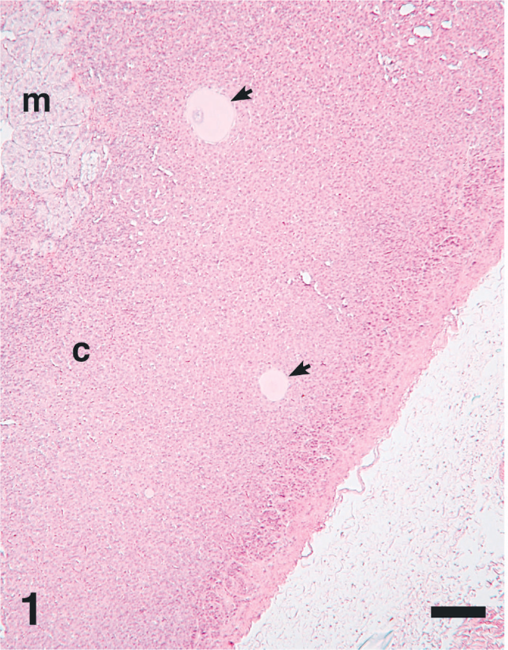
Adrenal gland; female porcupine with the presence of 2 large cells (trophoblast; arrows) in the cortex (c). m = medulla. HE. Bar = 90 μm.
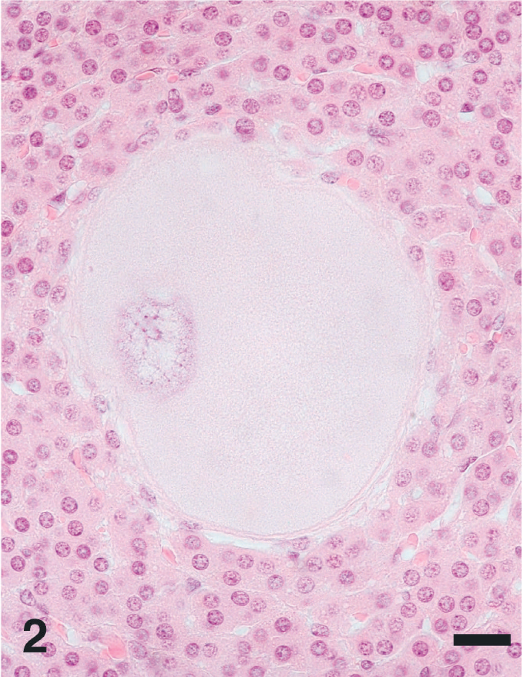
Higher magnification of Fig. 1 showing one of the large cells (trophoblast) with fine granular cytoplasm. HE. Bar = 12 μm.
The suspect trophoblasts were present only in the tissues of female porcupines, further supporting their proposed origin from the female genital organs (most likely the uterus). However, in only 2 of 4 cases were the uteri available for examination. In 1 case the animal was not pregnant, and in the other there was evidence of a recent pregnancy. No vascular changes were observed in these animals.
During early human pregnancy, trophoblasts cross the basement membrane of the uterine epithelium and vasculature to initiate successful implantation. 12 Similar mechanisms exist in other animals that have hemochorial placentation, and in such species, trophoblastic emboli have been reported in lungs and other organs. Such findings have been documented in nonhuman primates, 3 mice, 12 chinchillas, 6, 13 hamsters, 1 and guinea pigs. 2 The latter are animal models of tissue invasion by trophoblasts, and their uterine arteries have been extensively studied for structure and functional mechanisms. 2 In porcupines there has been only a single report of extrauterine trophoblastic giant cells identified within corpora lutea. 8 In humans the trophoblasts may transform into neoplastic lesions (choriocarcinoma) and have been known to metastasize to distant organs. 12 In this respect, 1 case of choriocarcinoma with metastasis to the lung has been documented in a rhesus monkey. 7
Footnotes
Acknowledgements
This project was funded by the Department of Pathobiology, University of Pennsylvania, and the Department of Agriculture, Commonwealth of Pennsylvania. Expert technical assistance was provided by Martha Church, Ginny Montgomery, Micky Fenneman, and Debora Moore (Iowa State University). Mention of trade names or commercial products in this article is solely for the purpose of providing specific information and does not imply recommendation or endorsement by the U.S. Department of Agriculture.