
Abstract
A uterine tumor, with histological and immunohistochemical features consistent with those of human choriocarcinoma, was identified in a 10-year-old unmated female pot-bellied pig (Sus scrofa). The tumor showed biphasic proliferation of cytotrophoblast-like cells and syncytiotrophoblast-like cells. Immunohistochemically, the syncytiotrophoblast-like cells were positive for human chorionic gonadotropin, and both types of cells were positive for cytokeratin and negative for vimentin, octamer-binding transcription factor 4, and α-fetoprotein. Because syncytiotrophoblasts are absent in the normal porcine placenta, the tumor was diagnosed as a choriocarcinoma-like tumor.
Choriocarcinoma is a highly malignant biphasic trophoblastic tumor composed of cytotrophoblastic and syncytiotrophoblastic cellular components. Spontaneous occurrence of this tumor is very rare in animals as well as in human beings. The known examples of choriocarcinoma in animals are limited to some cases identified in the uterus or ovary of macaques,4,7,11 rabbits, 6 and rodents.1,8 In addition, primary gastric choriocarcinoma has been reported in a dog. 9 To the authors’ knowledge, there has been no record of spontaneous choriocarcinoma in farm animals including pigs. Herein, a case of a choriocarcinoma-like tumor in a potbellied pig (Sus scrofa) is described, with histological and immunohistochemical features similar to those of human choriocarcinoma.
The case involved a female potbellied pig that was kept with other female pigs in the Toyohashi Zoo and Botanical Park (Aichi, Japan). The pig had been in the zoo since an early age and had never mated. At 10 years of age, the pig died. Vaginal bleeding, purulent vaginal discharge, and abdominal distention were present at the time of death. The onset of these symptoms occurred at different time points. Vaginal bleeding had been noted intermittently for over a year, and abdominal distention had become apparent approximately 4 months before death. Purulent vaginal discharge was observed for 3 weeks prior to death. For 17 days prior to the pig’s death, treatment with methenolone acetate, a amoxicillin and clavulanic acid, b lactobacillus preparations, c and vitamin complex d was orally administered twice a day.
The necropsy findings indicated prominent emaciation (38.2 kg body weight). Accumulation of clear yellow fluid containing fibrin was observed in the thoracic and abdominal cavities (more than 1,500 ml and 300 ml, respectively). The right uterine horn was irregularly enlarged and showed a large mass (approximately 20 cm in diameter; Fig. 1). The mass contained part of the ileum, rectum, and spleen and was tightly adhered to adjacent organs and the peritoneum by fibrous tissue. In the enlarged right uterine horn, the uterine wall was markedly thickened (maximal thickness of approximately 5 cm), and a large amount of purulent to mucopurulent exudate was present in the lumens. In the left horn and body of the uterus, wall thickening was not observed, but there was grayish white mucus in the lumens. Several white nodules (5–10 mm in diameter) were found in the liver and lungs.
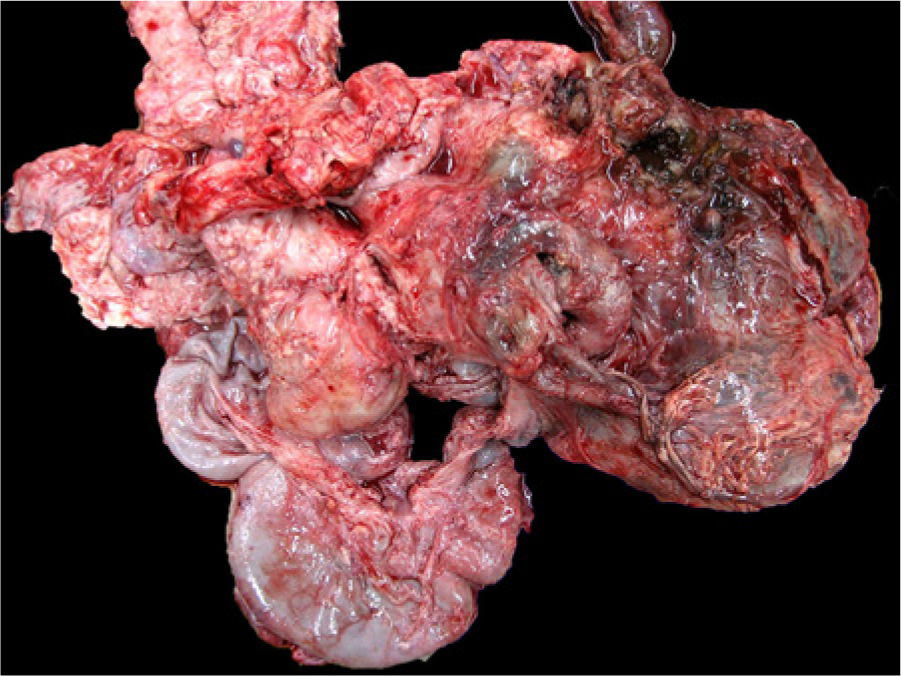
Macroscopic appearance of the tumor in the lower abdominal cavity of the potbellied pig. The size of the tumor mass is approximately 20 cm in diameter.
Tissue samples from the lung, liver, spleen, kidney, and uterus were fixed in 10% neutral buffered formalin and submitted for histological examination. Samples were routinely processed, and sections obtained from paraffin-embedded tissues were stained with hematoxylin and eosin. Serial sections were subjected to immunohistochemical staining using polyclonal antibodies for human chorionic gonadotropin (hCG; 1:300 dilution), e octamer-binding transcription factor 4 (OCT4; 1:100 dilution), f and monoclonal antibodies for pan-cytokeratin (clones AE1/AE3; prediluted), e cytokeratin 7 (clone OV-TL 12/30; 1:50 dilution), e α-fetoprotein (clone C3; 1:50 dilution), f and vimentin (clone V9; prediluted). e A standard avidin–biotin technique g was employed, using diaminobenzidine as the chromogen. Sections were counterstained with hematoxylin.
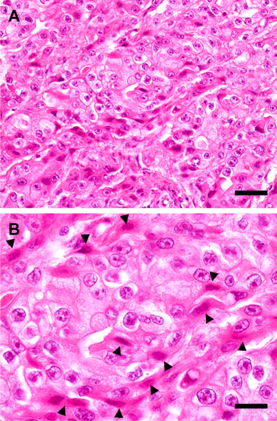
Microscopically, neoplastic cells proliferated most markedly in the endometrium of the uterus and were arranged in nests and sheets with scant fibrous connective tissues (Fig. 2). Neoplastic cells deeply invaded the myometrium and perimetrium (Fig. 2), and vascular invasion was frequently observed in these areas (Fig. 2). The tumor was composed of 2 distinct cell types. Their cellular characterization and arrangement in the current case formed a pattern consistent with biphasic proliferation of cytotrophoblasts and syncytiotrophoblasts observed in human choriocarcinomas. The major components were cytotrophoblast-like cells that had a round to oval nucleus containing prominent nucleoli, granular eosinophilic cytoplasm (Fig. 3), and moderate anisokaryosis. Bizarre giant cells were infrequently observed. The other type of tumor cells were syncytiotrophoblast-like cells that had hyperchromatic oval to spindle-shaped nuclei, dense eosinophilic cytoplasm, and frequently formed multinucleated cells (Fig. 3). The syncytiotrophoblast-like cells were sometimes arranged around foci of the cytotrophoblast-like cells (Fig. 3). Metastatic lesions were detected in the lungs and kidney and showed a similar biphasic proliferation pattern. Neoplastic cells were also observed in the blood vessels of the kidney, indicating hematogenous spread. In addition, microscopic analysis revealed that the hepatic and lung nodules represented a cholangiocarcinoma and its metastatic lesion, respectively. Therefore, metastatic foci of 2 different types of tumors were found in the pig’s lung.
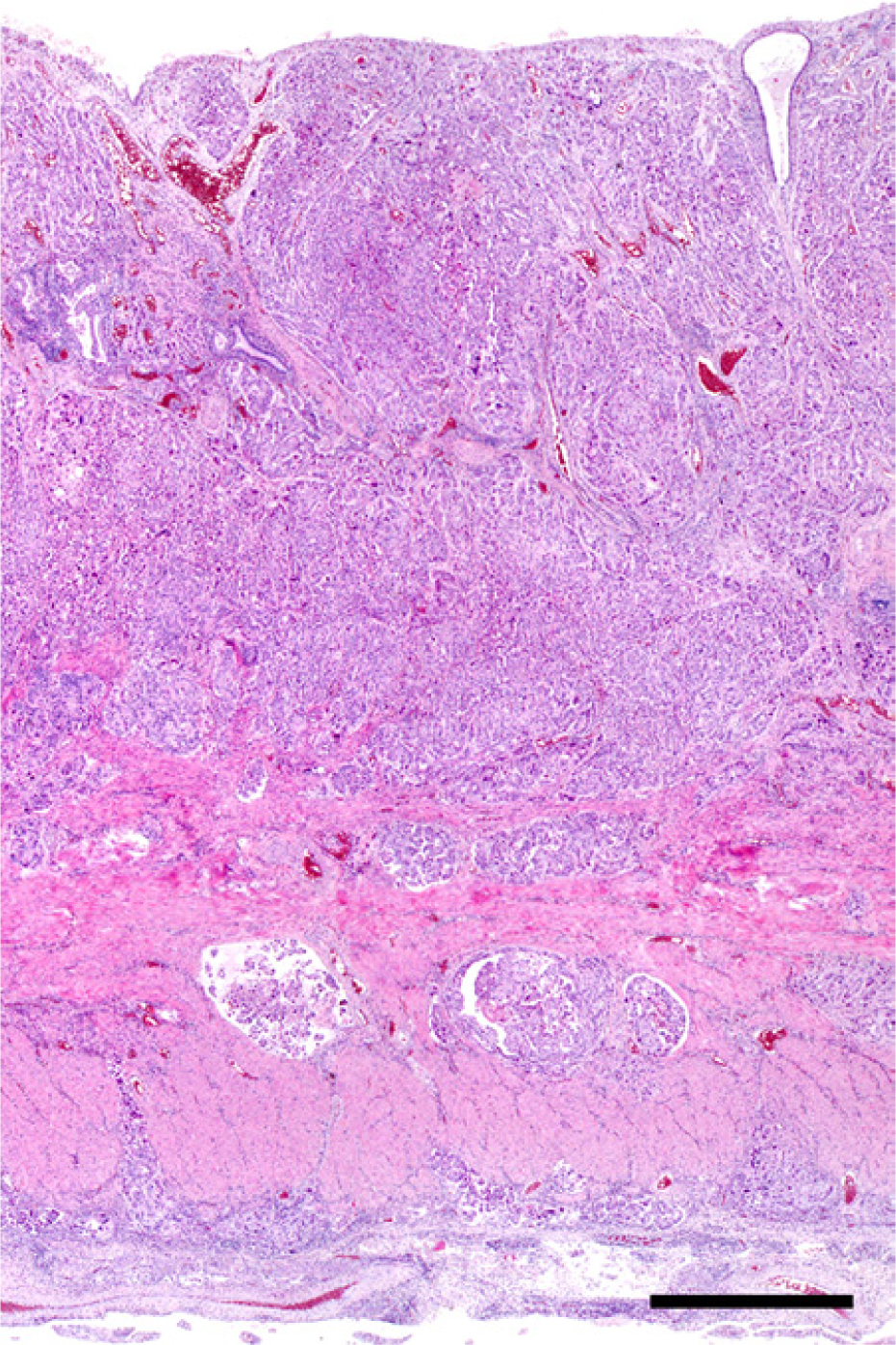
Hematoxylin and eosin staining of uterus of the potbellied pig. Neoplastic cells proliferated in all layers of the uterine wall and vascular invasion was noted. Bar = 1,000 µm.
Hematoxylin and eosin staining of the uterus of the potbellied pig.
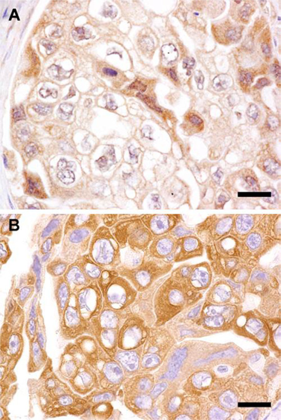
Immunohistochemically, as in the case of human choriocarcinoma, syncytiotrophoblast-like cells were positive for hCG (Fig. 4A). However, because hCG-positive syncytiotrophoblasts can be present in other types of germ cell tumors,12,13 additional immunohistochemical features were used for differential diagnosis (Table 1). Tumor cells were consistently positive for cytokeratin (Fig. 4B) and negative for vimentin, OCT4, and α-fetoprotein, a pattern that was distinctly different from that of dysgerminoma, embryonal carcinoma, and yolk sac tumors (Table 1). The absence of other germ cell components also ruled out the possibility of mixed germ cell tumors. Other trophoblastic tumors, including placental site trophoblastic tumors and epithelioid trophoblastic tumors, were excluded on the basis of histological characteristics, whereas epithelial-derived tumors were ruled out because of the presence of hCG-positive syncytiotrophoblast-like cells.
Immunostaining of the uterus of the potbellied pig.
Summary of the immunohistochemical findings of the tumor of the pot-bellied pig and comparison with those of germ cell tumors.*
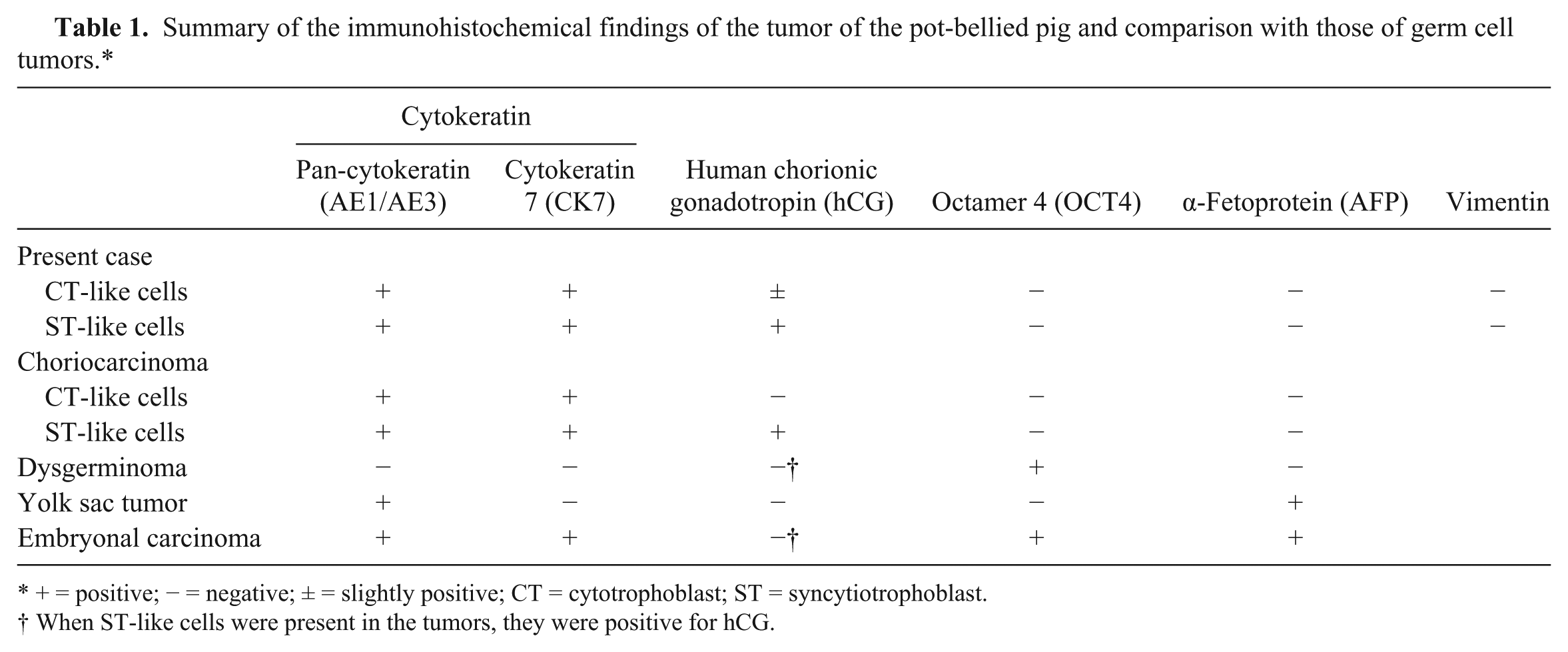
+ = positive; − = negative; ± = slightly positive; CT = cytotrophoblast; ST = syncytiotrophoblast.
When ST-like cells were present in the tumors, they were positive for hCG.
The histological and immunohistochemical findings of the tumor in the present case fulfilled the diagnostic criteria used for choriocarcinoma in human beings. However, there were some differences. Massive hemorrhage, which is one of the main histopathological features that characterize choriocarcinoma in human beings, 10 was not observed in the present case. Moreover, unlike its human counterpart, vascular channels surrounded by neoplastic trophoblasts were not present. 10 Furthermore, it should be noted that, although the presence of the syncytiotrophoblasts provided an important clue for diagnosis, the pig had an epitheliochorial placenta composed of a single layer of cytotrophoblasts without syncytiotrophoblasts. 3 To date, all cases of choriocarcinomas in animals have been reported in species with hemochorial or endotheliochorial placentas such as macaques,4,7,11 rodents,1,8 rabbits, 6 and dogs. 9 Therefore, because of the absence of syncytiotrophoblasts in normal porcine placenta, the tumor was diagnosed as a choriocarcinoma-like tumor.
Choriocarcinomas can be divided into gestational and nongestational types. It was clear that the current case was nongestational because the pig never mated. However, it was not possible to identify the origin of the tumor. In most cases, nongestational choriocarcinomas arise from germ cells in the ovaries. 2 Unfortunately, the ovaries could not be microscopically examined because they had not been successfully excised from the large tumor mass that involved the adjacent organs. In addition, although nongestational choriocarcinomas can rarely arise from trophoblastic differentiation within poorly differentiated carcinomas including endometrial carcinomas, 2 no component of endometrial carcinoma was found within the tumor.
In addition to the choriocarcinoma-like tumor in the uterus, a cholangiocarcinoma was found in the pig, which may have contributed to the emaciation. However, it is likely that the uterine tumor affected the animal’s outcome more severely. Whereas the cholangiocarcinoma formed relatively small nodules in the liver and lung, the uterine tumor formed a large mass that occupied the lower abdominal cavity, involved the surrounding organs, and spread to distant organs, possibly leading to impairment of organ function and circulatory failure. In addition, the purulent accumulation in the uterine lumen likely had a debilitating effect. It has been reported that potbellied pigs frequently have uterine lesions including endometrial and smooth muscle tumors. 5 However, to the authors’ knowledge, choriocarcinoma-like tumors have not been reported before in pigs.
Footnotes
Acknowledgements
The authors thank Ms. Chikako Iriyama (Laboratory of Veterinary Pathology, Gifu University) for expert technical assistance, Drs. Toshiya Kuno and Manabu Takamatsu (Department of Tumor Pathology, Gifu University Graduate School of Medicine) for their helpful advice, and Dr. Ruri Kondo (Yokkaichi City Food Inspection Center) for providing normal porcine placenta.
Declaration of conflicting interests
The author(s) declared no potential conflicts of interest with respect to the research, authorship, and/or publication of this article.
Funding
The author(s) received no financial support for the research, authorship, and/or publication of this article.
a.
Primobolan, Bayer Leverkusen, Germany.
b.
Augmentin, GlaxoSmithKline, Brentford, United Kingdom.
c.
Biofermin R, Biofermin Pharmaceutical, Kobe, Japan.
d.
Panvitan, Takeda Pharmaceutical, Osaka, Japan.
e.
Dako, Glostrup, Denmark
f.
Abcam, Cambridge, United Kingdom.
g.
Vectastain ABC kit, Vector Laboratories, Burlingame, CA.