
Abstract
The placenta, a fetal endocrine organ, is composed of subpopulations of trophoblasts, including cytotrophoblasts, and syncytiotrophoblasts. Trophoblastic populations can be distinguished based upon their expression of cytokeratin. The purpose of the current study was to develop an immunohistochemistry (IHC) method to identify trophoblasts selectively in frozen feline placental tissue using antibodies specific for cytokeratin. The mouse monoclonal antibody anti-human pan-cytokeratin AE1/AE3 and a commercial detection system were used. Nonspecific immunoreactivity was encountered that could not be eliminated with altered blocking methods. The nonspecific reactivity was attributed to the goat anti-mouse/rabbit immunoglobulin G (IgG) peroxidase polymer included in the commercial kit. Alternatively, a polyclonal rabbit anti-cow cytokeratin wide spectrum screening antibody with goat anti-rabbit IgG polyclonal secondary antibody was used to detect cytokeratin in feline placental tissue. The IHC procedure eliminated nonspecific immunoreactivity while specifically labeling cytokeratin. This new approach provides an IHC method to identify trophoblasts specifically in feline placenta.
Introduction
The placenta, an endocrine organ of fetal origin, is responsible for metabolic exchange between the mother and the developing fetus and produces hormones that are responsible for pregnancy maintenance and fetal growth. 13 Trophoblastic cells (cytotrophoblasts and syncytiotrophoblasts) are a population of cells that comprise the placental chorionic villi in mammals. These trophoblasts are fetal-derived cells of epithelial origin that cover the blastocyst and form the chorion, the fetal portion of the placenta. 13 Syncytiotrophoblasts comprise the outer trophoblast layer, contacting and forming attachment with the maternal endometrium. 13 The cytotrophoblasts form the inner layer of the trophoblast between the syncytiotrophoblasts and the chorionic villous capillaries. Human placental trophoblasts express unusual immunological markers, including HLA-G and other class I molecules that regulate immune suppression, 6,15 and cytokines that regulate immune function. 1,8 The immunological characteristics of trophoblastic cells contribute to the immunological privilege of the placenta, promoting fetal tolerance and successful pregnancy.
As a part of an ongoing study of Feline immunodeficiency virus (FIV) vertical transmission and virus-induced placental immunopathology, the impact of viral infection on gene expression by trophoblasts was investigated. The initial goal of the study was to develop an immunohistochemistry method to identify trophoblasts in feline placental tissue based on their expression of cytokeratin intermediate filaments. The ultimate purpose was to dissect trophoblasts from the tissues using laser capture microdissection for isolation of trophoblast RNA. To do so, it was important to identify trophoblasts specifically by immunolabeling. Attempts were made to detect cytokeratin antigens using the mouse anti-human pan-cytokeratin monoclonal antibody AE1/AE3 and a commercial horseradish peroxidase (HRP) kit, which utilizes a goat anti-mouse/rabbit immunoglobulin G (IgG) peroxidase-labeled polymer as a secondary antibody. However, in feline placentas, this system produced nonspecific reactivity that could not be eliminated with modification of protocol. Thus, an immunohistochemistry (IHC) procedure was developed utilizing a polyclonal rabbit anti-cow cytokeratin wide spectrum screening (WSS) antibody paired with goat anti-rabbit poly-HRP secondary antibody to detect cytokeratin in feline placental tissue. This procedure resulted in specific staining of cytokeratin in feline trophoblasts, eliminating nonspecific antibody binding.
Antibodies used in immunohistochemistry in the current study.*
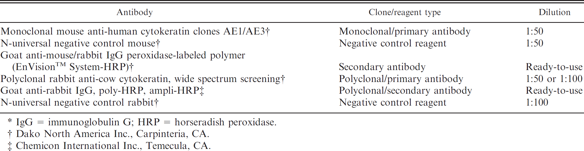
IgG = immunoglobulin G; HRP = horseradish peroxidase.
Dako North America Inc., Carpinteria, CA.
Chemicon International Inc., Temecula, CA.
Materials and methods
Feline placental tissues and antibodies
All procedures utilizing cats were performed with approval of the Mississippi State University Institutional Animal Care and Use Committee. Cats used in this investigation were part of an ongoing study of FIV-B-2542 12 transplacental transmission. Cats were reproductively mature, specific-pathogen-free (SPF) animals of less than 12 months of age when obtained from a commercial cattery. One group of cats was inoculated intravenously with FIV-B-2542; one group served as uninoculated controls. All queens were allowed to breed naturally with SPF toms. Breeding was observed, and pregnancy was confirmed by palpation and ultrasonography.
Kittens were delivered by cesarean section during week 8 of gestation (late-term). 17 Placentas were collected under sterile conditions using the following dissection procedure. The uterine horns were removed, and individual fetal membranes were collected. Following rinses with sterile phosphate buffered saline, membranes were incised with a sterile scalpel, and fetuses and placentas were collected. Placental tissues were snap frozen in liquid nitrogen and cryopreserved at −80°C. Tissues from 4 control cats only were used to develop the following assays. All antibodies used in these assays are shown in Table 1.
Histological evaluation of placental tissues
Optimum cutting temperature-embedded placental sections from late-term control and FIV-infected queens were fixed in acetone for 10 min and allowed to dehydrate for 1 min. Following dehydration, routine hematoxylin and eosin staining was performed to observe histology as follows. Placental tissues were stained with Mayer hematoxylin a for 15 min and washed under tap water for 20 min. Sections were counterstained with eosin for 2 min and dehydrated sequentially in 2 changes of 95% and 100% EtOH, 2 min each. After a 1-min dehydration at room temperature, sections were cleared twice with 2-min xylene rinses and mounted with slide mounting solution. b
Basic immunohistochemistry procedure
All IHC procedures were performed using the following basic staining protocol with modifications only to endogenous peroxidase quenching, serum pretreatments, and antibody incubations. Frozen placental tissues were cryo-sectioned to 4 μm and placed onto poly-L-lysine-coated slides. c Sections were allowed to air dry at room temperature overnight. Dried sections were fixed in acetone for 10 min at room temperature and allowed to air dry completely. To quench endogenous peroxidase activity, sections were blocked with a commercial peroxidase blocking reagent d for 10 min and washed in a bath of Tris buffer (TB; 0.05 M Tris-HCl, pH 7.0–7.6). The primary antibody or negative control reagent was diluted 1:50 in TB containing 1% bovine serum albumin. Sections were incubated with either the mouse anti-human pan-cytokeratin monoclonal antibody AEl/AE3 e or the mouse universal negative control reagent f for 10 min. Following the incubation of the primary antibody or the negative control reagent, sections were washed in separate baths of TB. Sections were then incubated with the goat anti-mouse/rabbit IgG peroxidase-labeled polymer g for 10 min and washed in a fresh bath of TB. Sections were incubated with the buffered substrate solution containing 3,3′-diamino-benzidine chromogen d,h for 10 min and washed in a bath of distilled water for 1 min. Sections were then counterstained with Mayer hematoxylin for 5 min and rinsed in fresh, distilled water for 1 min. The counterstain was blued by dipping sections 10 times in a bath of 37 mM ammonia and rinsing sections in a fresh bath of distilled water. Sections were dehydrated in 75%, 95%, and 100% EtOH for 30 sec and xylene for 5 min. Immunoreactivity was detected microscopically.
Endogenous peroxidase quenching and tissue blocking. Dehydrated sections were blocked by sequential pretreatment with both mouse and goat serum (1:100) for 10 min each. Blocking sera was removed from the slides, and unrinsed sections were quenched with peroxidase blocking reagent. Alternatively, dehydrated sections untreated with sera were quenched for 30 min with a solution of 2 ml 30% H2O2 a and 400 ml of methanol. 2 Quenched sections were blocked with 5% (wt/vol) nonfat dry milk in TB for 1 hr and 15 min.
Modified immunohistochemistry procedure. Sections quenched with peroxidase blocking reagent were incubated with mouse antibodies (as previously described) using the following conditions: no antibody, primary antibody only, and secondary antibody only. Sections quenched with H2O2 in methanol and blocked were incubated with the polyclonal rabbit anti-cow cytokeratin WSS antibody i or rabbit universal negative control reagent j and secondary goat anti-rabbit, poly-HRP. k
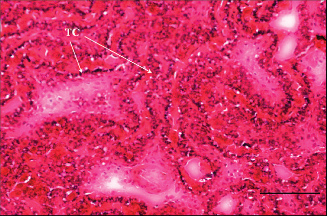
Hematoxylin and eosin staining of late-term frozen, acetone-fixed feline placenta. A placental section from a representative control queen is shown. The location of tropho-blastic cells (TC; arrows) is shown. Bar = 100 μm.
Results
Histological evaluation of placental tissues and immunohistochemistry
Hematoxylin and eosin staining of the placental tissue revealed the location of trophoblasts and provided confirmation that tissue integrity was not compromised during cryopreservation and frozen storage (Fig. 1). The results of all IHC methods are listed in Table 2.
Immunohistochemistry using AE1/AE3 mouse monoclonal antibodies and the commercial kit. To detect cytokeratin in acetone-fixed, frozen placental tissues, the mouse anti-human pan-cytokeratin monoclonal antibody AE1/AE3 and a negative control reagent with the commercial kit were used. Immunolabeling revealed similar immunoreactivity in tissues treated with the mouse monoclonal antibody AE1/AE3 and the negative control reagent (Fig. 2), indicating nonspecific labeling in the IHC procedure.
Parallel placental sections were stained for cytokeratin using serum pretreatments and modified antibody conditions. Cytokeratin labeling did not occur in sections subjected to the protocol in the absence of both primary and secondary antibody or sections that were treated with only the primary mouse monoclonal (Fig. 2C, 2D, respectively), suggesting that endogenous peroxidase activity was not the cause of nonspecific labeling. Sections that were treated with the goat anti-mouse/rabbit IgG peroxidase polymer alone had concentrated areas of nonspecific labeling (Fig. 2E), indicating that this secondary antibody was nospecifically reactive.
Novel immunohistochemistry technique using alternative antibodies. To overcome problems with nonspecific immunoreactivity, alternative antibody pairs were used to target placental tissue sections. Sections treated with the polyclonal rabbit anti-cow cytokeratin primary antibody (1:50 or 1:100) followed by secondary antibody developed strong, specific binding to cytokeratin (Fig. 3A). Sections treated with either secondary antibody alone (Fig. 3B) or with rabbit universal negative control antibody followed by secondary antibody (Fig. 3C) did not stain with chromogen.
Summary of methods used in the current study to detect cytokeratin antigens in feline placental tissues.*
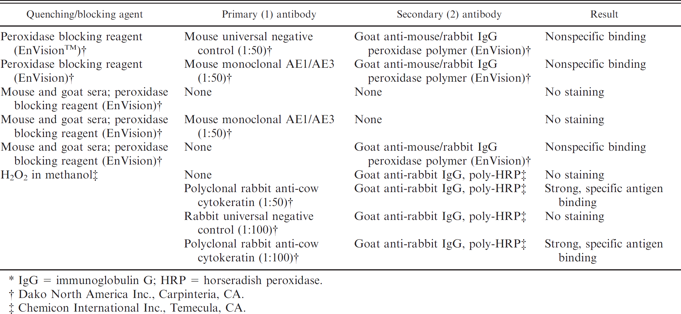
IgG = immunoglobulin G; HRP = horseradish peroxidase.
Dako North America Inc., Carpinteria, CA.
Chemicon International Inc., Temecula, CA.
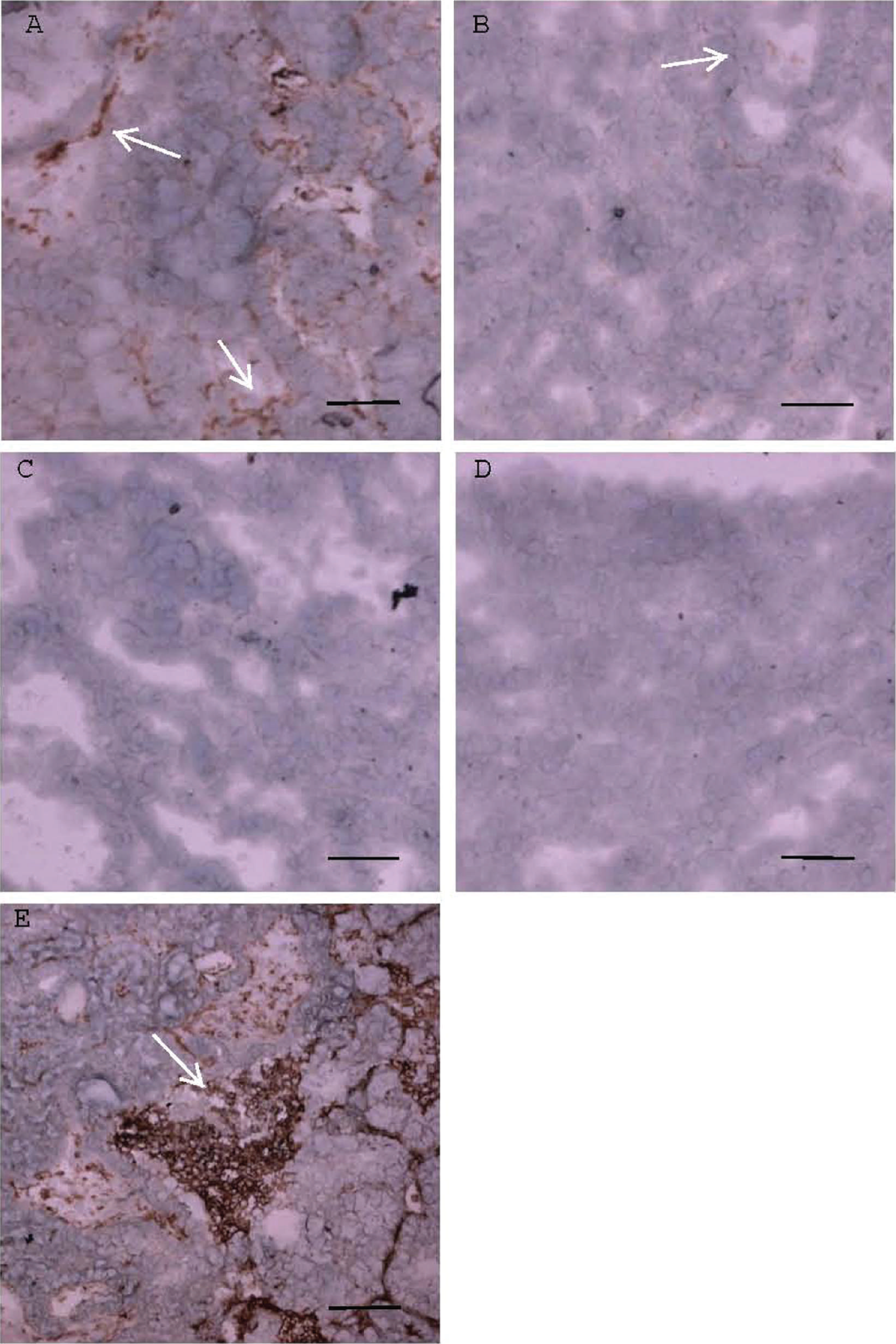
Immunostaining of late-term frozen feline placental tissue sections using mouse monoclonal antibodies and the commercial kit resulted in nonspecific immunoreactivity. Placental sections from a representative control queen are shown. Arrows show chromogen staining. Bars =100 μm. Sections labeled with the mouse universal negative control reagent (
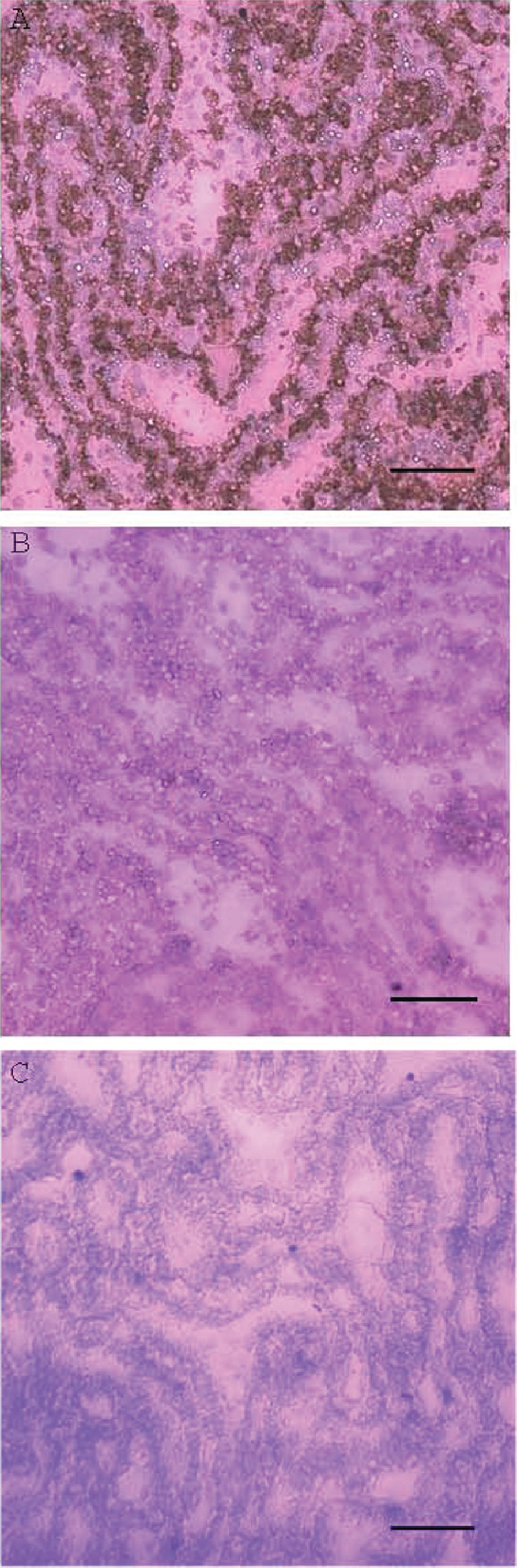
Immunohistochemical staining of late-term frozen feline placental tissue using polyclonal antibodies produced specific immunoreactivity to cytokeratin. Placental sections from a representative control queen are shown. Bars = 100 μm.
Discussion
To determine the role of trophoblasts in FIV-induced placental immunopathology, it was important to identify trophoblasts in feline placental tissue by IHC. The ultimate goal was the isolation of RNA from these cells for gene expression analyses. Therefore, snap-frozen tissues rather than formalin-fixed tissues were chosen, and no attempt was made to determine the quantity or distribution of these cells. To identify these cells, cytokeratin, a marker for trophoblasts, was targeted. The investigation began by using the mouse anti-human pan-cytokeratin monoclonal antibody AE1/AE3 with a traditional IHC kit. The use of anti-cytokeratin monoclonal antibodies to identify trophoblastic cellular populations with IHC has been described in detail in both the human and feline placenta. 4,5,9,16 Villous and extravillous human trophoblast populations were identified throughout pregnancy in frozen sections using monoclonal antibodies directed against cytokeratin. 9 Anti-cytokeratin monoclonal antibodies were used to evaluate the distribution of cytokeratin intermediate filaments in feline fetal chorionic lamellae. 16 However, immunostaining in the current study revealed nonspecific immunoreactivity in feline placental tissues labeled with the mouse monoclonal AE1/AE3 and goat anti-mouse/rabbit IgG peroxidase polymer, which was unable to be resolved with altered blocking techniques.
Nonspecific reactivity of the goat anti-mouse/rabbit IgG peroxidase polymer was demonstrated by reactivity to placental protein in a Western blot assay (data not shown). Likewise, nonspecific immunoreactivity in the IHC procedure was attributed to this secondary antibody. This kit component appears to be an unreliable IHC reagent for labeling feline placental tissue. Although the reliability of the monoclonal antibody AE1/AE3 was not specifically pinpointed in the feline placental tissues, this antibody failed to bind cytokeratin reliably in a majority of normal feline epithelial tissues in a previous report. 10
Nonspecific immunoreactivity occurred during IHC of human placental tissues when investigators attempted to identify dendritic cells by immunolabeling with mouse anti–cluster of differentiation (CD)83 monoclonal antibodies and HLA-G by immunolabeling with specific mouse monoclonal antibody. 3 In that study, nonspecific immunoreactivity occurred when mouse isotype IgG2b was used. Nonspecific binding of mouse antibody IgG2b isotype to endothelial cells in human chorionic villi was prevented by blocking tissues with purified human IgG, which contained a mixture of antibody isotypes. Although the reason for nonspecific immunoreactivity was unproven, it was attributed to inappropriate antibody binding to placental Fc receptors with an affinity for the mouse immunoglobulin Fc domain. Of interest, the placentas of both human beings and mice possess hemochorial placentation, similar mechanisms of IgG transport from mother to fetus, and an “antigen-independent” affinity for antibody. 3,7,14
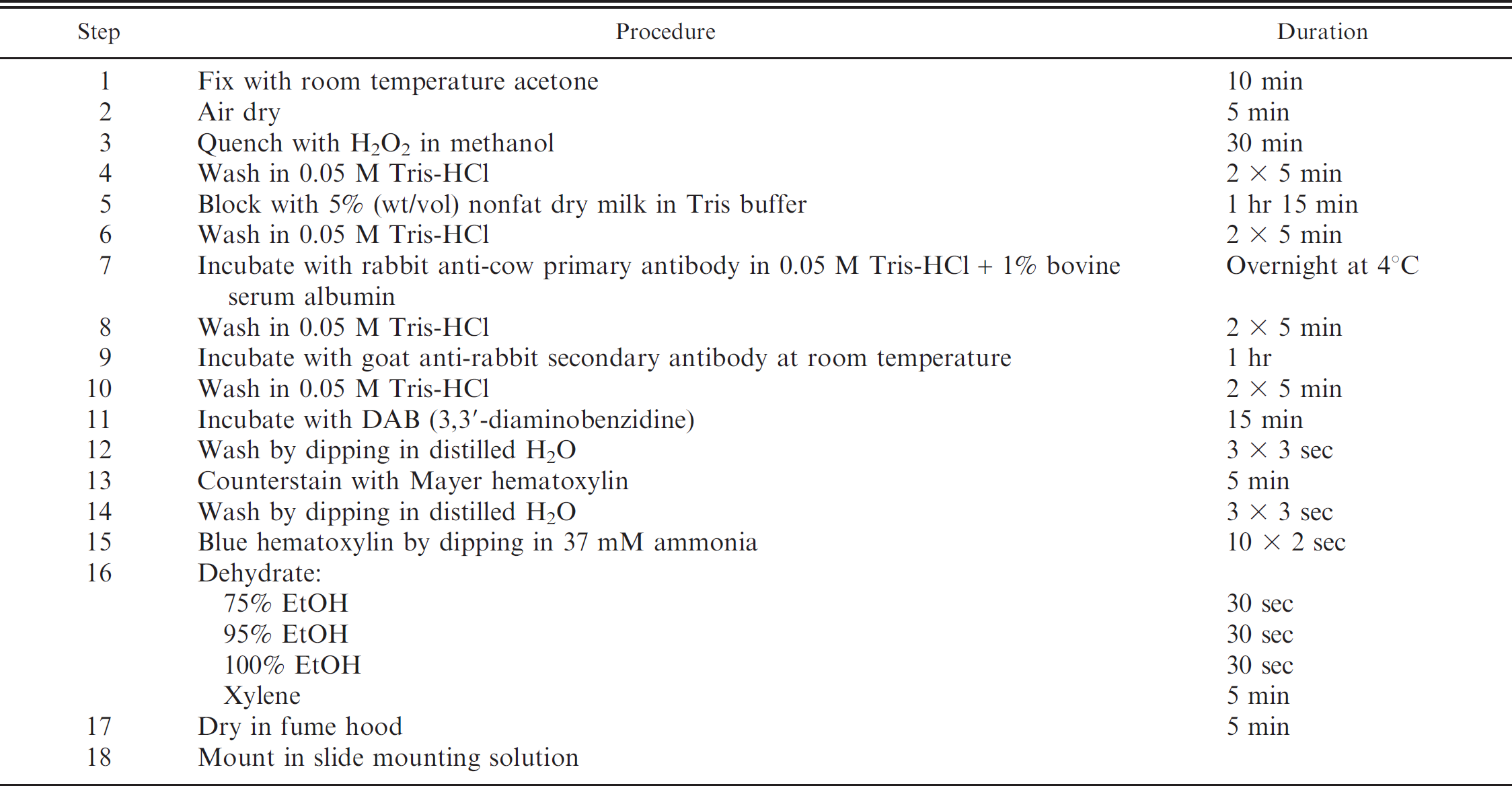
Immunohistochemistry protocol for trophoblast staining in frozen feline placental tissues.
Likewise, nonspecific immunoreactivity of a panel of mouse monoclonal and some polyclonal antibodies to feline placenta in IHC and immunofluorescence procedures has been encountered (data not shown). The approach described in the current study to eliminate this problem was to use a rabbit polyclonal antibody to bovine cytokeratin and secondary goat anti-rabbit, poly-HRP, along with adjusted quenching and blocking procedures. These modifications resulted in specific cytokeratin labeling. An alternative approach that has been shown effective in avoiding Fc receptor non-immune adherence is the use of Fab or F(ab')2 portions of the IgG molecule rather than the whole IgG molecule in immunoassays. 11 Immunohistochemistry in feline tissues with these fragmented antibodies was not attempted in the current study.
The data in the present study confirm that, like human placental tissue, the feline placenta presents a unique challenge to the use of immunolabeling for detection of placental antigens. The present approach (Table 3) to feline placental immunohistochemistry provides a useful alternative method for specifically targeting feline cytokeratin antigen and may provide a basis for targeting other placental antigens with appropriate immunoreactivity.
Acknowledgements
The authors thank Dr. Edward A. Hoover, Colorado State University, for generously providing the infectious plasma pool containing FIV-B-2542; Dr. William Bennett, University of Mississippi Medical Center, for providing technical advice, laboratory facilities, and antibodies; the histology staff in the Department of Pathobiology and Population Medicine at the College of Veterinary Medicine, Mississippi State University, for generously providing the polyclonal rabbit anti-cow cytokeratin WSS antibody and assistance with staining; Brittany T. Clay for assistance with histology; the laboratories of Dr. Jeffery Eells and Dr. Nikolay Filipov, College of Veterinary Medicine, Mississippi State University, for assistance with microscopy and photography; and Dr. Donna Gordon and Crystal Boudreaux, Department of Biological Sciences, Mississippi State University, for assistance with photography and photographic processing. This project was supported by the National Institutes of Health (2R15AI048419–02A1 and 3R15AI048419–02A1S1).
Footnotes
a.
Sigma-Aldrich, St. Louis, MO.
b.
Fisher Scientific, Waltham, MA.
c.
Lab Scientific Inc., Livingston, NJ.
d.
EnVision™, Dako North America Inc., Carpinteria, CA.
g.
Catalog no. N1590, Dako North America Inc., Carpinteria, CA.
f.
Catalog no. N1698, Dako North America Inc., Carpinteria, CA.
g.
Catalog no. K1392, Dako North America Inc., Carpinteria, CA.
h.
Invitrogen Corp., Carlsbad, CA.
i.
Catalog no. Z0622, Dako North America Inc., Carpinteria, CA.
j.
Catalog no. N1699, Dako North America Inc., Carpinteria, CA.
k.
Chemicon International Inc., Temecula, CA.