
Abstract
This report describes an invasive mammary carcinoma with a rare distinctive feature characterized by sebaceous differentiation of tumor cells. This tumor occurred in a 10-year-old female mixed breed dog. The patient had two masses in the left fifth mammary gland. Grossly, the masses were firm, whitish to light brown, and superficially ulcerated. On cut surface, they were multilobulated with foci of necrosis. Microscopically, the tumors were composed of two distinctive neoplastic components, intraductal papillary adenocarcinoma and sebaceous carcinoma. The regions of sebaceous tumor were clumped separately, contained well-developed sebaceous cells and keratinized epithelial cells, and were surrounded by few to several layers of basaloid cells. The cells with abundant foamy cytoplasm that resembled sebaceous cells were also found within the intraductal papillary-like nests of mammary carcinoma, providing evidence of sebaceous metaplasia. Sebaceous differentiation in a mammary gland tumor is possible, because skin appendages and ductal apparatus of the mammary gland share a common anlagen. This tumor had an aggressive behavior with lymphatic metastasis. Consequentially, the dog had a poor prognosis.
To our knowledge, sebaceous differentiation of mammary carcinoma has not been documented in veterinary literature yet, although there have been a few cases reported in humans. 5, 6, 10, 12, 13 In humans, sebaceous metaplasia has been reported in intraductal carcinoma of the breast, 5, 6, 10, 12, 13 or it has a concurrent presence of squamous differentiation. 5, 10, 12 However, due to the rarity of the peculiar variant, the prognosis of the sebaceous phenotype in mammary carcinoma remains unsettled. 13 Similarly, there is a variant exhibiting lipid-rich carcinoma of the mammary gland in dogs. 3, 8, 9 The lipid-rich tumor cells contain either multiple and small, or large and solitary vacuoles that pushed the nucleus to the periphery of the cell as the signet ring cell. 3, 7 Moreover, the lipid-rich carcinomas of the mammary gland in humans 11 and dogs 3 have a poor prognosis.
In this report, a rare case of sebaceous mammary carcinoma in a dog is described. A 10-year-old, mixed breed, female dog had a greasy mammary tumor. She was nulliparous and unspayed; the date of her last estrus was unknown. The mammary tumor was found 1 year prior by the owner. It grew gradually and developed some superficial ulcers in the last several weeks prior to registration at our teaching hospital. This ulcerated mass measured 5.2 × 4.8 × 3.7 cubic centimeters and was located in the left fifth mammary gland and inguinal area. There was another tumor of 2 × 1.8 × 1.5 cubic centimeters just cranially to the mass mentioned above. The regional (inguinal) lymph node was moderately enlarged. Radiographically, lateral and ventro-dorsal views of thorax and abdomen showed no significant disorder. Modified radical mastectomy (regional mastectomy) was performed to remove the left fourth and fifth mammary glands completely, including the adjacent lymph nodes. During the operation, the surgeon found tumor invasion into the fascia muscularis; consequently, the excised specimen was submitted for pathologic examination.
Grossly, the masses were firm, whitish to light brown, and superficially ulcerated. On cut surface, they were multilobulated with foci of necrosis. The inguinal lymph node was enlarged with whitish areas on the cut surface. Microscopically, the mammary mass consisted of two dominating types of tumor. The first was characterized by intraductal papillary-like nests with fibrovascular stroma (Fig. 1) and mild lymphoid cell infiltration. The tumor cells were pleomorphic with prominent nucleolus. Mitosis was frequent. The second showed sebaceous differentiation. The areas of this sebaceous tumor were characterized by multilobulated growth of sebaceous and keratinized epithelial cells, which were clumped with each other and surrounded by few to several layers of basaloid cells (Fig. 2). The sebaceous cells were characterized by small discrete cytoplasmic vacuoles that produced scalloping or rounded indentations in the nuclear membrane (Fig. 2, inset). Numerous mitotic cells were also found in the layers of basaloid cells. A few foci of clumped cells with an abundant foamy cytoplasm, resembling sebaceous cells, were also noticed within the intraductal papillary-like nests of mammary carcinoma, supporting the sebaceous metaplasia (Fig. 3). Large areas of invasive sebaceous carcinoma were commonly seen surrounding small-sized intraductal papillary tumor nests (Fig. 4). In the tumor cells with sebaceous differentiation, the periodic acid–Schiff (PAS) stain for glycogen and the mucicarmine stain for mucin were negative, whereas Oil Red O stain for lipid was positive (Fig. 5). Disseminated lymphatic spread to the lymph node was characteristic of the sebaceous carcinoma.
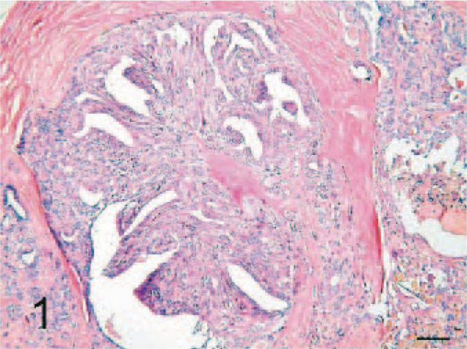
Mammary gland; dog. Intraductal papillary-like structures of mammary adenocarcinoma with prominent fibrous stroma. HE. Bar = 100 μm.
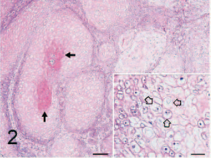
Mammary gland; dog. Regions of sebaceous carcinoma. Multilobulated growths of tumor cells are clumped and are surrounded by few to several layers of basaloid cells, with differentiation to distinctive sebaceous cells and/or keratinized epithelial cells in central zone (arrows). HE. Bar = 100 μm. The cytoplasmic lipid vacuoles produce scalloping or rounded indentations in the nuclear membrane (inset, open arrows). HE. Bar = 25 μm.
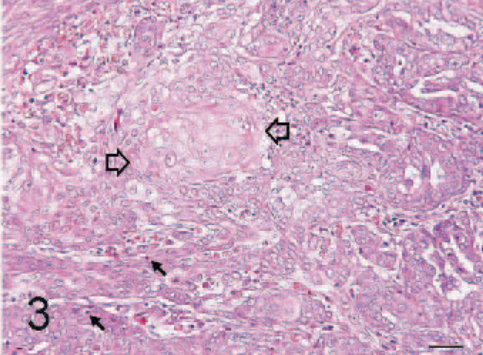
Mammary gland; dog. Intraductal papillary mammary carcinoma with a distinct area of sebaceous–squamous differentiation (open arrows). Mitotic figures (arrows) are common in the intraductal papillary nests. HE. Bar = 50 μm.
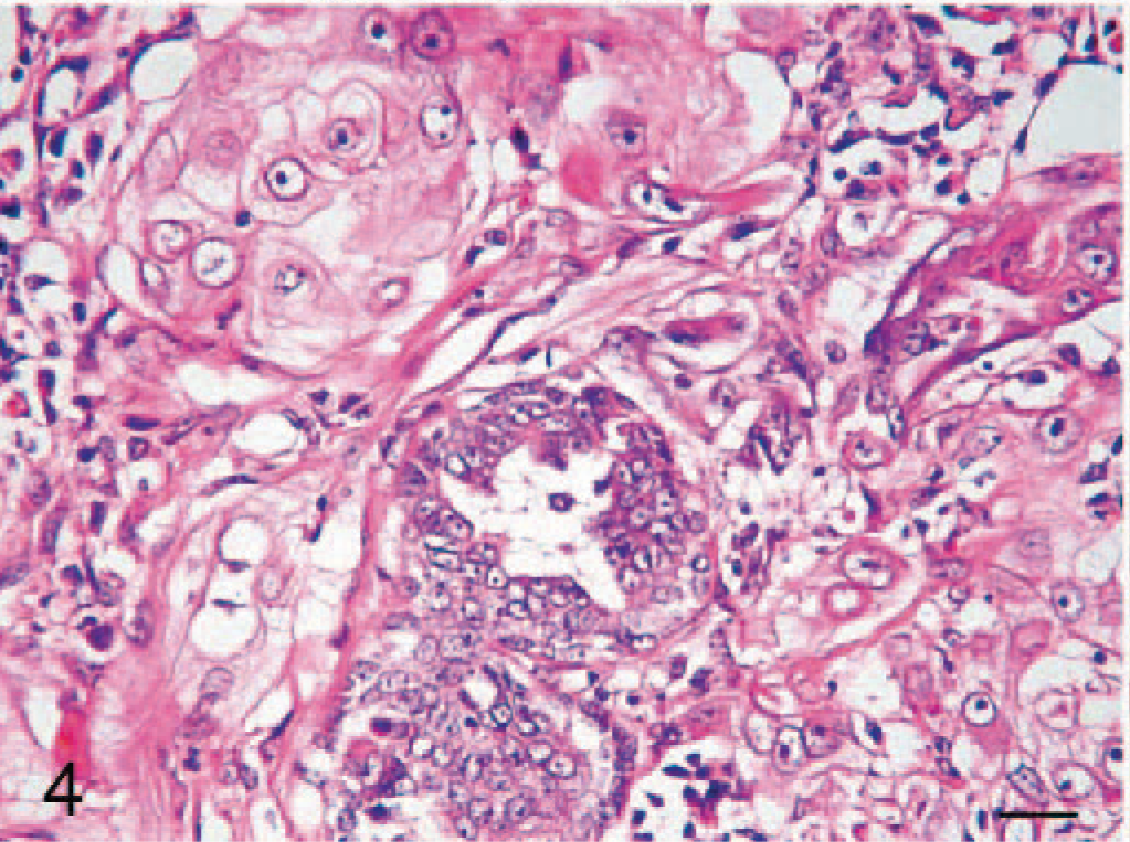
Mammary gland; dog. Small-sized intraductal tumor nests distributed among the large area of invasive sebaceous carcinoma. HE. Bar = 25 μm.
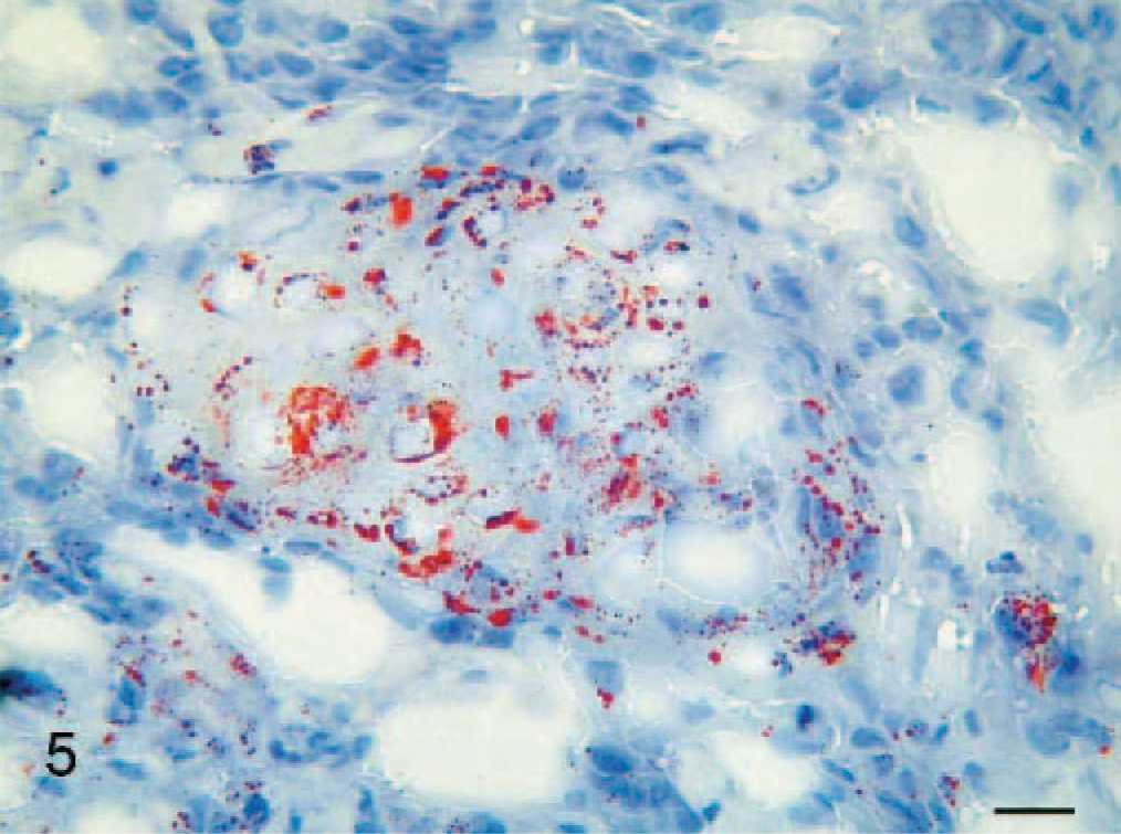
Mammary gland; dog. The frozen section shows positive result of Oil Red O stain. Note the varying sizes of granules, indicating the neutral lipid droplets within the cytoplasms of sebaceous-like tumor cells individually. Oil Red O. Bar = 25 μm.
No chemotherapy was conducted after surgical removal of mammary tumors. The prognosis was guarded. Two months later, the tumor recurred with metastasis to the iliac lymph node. The dog died 3 months after operation.
The mammary gland develops from a specialized sweat gland. 1 Both skin appendages and the secretory and ductal apparatus of the mammary gland share a common embryonic ectodermal anlagen. 13 The origin and pathway of the sebaceous metaplasia in mammary carcinomas remain unclear. It is suggested that there is an association between sebaceous gland metaplasia and squamous metaplasia. 5 However, in this case neoplastic glandular cells differentiated into cells that both contained lipid droplets within the cytoplasm and displayed scalloping or rounded indentations in the nuclear membrane. This is characteristic of sebaceous metaplasia, although initially the cells were erroneously interpreted as squamous metaplasia. Therefore, we propose that the sebaceous metaplasia probably was derived from mammary stem cells (basaloid cells) with a pluripotentiality of differentiation. Some previous studies also concluded that the mammary stem cells were converted into sebaceous cells or adenosquamous cells. 5, 12
Metaplastic sebaceous carcinoma of the mammary gland should be distinguished from primary sebaceous carcinoma of the skin adnexa. In fact, sebaceous carcinomas arising from skin sites are uncommon and seldom show widespread metastasis in the dog. 2, 4 The feature that distinguishes this case from a skin appendage tumor is the metaplastic transition of the intraductal papillary carcinoma from a small area of sebaceous differentiation to a large area of invasive sebaceous carcinoma with associated squamous differentiation.
Lipid-rich carcinoma, a variant of mammary carcinoma, has been documented recently in dogs. 3, 8, 9 It is characterized by tumor cells that 1) are arranged in solid nests and cords separated by a moderate amount of stroma and 2) have an abundant foamy cytoplasm containing a large amount of neutral lipid. 7 In contrast, the mammary carcinoma with sebaceous differentiation showed the characteristics of sebaceous tumor with multilobulated clumps and central areas of keratinized epithelial cells. These lipid vacuoles in sebaceous differentiation produce scalloping or rounded indentations in the nuclear membrane that are different from those found in lipid-rich carcinoma. Both sebaceous and lipid-rich mammary carcinomas may display positive staining results for Oil Red O stain from frozen sections.
In our patient, the mammary carcinoma with sebaceous differentiation had a poor prognosis. In humans, it has been suggested that the prognosis of this tumor is more favorable, with little propensity for metastatic spread. 5, 6, 13