
Abstract
Programmed death ligand 1 (PD-L1) is an immune checkpoint molecule that plays a crucial role in regulating antitumor immune responses. Canine mammary carcinomas (CMCs) are common tumors of dogs. Despite extensive studies on the heterogeneity of CMCs, there is still a lack of effective precision therapies for the treatment of CMCs. In this study, we aimed to investigate the correlation between PD-L1 mRNA and protein expression in CMCs and explore its association with histopathological grade and molecular markers, including the estrogen receptor, epidermal growth factor receptor 2, and cytokeratin 5/6 (CK5/6). Formalin-fixed paraffin-embedded samples were evaluated for PD-L1 mRNA expression using RNA in situ hybridization and PD-L1 protein expression using immunohistochemistry. We observed no substantial correlation between PD-L1 mRNA and protein expression in CMCs; however, PD-L1 mRNA levels were significantly higher in grade 3 than in grade 1 tumors (P = .001). In addition, we observed a positive correlation between PD-L1 protein expression and CK5/6 expression in CMCs (P = .032). These findings suggest that PD-L1 expression in CMCs is heterogeneous and may be regulated post-transcriptionally. Further studies are needed to explore the prognostic and therapeutic implications of PD-L1 expression in different molecular subtypes of CMCs and their potential as predictive biomarkers for immunotherapy.
Programmed death ligand 1 (PD-L1), a member of the B7 family, binds to programmed cell death protein 1 (PD-1) expressed on activated T-cells and inhibits antitumor immunity. PD-L1 is expressed in both hematopoietic and non-hematopoietic cells, including various tumor cell types. 24 Since immunotherapies targeting the PD-1/PD-L1 axis can block the suppression of T-cell activation and enhance antitumor immunity, the development of PD-1/PD-L1 inhibitors is actively underway. Atezolizumab, durvalumab, and avlumab have been approved by the Food and Drug Administration (FDA) as PD-L1 inhibitors for the treatment of human cancers, including melanoma, non-small cell lung cancer, Merkel cell carcinoma, and breast cancer. 1 However, biomarkers for predicting the therapeutic effects of these drugs remain unclear. Although using PD-L1 protein expression on tumor cells as a biomarker to select the patients for PD-1/PD-L1 immunotherapy and as a predictive biomarker for PD-1/PD-L1 immunotherapy has been proposed,10,25,33 the results were controversial due to variations in the PD-L1 antibody clones, cutoffs, and scoring systems. 30
PD-L1 mRNA expression is also being investigated as a biomarker for human cancers. RNAscope, an RNA in situ hybridization (RISH) method using formalin-fixed paraffin-embedded (FFPE) tissue, enables visualization and interpretation of mRNA expression in routine clinical samples. 37 PD-L1 mRNA expression detected by RNAscope was positively correlated with PD-L1 protein expression in head and neck squamous cell carcinoma, non-small-cell lung cancer, urothelial carcinoma, and meningioma in humans.11,12 However, some studies have reported inconsistencies between PD-L1 mRNA expression and PD-L1 protein levels in non-small-cell lung cancer.7,18
Canine mammary carcinomas (CMCs) are among the most common tumors in female dogs. Many factors, including age, breeds, sex hormones, and obesity, are known to act directly as risk factors for CMC progression. 13 Consequently, CMCs exhibit morphological and biological heterogeneity and can be classified based on various criteria, including histological and molecular subtypes. 15 Among those, molecular subtypes based on the expression of hormonal receptors (estrogen receptor and progesterone receptor [ER and PR]), ERBB2 (canine homolog of human epidermal growth factor receptor 2 [HER2]), and basal cell marker (cytokeratin 5/6 [CK5/6]) categorizes CMCs into 5 subtypes: luminal A, luminal B, ERBB2-overexpressing, basal-like, and triple-negative. 26 The most frequent subtype is the luminal A subtype, and the most malignant subtype is the basal-like subtype in CMCs. Molecular subtypes are associated with histopathological grade and lymphatic invasion. 15 Despite extensive studies on the heterogeneity of CMCs, the establishment of effective precision therapy for routine use in CMCs is still lacking, and surgical treatment remains the preferred approach to date. 35
Recently, studies on PD-L1 expression in CMCs have been conducted.3,20,21 Most CMCs show PD-L1 protein expression in tumor cells, 21 and high PD-L1 protein expression correlates with poor prognosis.3,20 However, PD-L1 mRNA expression in CMCs has not been investigated.
The aim of this study was to investigate the correlation between PD-L1 mRNA and protein expression and to determine whether PD-L1 expression in tumor cells is associated with the histopathological grade and molecules, such as ER, ERBB2, and CK5/6 in CMCs.
Materials and Methods
Ethical Statement
All samples were obtained from the FFPE tissue archives of the Department of Veterinary Pathology at Konkuk University, Seoul, Korea. All tissues in the archive were referred to the Department of Veterinary Pathology for histopathological examination from local animal hospitals in Korea after surgical excision from the dogs for curative purposes. All procedures were performed with informed consent from the owners of the dogs. No live animals were used for research purposes. This study did not require the approval of the Institutional Animal Care and Use Committee.
Sample Selection and Histopathological Evaluation
A total of 223 samples were obtained from the FFPE tissue archive of the Department of Veterinary Pathology. The dogs were diagnosed with mammary carcinomas from 2019 to 2021. Microsections were prepared from the samples, and hematoxylin and eosin staining was performed for histopathological evaluation. Simple carcinomas, according to histological classification of Zappulli et al, 38 were selected to exclude the differences according to the histological subtypes. The histopathological grades of the tumors were determined according to the criteria used by Clemente et al. 6 Three individual pathologists evaluated the histopathological grades, and only samples with grading consensus from all three pathologists were selected for the study. Among grade 1–3 samples, grade 2 samples were excluded because the difference in malignancy compared to grades 1 or 3 could be ambiguous. Ultimately, 63 grade 1 and 71 grade 3 samples were selected. Signalments and histopathological evaluations of samples are summarized in Supplemental Table S1.
RNA In Situ Hybridization
RISH was performed on 49 FFPE samples to evaluate PD-L1 mRNA expression in CMCs. To mitigate the potential degradation of mRNA owing to the storage period of the FFPE blocks, 22 grade 1 and 27 grade 3 samples were randomly selected from those obtained from late 2020 to 2021. The RNA quality can vary depending on the fixation process and storage conditions of the FFPE blocks; therefore, quantitative analysis was employed to correct for differences in mRNA retention among the FFPE samples. Canine PD-L1 (CD274, Advanced Cell Diagnostics, Hayward, CA, USA, Cat No. 488461) and POLR2A probes (Cat No. 310981) were used to detect target and housekeeping genes, respectively, on serially sectioned slides. RISH was performed using the RNAscope Assay Kit (Advanced Cell Diagnostics) following the manufacturer’s protocol.
Interpretation of RISH
To evaluate the expression levels of PD-L1 mRNA, five representative high-power (400X) fields showing PD-L1 mRNA signals in the tumor cells were selected for each sample. Digital images were acquired by locating the same fields on PD-L1 and POLR2A slides using an Olympus BX51 microscope with an ocular FN 22-mm objective and image transfer software (Olympus, Tokyo, Japan). The PD-L1/POLR2A ratio was then calculated using open-source image analysis programs, FIJI software (ImageJ win64; National Institutes of Health, Bethesda, Maryland, USA), and Icy software 8 to distinguish and count the mRNA dots expressed on tumor cells, following the methods described by Cho et al. 4
Immunohistochemistry
To evaluate the protein expression of estrogen receptor (ER), ERBB2, CK5/6, and PD-L1 in mammary carcinomas, immunohistochemistry (IHC) experiments were performed. Serial 4-μm-thick sections of FFPE tissues were prepared to minimize intra-tumoral heterogeneity. The deparaffinized slides were then treated with 2% H2O2 for 20 minutes to block endogenous peroxidase activity. Heat-induced antigen retrieval was performed using either a citric acid (pH 6) or Tris-ethylenediaminetetraacetic acid buffer (pH 9) in a pressure cooker, followed by cooling to room temperature. After washing 3 times in phosphate-buffered saline, the slides were blocked with a 5% goat serum solution for 30 minutes. Subsequently, the primary antibodies were applied at the appropriate dilution overnight at 4°C. Detailed information and protocols for each primary antibody are listed in Table 1. These antibodies have been validated and conventionally used for dogs in previous studies.3,16,29 After incubation, the slides were washed in phosphate-buffered saline 4 times, and secondary antibodies (Dako REAL EnVision kit; Dako) were applied for 40 minutes. Horseradish peroxidase and 3,3'-diaminobenzidine were used for labeling, and the slides were counterstained with Gill’s hematoxylin.
Primary antibodies and immunohistochemical protocols.
Abbreviations: PD-L1, programmed death ligand 1; ER, estrogen receptor; ERBB, epidermal growth receptor; CK5/6, cytokeratin 5/6.
For the PD-L1 antibody, a normal canine tonsil sample was used as the positive control. For the ER antibody, normal to hyperplastic canine mammary glands were used as positive controls. For the ERBB2 and CK5/6 antibodies, mammary carcinomas with ERBB2 overexpression and CK5/6-positive CMCs from our previous study were used as positive controls, respectively. 19 For negative controls, the antibody was replaced by isotype-matched immunoglobulins. All control tissues were selected from our FFPE tissue archive.
Interpretation of IHC
PD-L1 IHC expression was considered positive when more than 1% of the tumor cells exhibited labeling on the membrane and/or cytoplasm based on the study by Lopes-Neto et al. 20 The IHC results for ER, ERBB2, and CK5/6 were assessed following the method described by Sassi et al. 26 ER expression was considered positive when nuclear staining was observed in at least 5% of tumor cells. ERBB2 expression was considered positive when at least 10% of the tumor cells showed complete membranous labeling, and cytoplasmic CK5/6 labeling in at least 1% of the tumor cells was considered positive.
Statistical Analysis
Statistical analyses of RISH and IHC results were performed using the Statistical Package for Social Science (SPSS, Inc., IBM Company, Chicago, IL, USA) software. The correlation between the PD-L1/POLR2A ratio, histopathological grade, and IHC expression of PD-L1, ER, ERBB2, and CK5/6 were tested using the Mann–Whitney U test (P < .05). Fisher’s exact test (P < .05) was employed to evaluate the relationship between PD-L1 IHC expression, histopathological grade, and IHC expression of molecular markers (ER, ERBB2, and CK5/6).
Results
RISH Expression of PD-L1 in CMCs
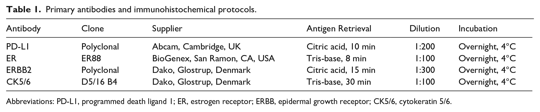
PD-L1 and POLR2A were detected as dots in both the epithelial and stromal regions of the tumor (Fig. 1). Only the dots located in the epithelial region were counted, and the dots in the stromal region including fibrous stromal cells and immune cells were excluded. The PD-L1/POLR2A ratios ranged from 0.05 to 2.31. The mean and median PD-L1/POLR2A ratios were 0.37 and 0.19, respectively. Dots were observed as clusters in the epithelial region. In the fields with high mRNA expression levels, the density of the clusters was higher.
Mammary carcinoma in dogs. RNA in situ hybridization (RISH). (a) Expression of programmed death ligand 1 (PD-L1) mRNA in grade 1 mammary carcinoma. PD-L1 is detected as few dots (arrows) in the epithelium. PD-L1 RISH. (b) Expression of RNA polymerase Ⅱ subunit A (POLR2A) mRNA in grade 1 mammary carcinoma. POLR2A is detected as abundant dots in the epithelium. POLR2A RISH. (c) Expression of PD-L1 mRNA in grade 3 mammary carcinoma. PD-L1 is detected as abundant and clustered dots in the epithelium. PD-L1 RISH. (d) Expression of POLR2A mRNA in grade 3 mammary carcinoma. POLR2A is detected as abundant dots in the epithelium. POLR2A RISH.
IHC Expression of PD-L1 and ER/ERBB2/CK5/6 in CMCs
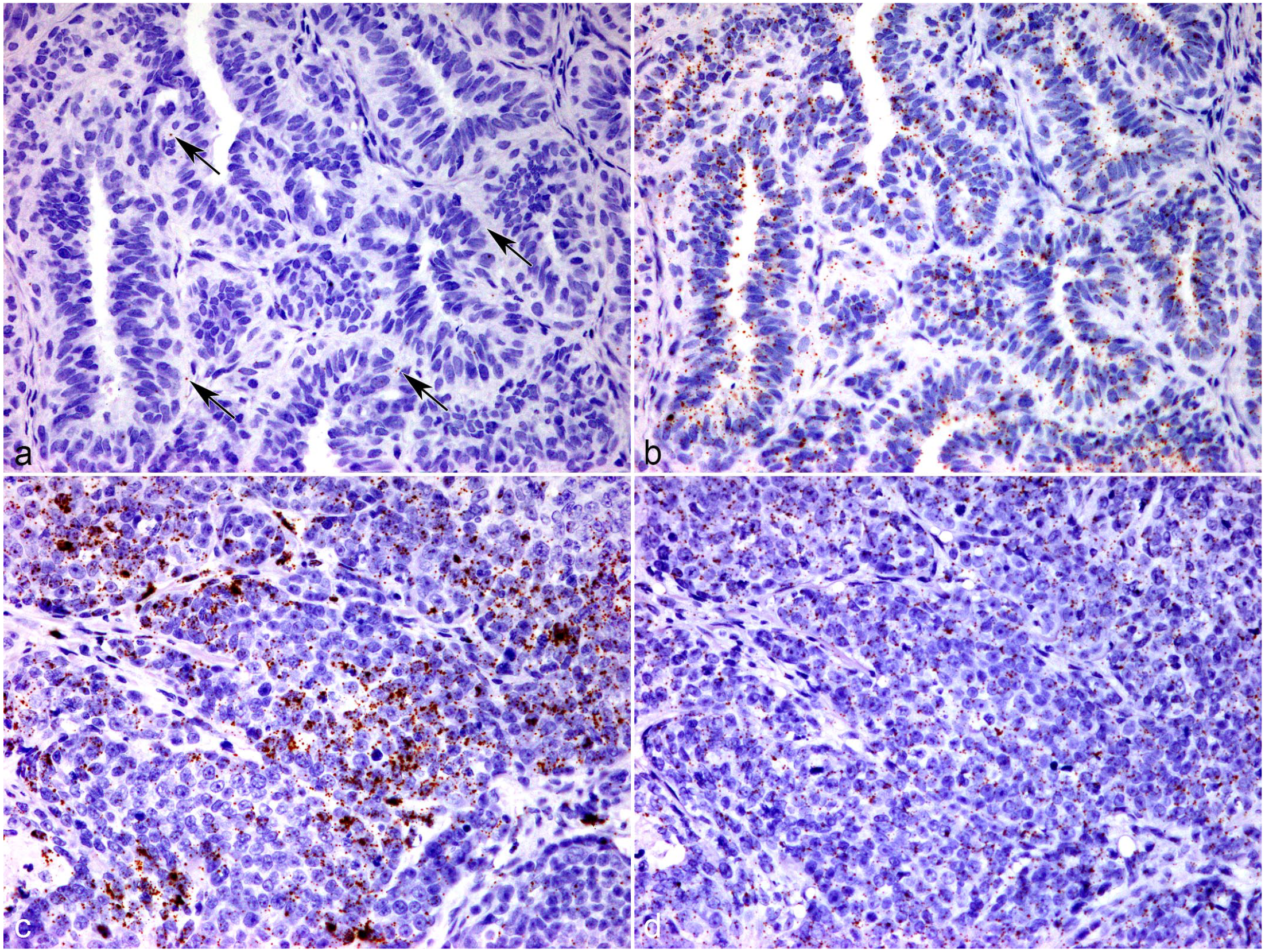
PD-L1 expression in tumor cells was positive in 61.94% (83/134) of mammary carcinomas. PD-L1 was labeled on the membrane and/or cytoplasm of the tumor cells (Fig. 2a). ER expression in tumor cells was positive in 48.51% (65/134) of patients, with nuclear labeling (Fig. 2b). ERBB2 labeling was observed in 75.37% (101/134) of cases, showing complete membranous labeling (Fig. 2c). The expression of CK5/6 by tumor cells was positive in 78.36% (105/134) of cases, showing cytoplasmic labeling (Fig. 2d).
Mammary carcinoma in dogs. Immunohistochemistry (IHC). (a) IHC for programmed death ligand 1 (PD-L1) shows cytoplasmic and/or membranous immunolabeling. PD-L1 IHC. (b) IHC for estrogen receptor (ER) shows nuclear immunolabeling. ER IHC. (c) IHC for epidermal growth factor receptor 2 (ERBB2) shows complete membranous immunolabeling. ERBB2 IHC. (d) IHC for cytokeratin 5/6 (CK5/6) shows cytoplasmic immunolabeling. CK5/6 IHC.
Correlation Between PD-L1/POLR2A Ratio and PD-L1 IHC Expression in CMCs
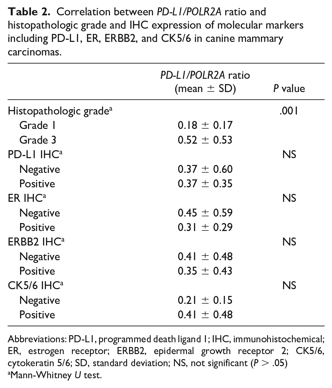
The correlation between the PD-L1/POLR2A ratio and PD-L1 IHC expression was investigated. The mean ± standard deviation of PD-L1/POLR2A ratios for PD-L1 IHC negative and positive tumors were 0.37 ± 0.60 and 0.37 ± 0.35, respectively. No association was observed between the PD-L1 mRNA and PD-L1 protein expression levels (P > .05, Table 2).
Correlation between PD-L1/POLR2A ratio and histopathologic grade and IHC expression of molecular markers including PD-L1, ER, ERBB2, and CK5/6 in canine mammary carcinomas.
Abbreviations: PD-L1, programmed death ligand 1; IHC, immunohistochemical; ER, estrogen receptor; ERBB2, epidermal growth receptor 2; CK5/6, cytokeratin 5/6; SD, standard deviation; NS, not significant (P > .05)
Mann-Whitney U test.
Correlation Between PD-L1/POLR2A Ratio and Other Parameters in CMCs
The association between the PD-L1/POLR2A ratio and other parameters, including the histopathological grade and IHC expression of ER, ERBB2, and CK5/6, is summarized in Table 2. The histopathological grade showed a significant association with the PD-L1/POLR2A ratio (P = .001, Fig. 3). The mean ± standard deviation of PD-L1/POLR2A ratios for grade 1 and grade 3 tumors were 0.18 ± 0.17 and 0.52 ± 0.53, respectively. The median PD-L1/POLR2A ratios were 0.15 and 0.32 for grade 1 and 3 tumors, respectively. The IHC expression of ER, ERBB2, and CK5/6 did not significantly correlate with the PD-L1/POLR2A ratio (P > .05).
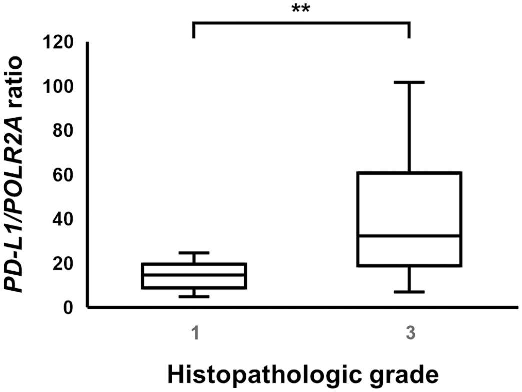
Comparison of the PD-L1/POLR2A ratio in canine mammary carcinomas (CMCs) according to the histopathological grade. The boxes show the median and quartiles. The whiskers show the highest and lowest values. The PD-L1/POLR2A ratio is significantly higher in grade 3 CMCs than in grade 1 CMCs. Mann-Whitney U test, **P = .001. PD-L1, programmed death ligand 1; POLR2A, RNA polymerase II subunit A.
Correlation Between PD-L1 IHC Expression and Other Parameters in CMCs
The associations between PD-L1 IHC expression and other parameters, including the histopathological grade and IHC expression of ER, ERBB2, and CK5/6, are summarized in Table 3. There was no statistical correlation between PD-L1 IHC expression and the histopathological grade. IHC expression of CK5/6 correlated with PD-L1 expression (P = .032). Neither ER nor ERBB2 expression were significantly associated with PD-L1 expression (P > .05).
Correlation between the expression of PD-L1 immunohistochemistry (IHC) and histopathologic grade and IHC expression of molecular markers including ER, ERBB2, and CK5/6 in canine mammary carcinomas.
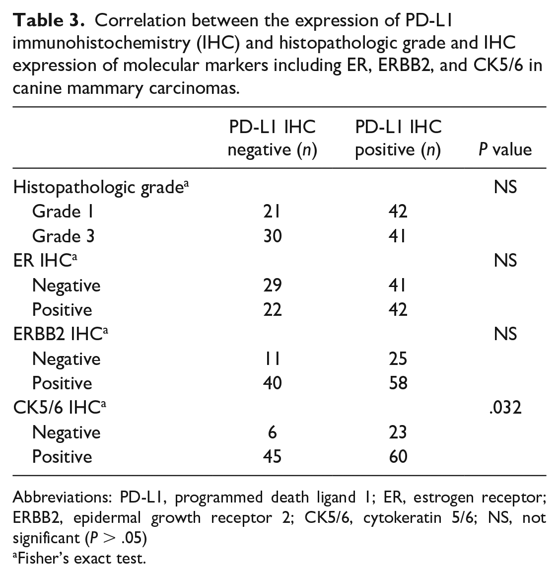
Abbreviations: PD-L1, programmed death ligand 1; ER, estrogen receptor; ERBB2, epidermal growth receptor 2; CK5/6, cytokeratin 5/6; NS, not significant (P > .05)
Fisher’s exact test.
Discussion
In this study, we demonstrated that PD-L1 mRNA expression was associated with histopathological grade. In addition, PD-L1 protein expression was evaluated, and PD-L1 mRNA and PD-L1 protein expression levels were compared. However, there was no considerable correlation between PD-L1 mRNA and protein expression levels. In addition, we found that PD-L1 expression and CK5/6 expression correlated at the protein level. To our knowledge, this is the first study to detect PD-L1 mRNA expression in CMCs using RISH.
The findings of this study revealed a substantial association between PD-L1 mRNA expression and the CMC histopathological grade. Specifically, higher levels of PD-L1 mRNA were observed in histopathological grade 3 tumors than in grade 1 tumors. These results differ from those of previous studies conducted on human breast cancer, in which no correlation between the PD-L1 mRNA expression and histopathological grade was reported.17,27 These conflicting results may be attributed to biological variations between species as well as the different research methods employed to detect mRNA expression. We calculated the PD-L1/POLR2A ratio expressed on tumor cells using RNA scope, correcting for the difference in mRNA retention between the samples. Schalper et al 27 also used RNAscope but measured mRNA levels by automated quantitative fluorescence detection. Kim et al 17 performed quantitative reverse-transcription (qRT) PCR using total mRNA isolated from tumor tissues.
In this study, no statistical correlation was found between PD-L1 mRNA and protein expression levels. This discrepancy may be attributed to post-transcriptional regulation. 36 Similar findings of weak concordance between PD-L1 mRNA and protein expression have been reported in previous studies on early human breast cancer. 39 Factors such as post-translational modifications and heterogeneity within tumor tissues may contribute to the lack of correlation between PD-L1 mRNA and protein levels in CMCs.
Furthermore, we observed that the PD-L1 expression was positively correlated with CK5/6 expression at the protein level in CMCs. CK5/6 is a basal cytokeratin marker used to classify the molecular subtypes of human breast cancer and CMCs. The positivity of CK5/6 has been associated with poor clinicopathological parameters in CMCs. 19 Interestingly, previous studies have demonstrated that basal-like subtypes (ER −, PR −, ERBB2 −, and basal marker +) frequently express PD-L1 protein, and higher PD-L1 gene expression has been shown to predict better survival outcomes in the basal-like subtype human breast cancer.2,23 In our study, PD-L1 expression was positively correlated with CK5/6 expression and was not associated with ER or ERBB2 expression. Whether the correlation between PD-L1 and CK5/6 IHC expression can be utilized as a predictive factor for the therapeutic effects of PD-L1 inhibitors in CMCs requires further study. The association between PD-L1 expression and the molecular subtype of CMCs should also be investigated.
Our study has some limitations. First, different clones of PD-L1 antibodies have been used in previous studies, including the FDA-approved clones 28-8, 22C3, and SP142. Although studies comparing the performance of different antibodies have demonstrated similar results in various human cancers,31,32,34 there may be variations in antibody specificity and sensitivity. 14 Second, the interpretation of PD-L1 IHC lacked standardized evaluation criteria among previous studies. Different studies have employed diverse evaluation methods, including varying scales for labeling intensity and cutoff values for determining PD-L1 positivity.3,20 In previous studies on CMCs, Ariyarathna et al 3 quantified PD-L1 IHC expression by considering the labeling intensity using a scale ranging from 0 to 3, and Lopes-Neto et al 20 evaluated PD-L1 IHC positive or negative cells when 1% of the tumor cells showed labeling.
At the early stage of this study, we categorized the PD-L1 IHC expression into scores from 0 to 3 (<1%: score 0, 1%–5%: score 1, 6%–50%: score 2, and >50%: score 3), according to the PD-L1 study on triple-negative breast cancer in humans. 9 The number of samples with scores of 0–3 was 51, 58, 14, and 11, respectively. However, no significant statistical correlations between PD-L1 IHC expression and other parameters (histopathological grade, molecular markers, and PD-L1 ISH expression) were determined with this classification. One of the limitations of this classification was the uneven numbers of samples in each class. Also, as the classification criteria were utilized from a PD-L1 study in human breast cancer, applying the criteria to dogs may require modification due to interspecies differences. To overcome these limitations, we modified the evaluation method to consider cases as PD-L1 positive when at least 1% of the tumor cells were immunolabeled, according to the study by Lopes-Neto et al. 20 We selected a polyclonal antibody used by Ariyarathna et al, 3 which showed a correlation between PD-L1 IHC expression and the histopathological grade of CMCs. The lack of correlation between PD-L1 IHC expression and histopathological grade in this study, in contrast to a previous study that used the same antibody, may be attributed to the different evaluation methods employed. Because canine-specific PD-L1 antibodies are under development,5,22 further studies are needed on standardized protocols and evaluation methods for PD-L1 IHC in dogs.
Lastly, when calculating PD-L1/POLR2A ratio, the dots in dense clusters may have been counted less accurately owing to limitations of the counting program. However, as our study and other previous studies utilizing similar counting methods have yielded meaningful results, the effect of the clustered dots may be insignificant.4,28
This study provides valuable insights into PD-L1 expression in CMCs by detecting PD-L1 mRNA expression using RISH. These findings highlight the association between the PD-L1 mRNA expression and histopathological grade of CMCs. Moreover, the correlation between PD-L1 and CK5/6 protein expression paves the way for future studies investigating the potential use of PD-L1 inhibitors in subsets of CMCs, such as those expressing CK5/6. Further research is warranted to explore the prognostic significance and therapeutic implications of PD-L1 expression and its association with different molecular subtypes of CMCs.
Supplemental Material
sj-pdf-1-vet-10.1177_03009858241226621 – Supplemental material for PD-L1 mRNA and protein expression in canine mammary carcinomas: Correlation with histopathological grade and molecular markers
Supplemental material, sj-pdf-1-vet-10.1177_03009858241226621 for PD-L1 mRNA and protein expression in canine mammary carcinomas: Correlation with histopathological grade and molecular markers by Min-Kyung Bae, Yeong-Ung Ko, Byung-Joon Seung, Jung-Hyang Sur and Nong-Hoon Choe in Veterinary Pathology
Footnotes
Acknowledgements
The authors thank Ms. E.-M. Yu for excellent technical assistance. This report represents part of a Ph.D. thesis by Min-Kyung Bae.
Supplemental Material for this article is available online.
Author Contributions
M-KB designed and performed the experiments; J-HS and N-HC contributed to the experimental design; M-KB, Y-UK, and B-JS performed the histological evaluations; M-KB performed the statistical analyses; and M-KB wrote the manuscript with contributions from other authors.
Declaration of Conflicting Interests
The author(s) declared no potential conflicts of interest with respect to the research, authorship, and/or publication of this article.
Funding
The author(s) received no financial support for the research, authorship, and/or publication of this article.
References
Supplementary Material
Please find the following supplemental material available below.
For Open Access articles published under a Creative Commons License, all supplemental material carries the same license as the article it is associated with.
For non-Open Access articles published, all supplemental material carries a non-exclusive license, and permission requests for re-use of supplemental material or any part of supplemental material shall be sent directly to the copyright owner as specified in the copyright notice associated with the article.