
Abstract
Sebaceous metaplasia in canine mammary tumors is a rare condition with only 1 case documented. The current study describes a case of sebaceous metaplasia in a mammary gland carcinoma of an 8-year-old intact, nulliparous female Poodle dog with a subcutaneous tumor located in the left fifth mammary gland. The lesion measured 0.7 cm × 0.5 cm × 0.6 cm in diameter, was firm, circumscribed, painless, non-haired, and non-ulcerated, and did not adhere to deep tissues. The cut surface was non-lobulated, non-encapsulated, whitish to gray, and opaque. Histological evaluation revealed 3 different populations of cells: the first was composed of columnar to cuboidal malignant epithelial cells arranged in intraductal papillary projections, the second of myoepithelial cells associated with a myxoid stroma, and the third presenting sebaceous metaplasia similar to those previously described in both human and veterinary medicine.
Mammary tumors are the most common tumors among both dogs and human beings 9 and can exhibit different biological behavior and prognosis, depending on histological classification.17,22 Malignant tumors represent 41–53% of all mammary neoplasms in female dogs. 1 These neoplasms can be characterized according to their cellular components as carcinomas and/or sarcomas, and includes those with sebaceous differentiation.2,18 Despite some morphological and developmental links between the mammary gland and skin appendances, sebaceous metaplasia is only rarely encountered in benign or malignant epithelial lesions of this gland. 6
Mammary carcinoma with sebaceous metaplasia refers to the presence of mature sebocytes interspersed between malignant ductal or glandular epithelium.2,18 Although the origin of metaplastic sebaceous gland cells remains unclear, some authors suggest that because breast glands are modified skin glands, the metaplastic cells may originate from mammary stem cells that present pluripotent differentiation. 7
In human medicine, a recent World Health Organization (WHO) classification for breast tumors has recognized mammary sebaceous carcinoma as a distinct subtype of invasive breast carcinomas, 17 although few cases are described in the literature.8,12 However, in veterinary medicine, no such subtype has been recognized by WHO, 11 probably due to its rarity, with only 1 case having been reported in current literature. 2 The present case report describes a female dog with sebaceous metaplasia in a mammary carcinoma.
An 8-year-old female intact Poodle dog with a subcutaneous mammary tumor located in the left fifth mammary gland was presented for excisional biopsy at the Department of Clinical Medicine at São Paulo State University (Botucatu, São Paulo, Brazil). The lesion measured 0.7 cm × 0.5 cm × 0.6 cm, was firm, circumscribed, painless, non-haired, and non-ulcerated, and did not adhere to deep tissues. Based upon physical examination, thoracic radiography, abdominal ultrasound, and clinical TNM (tumor, node, metastasis) staging, 13 the biopsy was classified as T1, N0, and M0. Hematological as well as biochemical parameters were within the normal ranges.
The specimen was submitted to the Veterinary Pathology Service (Botucatu, São Paulo, Brazil) for further histological analysis. Grossly, the mass was firm, well demarcated, and covered by hairless skin. The cut surface was non-lobulated, non-encapsulated, whitish to gray, and opaque. The lesion was located in subcutaneous tissue with no connection to overlying skin or the nipple. The tissue was fixed in 10% neutral buffered formalin, trimmed, routinely processed, and embedded in paraffin. Routine histological sections 4 µm in thickness were stained with hematoxylin and eosin, periodic acid–Schiff (PAS), mucicarmine, and Alcian blue, and evaluated by light microscopy. Immunohistochemistry for cytokeratin 14, which labels myoepithelial cells, was also performed. Histopathological classification was conducted according to the WHO International Histological Classification of Tumors of Domestic Animals. 11
Histologically, within the mammary tumor, 3 dominant types of tumor cells were observed (Fig. 1). The first type was characterized by intraductal papillary projections composed of columnar to cuboidal epithelial cells with indistinct acidophilic cytoplasm. Nuclei were round to oval with a finely stippled chromatin pattern and 1 or more conspicuous nucleoli. Mitotic figures were rare. The second type was characterized by myoepithelial cells associated with a myxoid stroma located in the central core of papillary projections or between acinar units. The cells were spindle-shaped and arranged in a stellate pattern with a discrete acidophilic cytoplasm and fusiform hyperchromatic nuclei (Fig. 2). The third cellular population showed sebaceous differentiation and was characterized by foamy, moderately acidophilic cytoplasm and small round hyperchromatic nuclei located peripherally. These cells were interspersed within myxoid stroma or between epithelial cells in papillary projections. Based upon gross and histopathological findings, a diagnosis of sebaceous metaplasia in a mammary gland non-infiltrative carcinoma with myoepithelial component was made. Alcian blue yielded positive staining of the myxoid stroma. Periodic acid–Schiff and mucicarmine stains were both negative. Immunohistochemical labeling of myoepithelial cells for CK14 yielded a positive and diffuse reaction (Fig. 3).
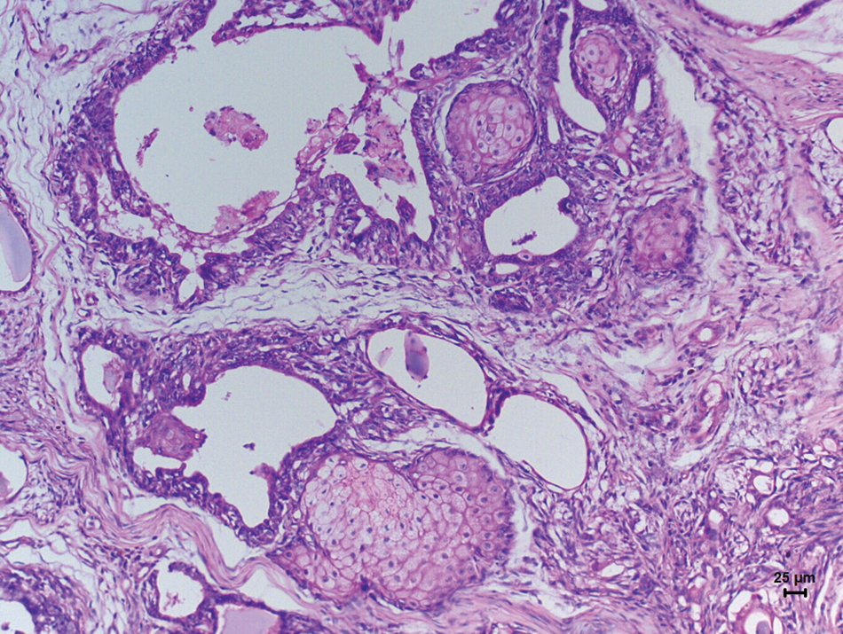
Photomicrography of mammary carcinoma in a female dog. Note multiple foci of sebaceous differentiation composed predominantly of mature sebocytes. Hematoxylin and eosin. Bar = 25 µm.
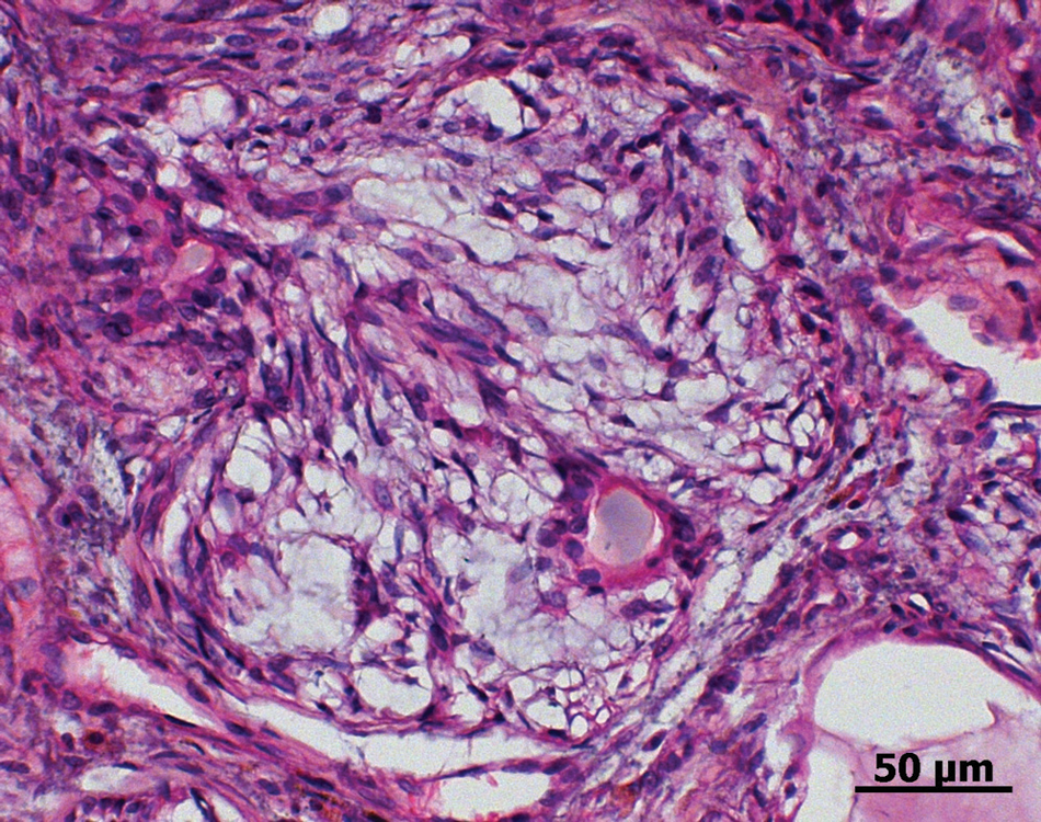
Photomicrography of mammary gland carcinoma in a female dog. Note myoepithelial component characterized by spindle to stellate cells associated with a myxoid stroma. Hematoxylin and eosin. Bar = 50 µm.
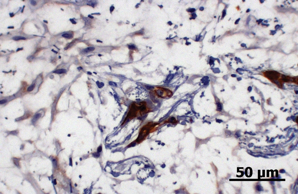
Photomicrography of mammary gland carcinoma in a female dog. Note positive and marked diffuse immunolabeling of myoepithelial cell at center. Some myoepithelial cells showed a weak and diffuse cytoplasmatic staining. Cytokeratin 14 and 3,3’-diaminobenzidine chromogen. Bar = 50 µm.
Mammary gland carcinomas in domestic animals are classified as non-infiltrating carcinoma, complex carcinoma, simple carcinoma, special carcinoma types, carcinosarcoma, and carcinoma in a benign tumor. 11 However, sebaceous metaplasia is not categorized as a carcinoma subtype by the WHO. 11 In human medicine, it is considered a subtype of invasive ductal carcinoma. 17
Sebaceous metaplasia in mammary tumors is a condition described as rare in human medicine with only a few documented cases.8,12 In veterinary medicine, the only previously documented case of such a peculiar mammary carcinoma with sebaceous differentiation is described in a 10-year-old mixed-breed female dog. 2 Thus, information on its clinicopathology, treatment, and prognosis remains very limited in both human and veterinary medicine, which makes the current case report an important contribution in terms of comparative pathology.
The morphological characteristics of the sebaceous component observed in the present case were similar to those described in the literature, in which the cytoplasm has 1 or more clear vacuoles and a small eccentrically located nucleus.2,7,10,15,18 Similar to the present case, a few foci of clumped sebaceous cells within papillary projections have been observed in a mammary carcinoma in a female dog 2 and an intraductal papilloma in a female patient. 7 Most sebaceous carcinomas in human beings show sebaceous units predominantly arranged in a more solid appearance characterized by small nests or lobules that sometimes present additional basal cells,6-8,10,12,15,18,20 which cannot be seen in the current case. The latter pattern was also observed previously. 2 Although the histogenetic origin and pathway of the sebaceous component in mammary carcinomas remain unclear, previous findings suggest that it can be derived from mammary pluripotent stem cells or embryonic displacement of epidermal anlage into the breast parenchyma.2,20 In addition, the present case showed sebaceous units interspersed within myxoid stroma, a finding that suggests another possible source of sebaceous cells by means of reverse mesenchymal–epithelial transition. 3
It was not possible to classify the mammary carcinoma described herein because there is no such variant in the WHO classification for domestic animals, and according to the most recent WHO human classification, this type of carcinoma is characterized by a lobulated or nested proliferation of an admixture of clear cells with cytoplasmic lipid vacuoles of sebaceous type and smaller dark or eosinophilic cells without vacuolation. 12 The morphological characteristics observed in the present case are similar to those described for complex carcinomas, in which epithelial and myoepithelial components are arranged in a tubulopapillary or solid fashion and a reticulated myxoid pattern, respectively. 11
Sebaceous carcinoma of the mammary gland must be differentiated mainly from lipid-rich carcinomas, primary sebaceous carcinomas of the skin adnexa, mucin-rich carcinomas, and glycogen-rich clear-cell carcinomas. Lipid-rich carcinomas are uncommon mammary gland neoplasms described in dogs and human beings. The main cytomorphological criterion used to recognize lipid-rich carcinomas is the presence of at least 80% of the tumor cells that have a vacuolated cytoplasm with a large amount of neutral lipid, generally with a signet ring appearance.4,14 Tumor cells in the current case did not display the classical features needed to be classified as a lipid-rich carcinoma. The neoplasm had sparse transitional areas presenting sebaceous morphology and no signet ring appearance. Although, some lipid-rich carcinomas can display sebaceous cells with a fine vacuolated cytoplasm, 14 similar to the present case, the great majority of tumor cells did not display this. In addition, canine mammary lipid-rich carcinomas generally show a lobular growth pattern, which was not observed in the present case. 14 Moreover, an intraductal proliferation of sebaceous cells both in human beings16,19,21 and dogs 14 have been described in literature.
Sebaceous carcinoma of skin adnexa is an uncommon malignancy observed in dogs that can also affect the mammary gland region, making the differential diagnosis very challenging. However, the majority of cases occur on the head. 5 The main feature that distinguishes the current case from a skin appendage tumor is the abrupt metaplastic transition between the intraductal mammary component into fully developed sebaceous cells, similar to that described previously. 2 Additionally, sebaceous carcinomas of the skin do not present a myxoid component, as demonstrated by positive staining with Alcian blue inter-spread between the tumor lobules or inside papillary projections. Plus, the absence of continuity to underlying skin corroborates the assumption that the neoplasm in the present case originated from the mammary gland.
Tumor cells displaying sebaceous characteristics were negative for mucicarmine and PAS stains, similar to those described previously. 2 Thus, the authors were able to exclude mucinous and glycogen-rich clear-cell carcinomas because, in these types of tumors, the predominant feature is the presence of large amounts of mucinous material that stains positively with PAS or Alcian blue stain. 11
In summary, based on the histological findings, the current tumor is analogous to those previously described in the literature as mammary carcinoma with sebaceous differentiation and sebaceous metaplasia with the extent of sebaceous morphology differing among the reported cases.
Footnotes
Acknowledgements
The authors are grateful to Maria Valéria Dalanezi for histological processing techniques.
The author(s) declared no potential conflicts of interest with respect to the research, authorship, and/or publication of this article.
The current study was financially supported by FAPESP (process 2009/14701-5).