
Abstract
An 11-year-old, male, neutered Cavalier King Charles spaniel was euthanatized because of recurrent seizures and inflammatory bowel disease. An incidental finding at necropsy was the presence of bilateral, firm, white nodules across the petrosal crest of the skull. Microscopically, the nodules were composed of normal myelinated nerve fibers within a mucinous stroma. A diagnosis of cranial nerve hamartoma was made.
Hamartoma is an excess growth of normal cells and tissue native to the organ in which it occurs. Hamartomas reported in animals include vascular,9 mesenchymal,12 pulmonary microcystic,11 muscle,7,13 melanin,5,8 bile duct,2 ovarian interstitial cell,6 and cutaneous adnexa.5 In the nervous system, vascular hamartomas are the most commonly reported,10 and there are single case reports of a melanotic8 and a hypothalamic hamartoma.10 Hamartoma of nerves has not been reported.
An 11-year-old, neutered, male Cavalier King Charles spaniel was euthanatized after a 9-month course of inflammatory bowel disease and a history of seizures since 1 year of age. Necropsy findings included the presence of bilateral, firm, white, glistening nodules, 2 cm × 1 cm × 1 cm, lying on the dorsal surface of the temporal bone medial to the petrosal crest (Fig. 1). The nodules compressed the cerebellum at the cerebellopontine angle on both sides, leaving indentations in the cerebellum. The cause of the seizures was presumed to be secondary to the indentations; the brain was normal microscopically.
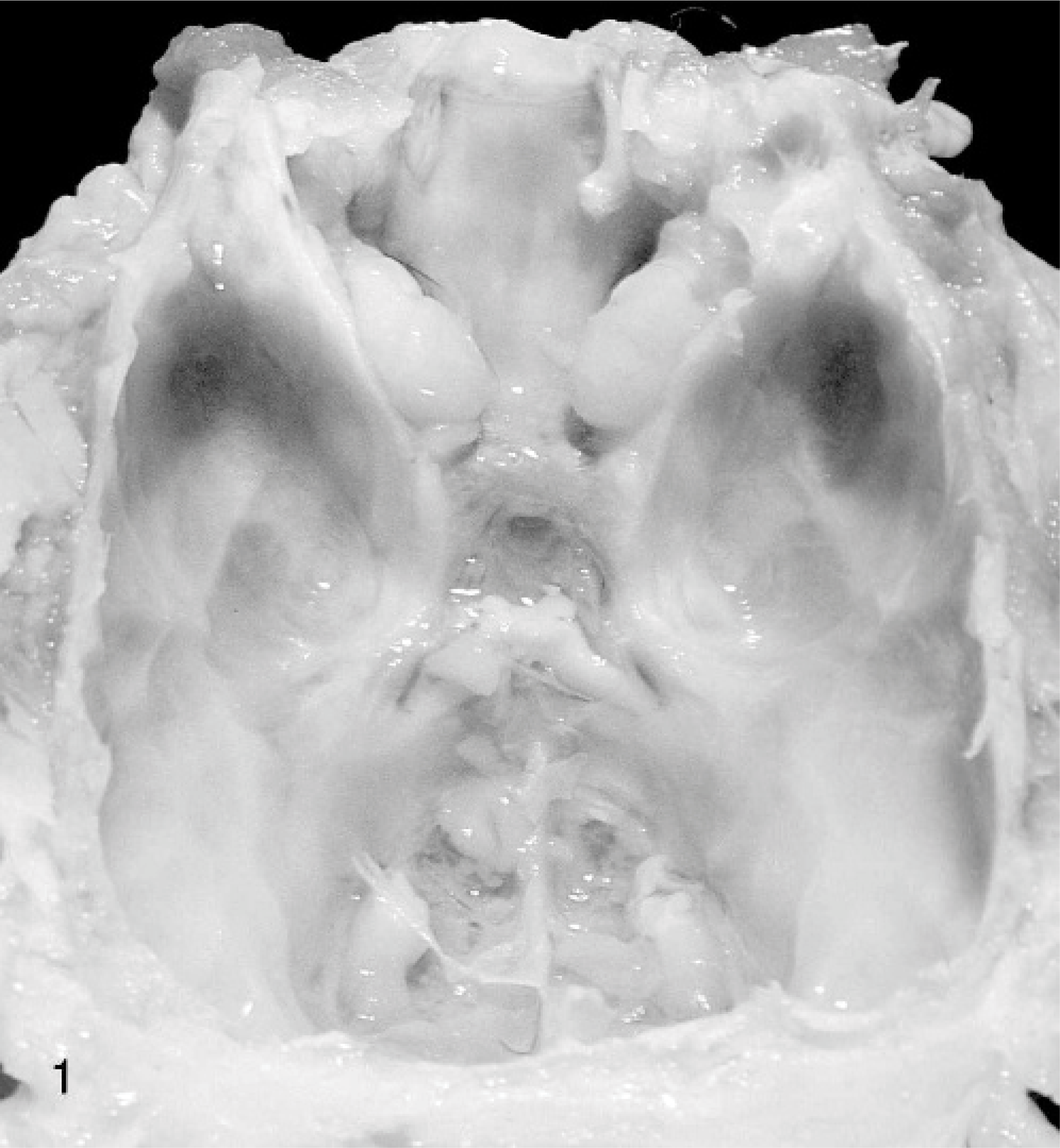
Skull; dog. Bilateral nodules of cranial nerve hamartoma located over the temporal bones medial to the petrosal crest.
One of the bilateral tumors was examined histologically. The mass contained normal myelinated nerve fibers within a mild, mucinous stroma (Fig. 2). Each nerve fiber was composed of a normal axon surrounded by a thick myelin sheath. These normal nerve fibers were assumed to be part of the vestibulocochlear or facial nerves, and a diagnosis of cranial nerve hamartoma was made.
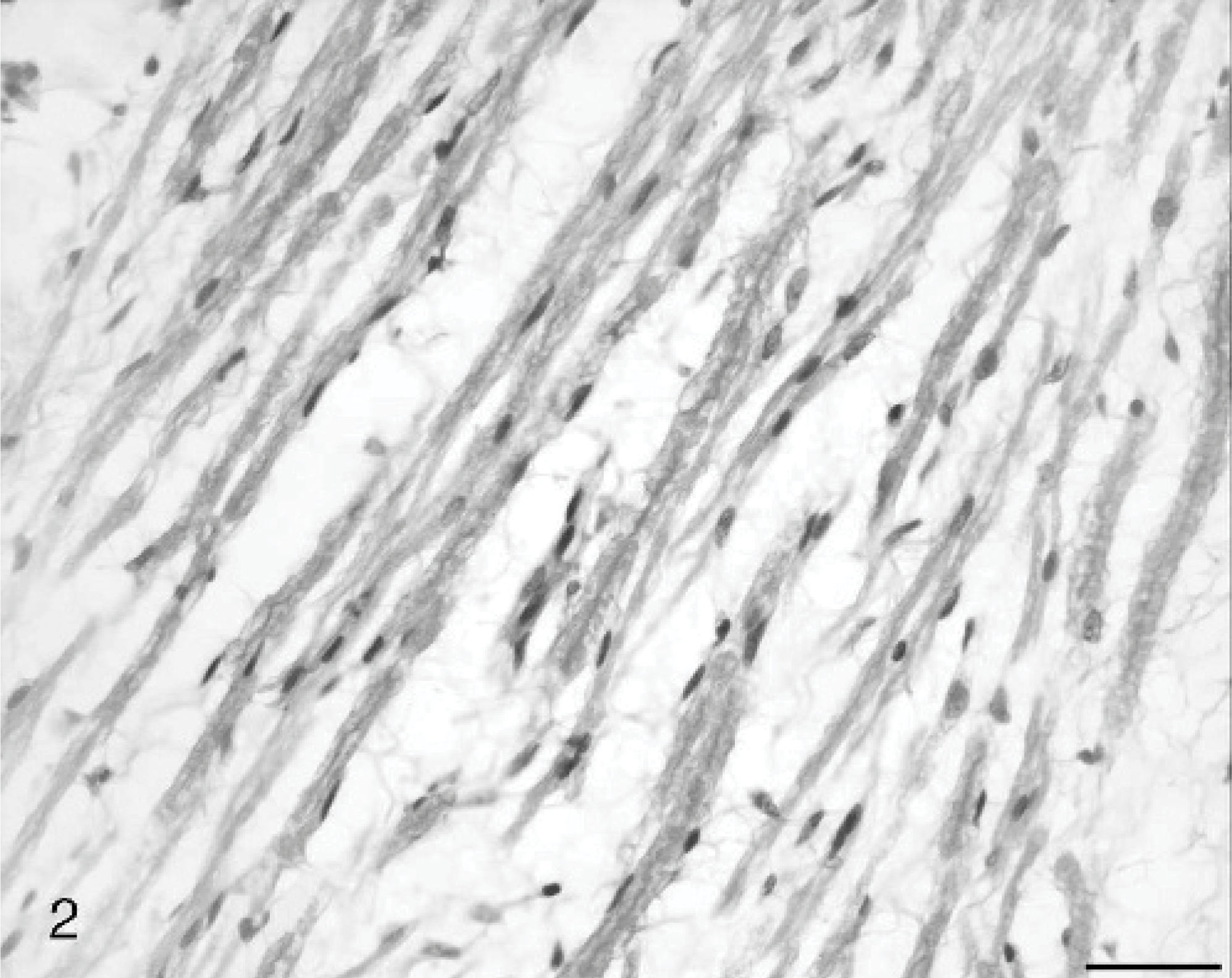
Skull mass; dog, Normal nerve fibers within a mucinous stroma. HE. Bar = 25 μm.
The gross appearance and location of the tumors in this dog are typical of acoustic schwannoma and neurofibromatosis reported in humans.1,3 Because the tumor in this dog is composed of normal nerve fibers and is not a neoplasm, a diagnosis of hamartoma is preferred. Hamartomas of the nervous system in people usually involve blood vessels or nerve cells. A nerve cell hamartoma of the internal auditory canal has been reported in humans4 but a hamartoma of nerve has not been reported in human or animals.