
Abstract
A 9-year-old, female German Shepherd Dog presented with a firm bulging lesion at the tip of the tail. Histologically, the lesion was characterized by numerous poorly circumscribed clusters of vascular structures resembling capillaries that were separated by normal or mucinous connective tissues. The capillary-like vascular structures were composed of several layers: a single von Willebrand factor–positive endothelial cell layer with round, oval, or flattened hyperchromatic nuclei; and 1 or 2 surrounding spindle-shaped smooth muscle actin–positive pericyte layers. In the deep portion of the lesion, there were large vessels that showed morphological aberrations. These thin-to-thick–walled blood vessels corresponded to arteries and veins of varying diameters and were surrounded by fibrosis. The present case report describes a rare cutaneous vascular hamartoma, accompanied by aberrant arteriovenous structures.
Angiomatosis is a heterogeneous group of vascular proliferations in human beings, 2 but has not been well documented in dogs and cats.1,8 This syndrome is considered potentially progressive and possibly represents a congenital proliferation of vessels rather than a true neoplasm. 1 Based on morphological variations, locations of lesions, and clinical behaviors, canine and feline angiomatosis are categorized as progressive angiomatosis or scrotal-type vascular hamartoma (STVH). 8 STVH is a rare condition affecting middle-aged or older dogs1,6,7 that develops on pigmented scrotal skin, the tail base, or the caudal trunk, and a single case has been described on the medial carpus.1,8 Spontaneous regression has not been described, and the lesion progresses and enlarges with time. A predilection to develop this lesion has been reported for certain dog breeds (Scottish Terriers and Airedale Terriers) that have pigmented scrotal skin. 7 Of the clinical and histopathological features of STVH described in the veterinary literature, a single case has been reported as hemangioma of the scrotum in dogs. 12 The current report describes a vascular hamartoma, which developed on the tail tip, that histologically resembled STVH and was characterized by aberrant arteriovenous structures.
A 9-year-old, female German Shepherd Dog examined at a veterinary clinic exhibited a firm, bulging lesion at the tip of the tail (Fig. 1a). The swollen area was approximately 6 cm from the tip and showed mild hemorrhage and ulceration. A punch biopsy was performed, and the bulging lesion was then surgically excised by docking and submitted for histopathological examination. Upon sectioning, the lesion was gray–white and appeared poorly demarcated (Fig. 1b) from the surrounding tissues. Additionally, a 3 cm in diameter cavernous hemangioma was present at the base of the tail. No continuity was evident between this hemangioma and the tail tip lesion, as the mass was separately biopsied from the bulging area. During the 7 months since surgical excision of the 2 lesions, no local recurrence has been recognized, but a mast cell tumor developed on a digit of the right anterior limb.
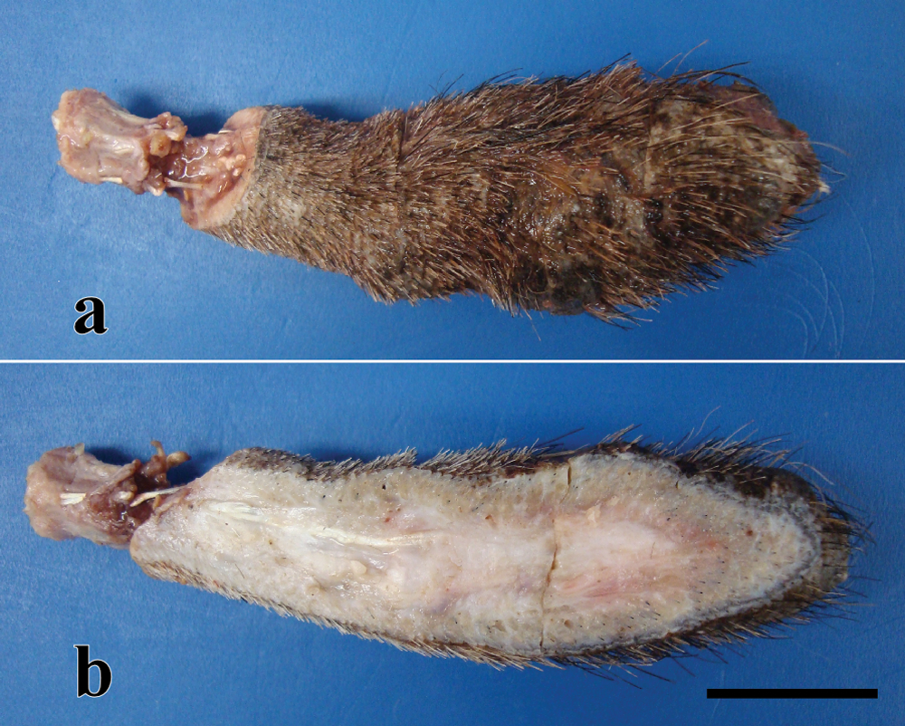
German Shepherd Dog. a, bulging lesion at the tip of the tail. b, lesion sectioned. The hair covering the lesion has been clipped for surgery. Bar = 2 cm.
The excised tail lesion was fixed in 10% neutral buffered formalin, embedded in paraffin, sectioned, and stained using hematoxylin and eosin, periodic acid–Schiff (PAS), Alcian blue (pH 2.5), and elastica van Gieson (EVG) stains. Immunohistochemical staining was performed using an immunoenzyme polymer method with rabbit anti–human von Willebrand factor (vWF) antibody a and mouse anti–human smooth muscle actin (SMA) antibody (clone 1A4) a as primary antibodies. Peroxidase-conjugated anti-rabbit immunoglobulin (Ig)G b or peroxidase-conjugated anti-mouse IgG b were used as secondary antibodies. Cell proliferative activity was also examined immunohistochemically, using mouse anti–proliferating cell nuclear antigen (PCNA) antibody (clone PC10) a along with peroxidase-conjugated anti-mouse IgG b as a secondary antibody. Vascular endothelial cells, vascular smooth muscle cells, and proliferating epidermal basal cells were used as internal positive controls for vWF, SMA, and PCNA, respectively. Sections were also stained under identical conditions with normal rabbit or normal mouse IgG to serve as negative controls. Mayer hematoxylin was used for counterstaining. For comparison, canine hemangiomas (n = 4), hemangiosarcomas (n = 4), and granulation tissue (n = 4) were examined, with particular attention to development of pericytes around vascular structures.
Histologically, the lesion was characterized by numerous poorly circumscribed clusters of vascular structures resembling capillaries or capillary buds that were separated by collagen fibers or Alcian blue–positive mucinous substances (Fig. 2). Clusters of vascular structures were seen throughout the subcutis and deep dermis (Fig. 2a), and several vascular clusters were seen attached to the caudal vertebra (Fig. 2c). Characteristically, these redundant vascular structures often surrounded cutaneous adnexa (epitrichial glands and follicles; Fig. 2b). Capillary-like vasculature were composed of several layers, with a single vWF-positive endothelial cell layer showing round-oval or flattened nuclei (Fig. 2b inset) surrounded by 1–2 layers of SMA-positive pericytes (see below). The basement membrane present in each vascular structure was delineated by PAS staining, and these vascular structures resembled the proliferating capillary vessels observed in granulation tissues. Mitotic figures were extremely rare, and few PCNA-positive endothelial cells or pericytes were observed within the vascular structures. In the deep dermis of the lesion, abnormally shaped larger vessels were frequently observed (Fig. 2a), and these thin-to-thick–walled blood vessels that resembled poorly formed, variably sized arteries and veins were surrounded by fibrosis. The artery-like vessels were characterized by an irregular thick intima, variable amounts of elastic and collagen fibers, and irregularly shaped luminal spaces (Fig. 3). Irregularities in the amount and orientation of smooth muscle bundles in the vessel walls were often observed (Fig. 4). The lumina of the dysplastic vascular structures were lined by vWF-positive endothelial cells. In the overlying epidermis, there was acanthosis accompanied by mild amounts of melanin pigment accumulation. Ulceration, hemorrhage, and inflammation were also present in the dermis.
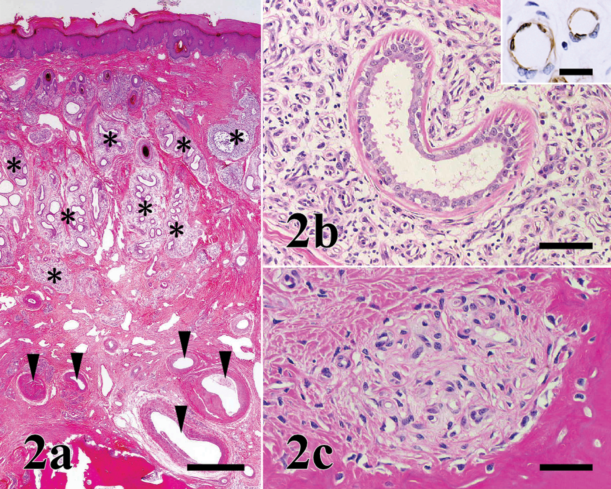
German Shepherd Dog. Histological appearance of the tail tip lesion. a, clusters of vascular structures are seen throughout the dermis (*). The deep dermis contains large vessels (arrowheads). Hematoxylin and eosin (HE) stain. Bar = 1,000 µm. b, proliferating capillaries surround a sweat gland. HE. Bar = 200 µm. Endothelial cells of capillary-like structures are von Willebrand factor–positive. Immunostain. Inset: bar = 50 µm. c, clusters beside the caudal vertebra. Proliferative vascular clusters are attached to caudal vertebrae. HE. Bar = 50 µm.

German Shepherd Dog. Artery-like vessels showing an irregularly thick tunica media that is completely absent in some parts of the wall. A partial internal elastic lamina (arrowheads) is present in some areas, but is almost absent in others. Collagen fibers are well developed throughout the vessel. Elastica von Gieson stain. Bar = 100 µm.
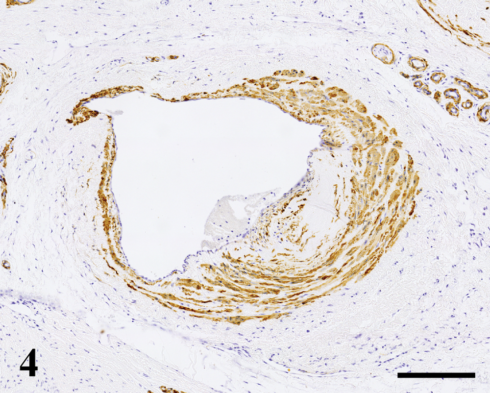
German Shepherd Dog. Thick layers of smooth muscle are seen in some segments of the vessel wall, but these cells are nearly absent in other areas. Smooth muscle cells are often aberrantly oriented. Immunolabeling by α-smooth muscle actin antibody. Bar = 100 µm.
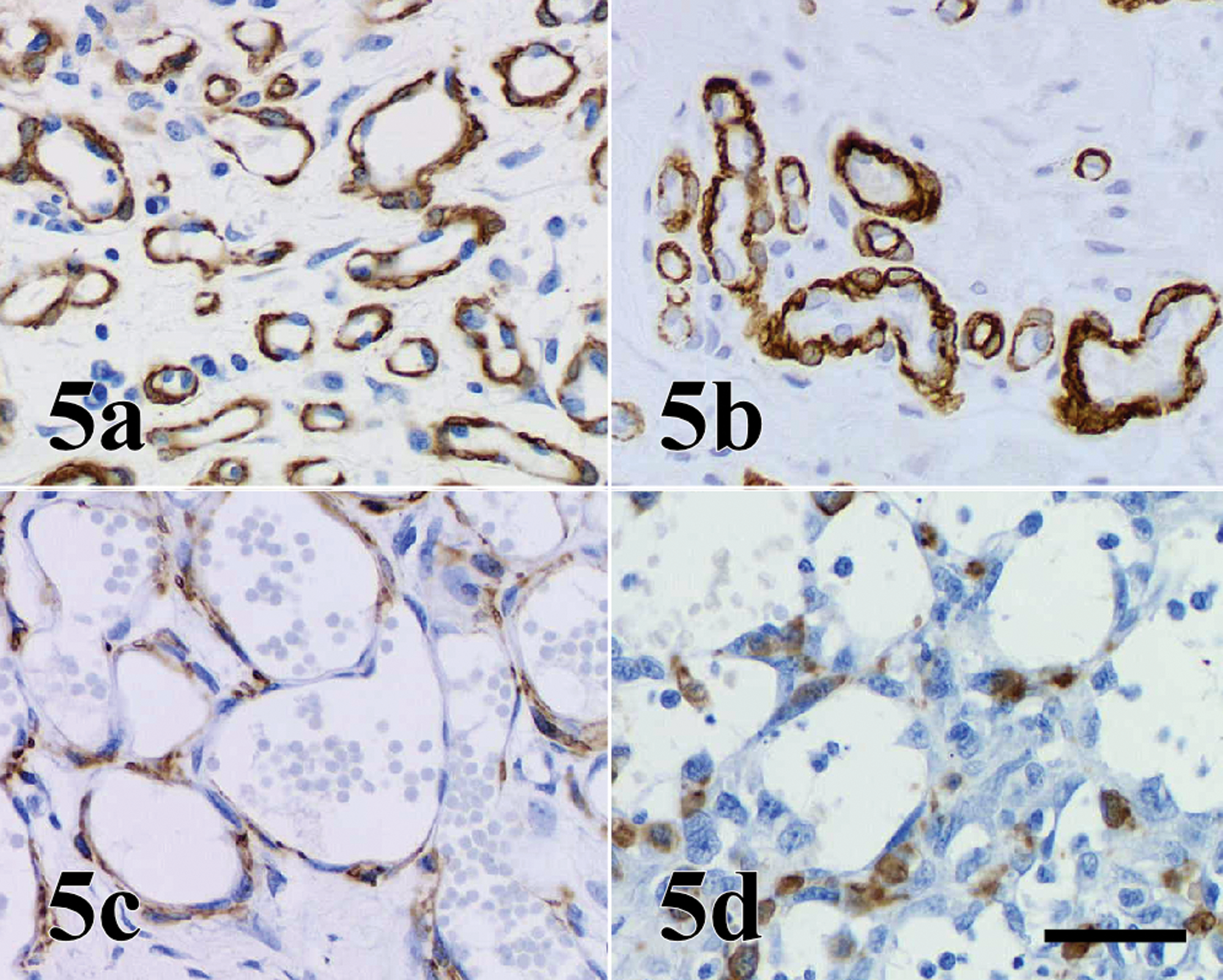
Immunostaining of smooth muscle actin (SMA). a, present case; b, granulation tissue; c, hemangioma; and d, hemangiosarcoma. Prominent SMA-positive mature pericytes layers are seen in each capillary-like structure in the current case (a) and proliferating capillary vessels in granulation tissue (b), but not in hemangiomas (c) or hemangiosarcomas (d). Bar = 50 µm.
On the basis of histopathological findings, the lesion was diagnosed as STVH. As a characteristic histological feature in STVH, larger preexisting dermal blood vessels are surrounded by clusters of capillary-like structures in the subcutis.1,7,8 In the tail mass described herein, these structures were centered on epitrichial sweat glands rather than vessels, a finding also reported in one other case. 8 In addition, the capillary-like structures were scattered throughout the dermis, and several vascular clusters were attached to the bone, although no signs of infiltration were apparent. This feature of noninvasive proliferation allowed differentiation from progressive angiomatosis. Progressive angiomatosis shows extensive infiltrative and expansile growth of vascular structures.8,10 The benign nature of the lesion was also supported by the organized structure of the vessels as demonstrated by immunohistochemical stains. The SMA-positive pericytes layers and basement membrane observed in each capillary-like structure resembled the proliferating capillary vessels seen in granulation tissues, but these well-defined mature pericyte layers and basement membrane are not seen in each vascular structure in hemangiomas or hemangiosarcomas (Fig. 5). According to the morphological features of the present case, there may have been a polyclonal proliferation of the capillaries indicating a hamartomatous nature. The differential diagnosis of this lesion might be angiomatosis secondary to lymphedema and capillary hemangiomas. However, angiomatosis secondary to lymphedema are accompanied by characteristic hypoplasia of the deep lymphatic vessels and markedly dilated lymphatics,7,8 which were not seen in the current case. The small vessels of capillary hemangiomas do not show orientation of the vascular structures around the larger blood vessels and cutaneous adnexa. 8 In addition, hemangiomas of the subcutis are usually solitary, well-circumscribed masses,6,8 while the lesion in the current case was widespread and poorly demarcated from the surrounding tissues.
To the authors’ knowledge, the aberrant arteriovenous structures observed within the deep dermis in the present case have not been reported in any previous cases of STVH in dogs. The histological features of these arteriovenous structures resembled those in arteriovenous hemangioma (arteriovenous malformation), a type of human vascular tumor. Arteriovenous hemangioma is usually present in the skin of the head, neck, and limbs,3,5 but may also occur on the scrotum. 9 The histological features are variable, but include a mixture of arteries and veins of varying calibers, with a predominance of the latter. 3 The morphological characteristics of the arteriovenous proliferations of the tail mass seen in the dog in the current report resembled those of arteriovenous hemangioma, but a simultaneous proliferation of capillary-like vessels has not been mentioned in human cases.
Abnormal vascular structures of vascular hamartomas in dogs have been classified into the following 2 morphological types: 1) redundant vessels ranging from capillaries or capillary buds to large arteries,6,8,11,12 and 2) vessels showing dysplastic elements such as tortuous, thick-walled, vascular structures containing smooth muscle, and mature arteries and veins in a tortuous pattern surrounded by fibrous tissue.4,11 The present report describes a rare case sharing both morphological types. Involvement of a common factor involved in vasculogenesis such as basic fibroblast growth factor or vascular endothelial growth factor is likely to be involved in the pathogenesis of these lesions.
Footnotes
a.
Dako Denmark A/S, Glostrup, Denmark.
b.
Histofine Simple Stain MAX-PO(R), Histofine Simple Stain MAX-PO(M); Nichirei, Tokyo, Japan.
The authors declared that they had no conflicts of interests in their authorship and publication of this contribution.
This study was partially supported by a project grant (Creative Research Project, 2009) awarded by the Azabu University Research Services Division.