
Abstract
Deciduosarcoma is a rare, hormonally dependent neoplasm with features of malignancy, previously reported only in rabbits enrolled in chronic toxicology studies involving estrogens with or without progestins. An exploratory laparotomy was performed on a 6-year-old pet Dutch dwarf rabbit following palpation of a 6-cm-diameter abdominal mass. Grossly, the mass was fleshy and nodular, adhered to but not appearing to originate from the small intestine, with a smaller mass of similar appearance involving the uterus, and an effaced mesenteric lymph node. Histologically, the mass was characterized by spindloid cells and large epithelioid cells with abundant pale eosinophilic vacuolated cytoplasm and an infiltrative pattern of growth. Giant cells with large, bizarre, hyperchromatic nuclei were common. Cells were positive by immunohistochemistry for vimentin and progesterone and estrogen receptors and negative for pancytokeratin (AE1/AE3), cytokeratin 18, desmin, alpha-smooth muscle actin (SMA), and CD10. Based on histologic and immunohistochemical findings, a diagnosis of deciduosarcoma was made.
Deciduosarcomas are unusual and rare neoplasms unique to rabbits, first reported in toxicology studies involving chronic vaginal silastic implants delivering estrogen and levonorgestrel (a progestin). 5, 7 Tumors were composed of anaplastic decidualized endometrial stromal cells with invasive growth and neoplastic emboli and involved the uterus and draining lymphatics, with occasional metastasis to the lungs. Some tumors appeared to be primary tumors of the spleen or abdominal viscera. Deciduosarcomas were later induced in female rabbits and in the spleens of castrated male rabbits given estrogen and levonorgestrel for 30 days. 6 Lesions were shown to regress following steroid hormone withdrawal. Exogenous estrogen was determined to be necessary for deciduosarcoma development, while exogenous progestins promoted the process. 1
An approximately 6-year-old female Dutch dwarf rabbit was presented for anorexia and lack of stool production. Abdominal palpation revealed a 6-cm-diameter mass in the abdomen caudal to the stomach. Haircoat was normal, mammary glands and vulva did not appear enlarged, and there was no clinical evidence of hyperestrinism or a pituitary disorder. An exploratory laparotomy was performed, revealing a large 6-cm-diameter mass in the left cranial abdomen with adhesions to a loop of small intestine and abdominal wall. The mass did not appear to be of intestinal origin. A smaller 2-cm-diameter mass was present in the wall of the uterus. The masses were firm, nodular, fleshy, pale tan, with a smooth irregular nodular surface and areas of congestion with foci of necrosis. The ovaries were grossly normal. An intestinal resection and anastomosis was performed to remove the larger mass, and the animal was routinely spayed. An enlarged mesenteric lymph node was removed as well. The animal recovered uneventfully, and was asymptomatic until it died suddenly 40 days later. Death was thought to be unrelated to neoplastic disease, and the owners declined postmortem examination.
Tissues were fixed in 10% neutral buffered formalin, paraffin embedded, sectioned at 4 µm, and stained with hematoxylin and eosin, and periodic acid–Schiff (PAS) with and without diastase digestion. For immunohistochemistry examination, sections were deparaffinized and steamed in a citrate buffer for 5 minutes for antigen retrieval. The slides were then incubated with the following antibodies using the avidin-biotin peroxidase method: cytokeratin AE1/AE3 (prediluted, Ventana Medical Systems Inc., Tucson, AZ), desmin (1 ° 100, Dako Corp., Carpinteria, CA), alpha-smooth muscle actin (prediluted, Ventana), vimentin (prediluted, Ventana), CD10 (prediluted, Ventana), estrogen receptor (prediluted, clone 6F11, Ventana), and progesterone receptor (prediluted, Ventana). The reaction product was visualized with 3,3′-diaminobenzidine chromagen and the sections were counterstained with 0.1% hematoxylin (Sigma, St. Louis, MO), dehydrated, and mounted. Immunoperoxidase staining was performed with the automated Bio Tek-1000 immunostainer system (Bio Tek Solutions Inc., Santa Barbara, CA). All procedures were performed according to the manufacturers' protocols. The staining of nonspecific controls was the same except for substitution of the primary antibody with preimmunization mouse serum.
Histopathology showed an invasive mass originating in the mesometrial aspects of the endometrial stroma in the uterine body and invading the myometrium and mesometrium with an infiltrative growth pattern, forming a large nodular mass in the mesometrium (Fig. 1). Cells were arranged in sheets and streaming bundles, often appearing as finger-like cords dissecting between smooth-muscle cells or arranged concentrically around large, dilated blood vessels with thin walls (Fig. 2). Cells were anaplastic and consisted of two intermingling populations. There was a smaller population of spindloid cells with scant eosinophilic cytoplasm and cigar-shaped central nuclei with finely stippled chromatin and a single indistinct nucleolus. More frequently, cells were epithelioid with moderate to abundant amounts of pale eosinophilic or vacuolated cytoplasm, and large oval eccentric nuclei with finely stippled to vesicular chromatin and 1–2 distinct nucleoli (Fig. 3). Vacuolization varied from numerous fine vacuoles to multiple large (5 µm) clear vacuoles surrounding a central nucleus. Vacuoles did not stain with PAS, but tissues were not fixed in formalin alcohol. Binucleate and multinucleate cells were common, as well as frequent giant cells with large, bizarre, hyperchromatic nuclei. Anisocytosis and nuclear pleomorphism were striking, and mitoses averaged 400 × magnification. Multiple foci of necrosis were present. Neoplastic cell borders were enhanced by fine PAS-positive, diastase resistant fibers. The intestinal mass consisted of identical cells forming a discrete mass in the mesentery, apposing but not invading the muscularis. The mesenteric lymph node was completely effaced by neoplastic cells.
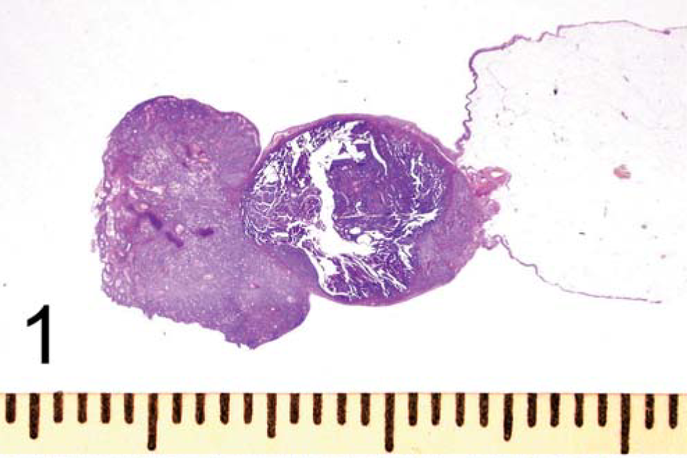
Uterine body, deciduosarcoma; rabbit. Proliferations of pale cells within the endometrial stroma can be seen near the attachments of the broad ligaments, with invasion into myometrium and mesometrium. An endometrial adenocarcinoma is present as well. PAS stain. Scale is in centimeters.
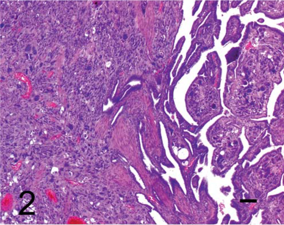
Uterus, deciduosarcoma; rabbit. Large, pale cells invade myometrium, often resulting in islands of dissected myofibers. Vacuolated cells and karyomegalic giant cells can be seen as well. Atypical endometrial glands have invaded myometrium. HE stain. Bar = 100 µm.
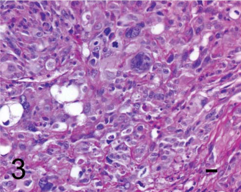
Uterus, deciduosarcoma; rabbit. There is marked atypia with frequent giant cells containing large, bizarre, hyperchromatic nuclei, as well as frequent cytoplasmic vacuolization. HE stain. Bar = 20 µm.
Neoplastic cells showed strong cytoplasmic staining for vimentin and strong nuclear staining for estrogen and progesterone receptors (Fig. 4). Neoplastic cells did not stain with desmin or alpha-smooth muscle actin (SMA), although myometrial smooth-muscle stained strongly. Neoplastic cells did not stain for pancytokeratin, although endometrial epithelium showed strong cytoplasmic staining. Likewise, neoplastic cells were negative for CD10, a marker for nondecidualized endometrial stroma, 4 but endometrial stromal cells showed positive cytoplasmic staining. Additionally, there was an invasive endometrial adenocarcinoma present in the uterus that collided with, but was easily distinguishable from, the deciduosarcoma. The adenocarcinoma stained positively for pancytokeratin and progesterone and estrogen receptors and negatively for all other markers.
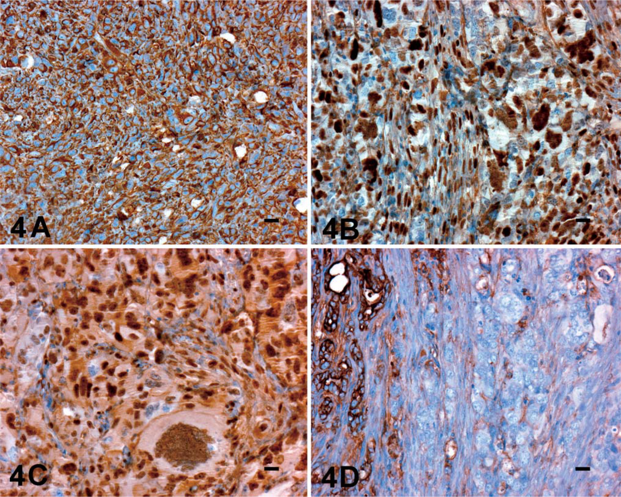
Uterus, deciduosarcoma; rabbit. Immunohistochemistry shows positive reactions for vimentin (A), progesterone receptor (B), and estrogen receptor (C). Decidual cells are negative for pancytokeratin (D), although invasive endometrial adenocarcinoma cells show positive reaction. Bar = 20 µm.
The diagnosis of deciduosarcoma in this animal is based on distribution and characteristic histology and is supported by immunohistochemical findings. The cellular morphology of proliferating spindle cells and large polygonal cells with pale or vacuolated cytoplasm and large, bizarre nuclei is consistent with previously reported deciduosarcomas. Involvement of the mesometrial endometrium, with invasion into myometrium, and subserosal growth on abdominal viscera have been reported as well. Immunostaining for vimentin and progesterone and estrogen receptors support myometrial or endometrial stromal differentiation. The lack of immunostaining for desmin or SMA makes epithelioid or pleomorphic leiomyosarcoma unlikely. Additionally, tumor-cell morphology is more consistent with decidualized endometrial stroma, as poorly differentiated leiomyosarcomas generally have closely packed nuclei with scant cytoplasm. A positive reaction for vimentin and failure to stain for cytokeratins preclude trophoblastic origin for the mass. The neoplastic decidualized stromal cells are clearly distinct from the carcinomatous endometrial epithelial cells, although they do occupy the same space. A diagnosis of malignant mixed mesodermal tumor was considered but rejected because of the high incidence of spontaneous endometrial adenocarcinomas in aged does, as well as the predominantly decidual nature of the neoplasm and limited size of the adenocarcinoma.
Deciduosarcoma is a phenomenon unique to rabbits, although hyperplastic lesions of decidualized stromal cells have been reported to occur in the uterus and subserosal abdominal locations in mice, rats, guinea pigs, and humans. 2 Both the endometrium and peritoneum are derived from juxtacoelomic tissue in the embryo, and lesions may be induced in response to endocrine and/or mechanical stimulation. However, although these hyperplastic lesions may appear malignant by virtue of their invasive growth and cellular atypia, metastases do not occur in any other species except the rabbit, where pulmonary metastases have been reported. It is this metastatic nature that earns the rabbit lesion its malignant classification. 2, 6 Although the lungs were not examined in this animal, the tumor described herein had effaced a mesenteric lymph node, consistent with a metastatic nature.
Although the rabbit was free roaming indoors only, questioning of the owners failed to reveal any obvious potential sources of estrogenic exposures, such as contraceptives or vaginal topical creams. Diet consisted of commercial alfalfa hay fed ad libitum, with no soy-based pellets. Alfalfa fodder may contain variable amounts of the estrogenic compound coumestrol. 3 It is interesting to speculate that the deciduosarcoma arose secondary to mechanical irritation from the invasive endometrial adenocarcinoma in combination with dietary phytoestrogens, but the high incidence of endometrial carcinomas in this species, absence of clinical signs of hyperestrinism, and widespread feeding of alfalfa hay does not support this. Considering the age of this animal and in comparison with the younger ages used in toxicity studies, deciduosarcoma may be a rare spontaneous tumor, the incidence of which is modulated by treatment with exogenous estrogens. Previously reported deciduosarcomas did not have demonstrable mitotic figures and regressed when exogenous estrogen was withdrawn. This particular tumor may have acquired additional mutations, resulting in a more malignant phenotype.