
Abstract
A rabbit (Oryctolagus cuniculus) with a homologous malignant mixed müllerian tumor (MMMT) of the uterus with decidualization in the sarcomatous components is described. On histologic examination, the neoplasm was characterized by a carcinomatous and a sarcomatous component with invasion of the myometrium. The epithelial component was a well-differentiated carcinoma, and the nonepithelial component contained large amounts of intracytoplasmic glycogen. The changes in stromal cells were morphologically similar to changes usually found in decidual cells in the pregnant uterus or in deciduosarcomas in rabbits. Results of immunohistochemical analysis indicated that the epithelial components stained positive with cytokeratin (CK7, AE1/3) and the decidual-stromal cells stained positive for vimentin, but did not stain with α-SMA, actin, and desmin. This case fulfills all the criteria of an MMMT in having a carcinomatous and a sarcomatous component, but differs from cases of MMMT in women in that the sarcomatous component had decidualized. To the authors' knowledge, this is the first report of a malignant mixed müllerian tumor in rabbits.
Uterine neoplasms are the most common neoplasms in aged rabbits, and most cases are uterine adenocarcinomas. 11 In women, uterine adenocarcinomas are common, but mixed epithelial-nonepithelial tumors of the uterus are rare and develop principally in postmenopausal women. They are classified according to the histologic appearance of the epithelial and nonepithelial components which, on light microscopy, are intimately admixed. The terms malignant mixed mesodermal tumor and carcinosarcoma are synonyms for this neoplasm. The neoplasms are further differentiated into homologous subtypes (stromal components show differentiation to tissues seen in normal endometrium) and heterologous subtypes (stromal components show differentiation to tissue types not seen in normal endometrium [e.g., osteosarcoma, chondrosarcoma]). 9, 10 A small number of malignant mixed müllerian tumors (MMMTs) in cats and rodents have been described. 3– 5, 7, 8
The rabbit has a discoid labyrinthine hemodischorial placenta with countercurrent fetomaternal blood flow, which is similar to the placenta found in rodents and primates. 2 The endometrial change that occurs in response to blastocyst implantation is referred to as decidualization. When the stimulus is physiologic, the transformed endometrial stromal cells are called decidual cells and the resulting tissue is the decidua; 1 however, the tissue resulting from experimental or artificial stimulation of the endometrial stroma is called a pseudodecidua or deciduoma. 1
The stimulus that induces the change from endometrial stromal cells to decidual cells is progesterone, which is attributable to either pregnancy or administration of the hormone. The morphologic change associated with decidualization occurs principally in the endometrial stromal cells and includes cellular hypertrophy and marked accumulation of glycogen in the cytoplasm of the cells. The decidual cells have a euchromatic nucleus and prominent nucleoli and are embedded within an extensive extracellular matrix that contains collagen and heparan sulfate proteoglycans. 1 At the onset of decidualization, the endometrial glands initially enlarge, but with advancing pregnancy, the glands undergo atrophy. The endometrial blood vessels are lined by endothelial cells that are large and have hyperchromatic nuclei. 13
Uterine tissue from a female rabbit of unknown age was submitted to the surgical pathology service of the Laboratory of Pathology and Toxicology, University of Pennsylvania, School of Veterinary Medicine. Prior history was not available as the rabbit had been a rescue case. The entire uterus was removed, and the enlarged portion of the body of the uterus was submitted for histologic examination. Three pieces of the uterus were fixed in neutral-buffered 10% formalin, processed in routine manner, and embedded in paraffin. Sections (4–5 µm thick) were stained with HE. The initial diagnosis was uterine adenocarcinoma. However, on review of this case as part of a retrospective study of uterine adenocarcinomas in rabbits, further evaluation of the neoplasm was undertaken because of its unusual histologic appearance. Additional sections were stained with periodic acid–Schiff (PAS) and PAS with diastase digestion. Immunohistochemical staining was performed using primary mouse monoclonal antibodies to cytokeratin (CK; AE1/3; Boehringer Mannheim Biochemica, Mannheim, Germany), CK 7 (OV-TL 12/30; Dakocytomation Denmark A/S, Glostrup, Denmark), vimentin (VIM, v9; Dakocytomation Denmark A/S), human smooth muscle actin (α-SMA, 1A4; Dakocytomation Denmark A/S), human muscle actin (actin, HHF35; Dakocytomation Calfornia Inc., Carpinteria, CA), human desmin (desmin, D33; Dakocytomation Denmark A/S), and human CD31 endothelial cells (CD31; JC70A; Dakocytomation Denmark A/S). Microwave antigen retrieval was performed at 90°C for 9 minutes (CK, VIM). The tissues stained with CD31 were incubated with pronase (Roche Diagnostic, Indianapolis, IN) at 37°C for 30 minutes. The tissues were incubated with primary antibodies at 4°C overnight for CK, CK7, VIM, α-SMA, desmin, and CD31 or at room temperature for 1 hour for actin. The secondary antibody, which was a labeled polymer prepared by combining amino acid polymers with peroxidase and goat antimouse immunoglobulin reduced to Fab fragment (Histofine, mouse stain kit, Nichirei, Tokyo, Japan), was incubated for 30 minutes at room temperature. 3′3-Diaminobenzidine (CK, CK7, VIM, α-SMA, actin, desmin) and 3-amino-9-ethylcarbazole (CD31) were used as the detection system. The slides were counterstained with Mayer's hematoxylin.
Compared with the contralateral uterine horn, which acted as an internal control, the endometrium of the neoplastic mass was markedly thickened, whereas the myometrium on the affected side was attenuated. There were multifocal areas of necrosis within the neoplastic lesion. The surface epithelium was cuboidal, but became attenuated in some areas. The endometrial glands were present as individual glands or in small clusters, and the lining epithelial morphology varied from cuboidal to columnar. The neoplastic epithelial cells had large, ovoid, vesicular nuclei; small nucleoli; dispersed chromatin; and an abundant amount of eosinophilic cytoplasm with distinct cell margins. A small amount of amorphous eosinophilic material was present within some of the glandular lumina. An average of 1.6 mitoses/high-power field (range, 0–4 mitoses/400× field) was observed.
The stromal/decidual cells were large, with centrally to peripherally located nuclei. The nuclei were ovoid and varied from euchromatic to hyperchromatic. The cells had an abundant amount of pale eosinophilic cytoplasm, and cells margins were distinct. There was only modest nuclear and cellular pleomorphism, and <1 mitotic figure/high-power field was observed. The stromal/decidual neoplastic cells stained positive with PAS, with large amounts of clumped PAS-positive granular material in the cytoplasm of the cells. The cell margins were readily evident, but were not accentuated by use of this stain. PAS-Positive cells were not seen lining the blood vessels. Treatment with PAS with diastase completely removed the staining of the stromal/decidual cells, and the cell membranes were more prominent. The basement membrane surrounding the glandular component and the retained secretion within the glandular lumina stained with PAS/diastase. At the junction of the endometrium and myometrium, the neoplastic stromal/decidual cells had more fusiform hyperchromatic nuclei with less cytoplasm. Greater numbers of mitoses were observed in this portion of the neoplastic mass. The epithelial and stromal/decidual portions of the neoplastic mass had invaded the myometrium, but involvement or extension to the serosal surface was not observed.
The vascular component in the stromal/decidual portion of this tumor consisted of large vessels with erythrocytes and granulocytes in the lumina. The vessel walls were lined by hypertrophic endothelial cells that bulged into the vascular lumina. The endothelial cells had single or multiple hyperchromatic or euchromatic nuclei with an abundant amount of eosinophilic cytoplasm and indistinct cell margins. Granulocytes, some of which had degenerative changes, were observed in the cytoplasm of some of the endothelial cells. These vessels were only 1–2 cells thick, and external to the vessels were sheets of stromal/decidual cells.
The results of the histochemical staining and the immunohistochemical analyses are shown in Table 1. The epithelial components stained positive with cytokeratin (CK7, AE1/3) and did not stain with vimentin. The decidual-stromal cells stained positive for vimentin and did not stain with α-SMA, actin, and desmin. The hypertrophic endothelial cells stained with CK AE1/3, vimentin, and CD31, but did not stain with CK7.
Histochemical and Immunohistochemical results
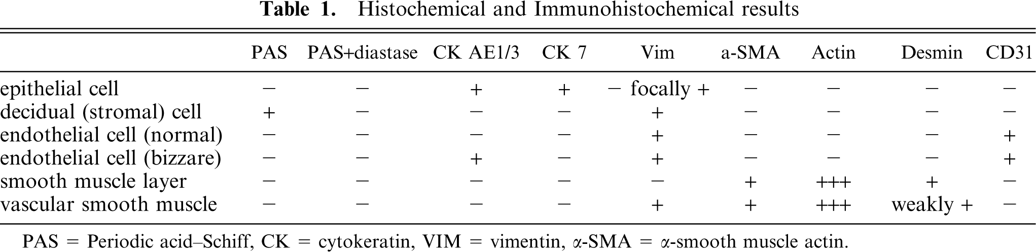
PAS = Periodic acid-Schiff, CK = cytokeratin, VIM = vimentin, a-SMA = α-smooth muscle actin.
Endometrial adenocarcinomas are the most commonly encountered neoplasms in Oryctolagus spp., particularly New Zealand White rabbits. Most neoplasms arise in the uterine body, and size varies from 1 to 5 cm. The rate of tumor growth is variable, and many affected rabbits die of metastatic disease 1–2 years after the onset of clinical signs of disease. The adenocarcinomas develop from the glandular epithelium and usually are initially situated in the mucosal folds adjacent to the mesometrial insertion. They are characterized histologically by irregular aggregates of glandular tissue embedded in a myxoid stroma that is extremely well vascularized. It has been suggested that there is a progression of lesions: from cystic hyperplasia to dysplasia to carcinoma-in-situ to carcinoma. 11
Decidualization of endometrial stromal cells is found in mammals with a hemochorial placentation. Decidualization is attributable to the effects of progesterone on the endometrial stromal cells, and is the response of the endometrium to trophoblast invasion. 1 To the authors' knowledge, there are no reports of spontaneously developing neoplasms with decidualization in rabbits. Malignant neoplasms of decidual origin (deciduosarcomas) have been experimentally induced in rabbits by intravaginal implantation of devices that released estrogen and progestin over a prolonged period. Those neoplasms were characterized by diffuse endometrial proliferation of large, vacuolated, polygonal cells that contained extensive amounts of glycogen (PAS positive), necrosis or loss of the surface endometrial epithelium, and absence of endometrial glands in the neoplastic lesions. Within the decidualized endometrium, the vessels were often lined by bizarre endothelial cells. 13 In some rabbits, deciduosarcomas were also found in the spleen and in abdominal organs, but they were not thought to be metastatic neoplasms. 12
There are a few reports of spontaneous MMMTs in cats and rats. Rhabdomyosarcomatous and cartilaginous differentiation in sarcomatous components of MMMTs were reported in rats. To our knowledge, there are no reports of decidualization in the sarcomatous components of MMMTs in these species. In women, the most common uterine mixed tumor is the MMMT that consists of an admixture of epithelial and mesenchymal elements. Less common are müllerian adenosarcomas, adenofibromas, and carcinofibromas. 9, 10
In women, MMMTs are usually soft, broad-based, polypoid tumors that fill the endometrial cavity and involve myometrial invasion, similar to the gross appearance of the tumor in this rabbit. On microscopic examination, there is an intimate admixture of carcinomatous and sarcomatous components. The sarcomatous component typically has the appearance of a spindle cell sarcoma resembling high-grade endometrial stromal sarcoma, leiomyosarcoma, fibrosarcoma, malignant fibrous histiocytoma, undifferentiated sarcoma, or combinations thereof . 9, 10 To our knowledge, decidualization of the malignant stromal cells in women has not been described. Myometrial lymphatic and vascular invasion is found in almost all MMMT cases.
This case fulfills all the criteria of a MMMT in having a carcinomatous and a sarcomatous component, but differs from human cases of MMMT in that the sarcomatous component has decidualized. The neoplastic decidual cells are large, vacuolated, polygonal cells rather than the spindle cells that make up the sarcomatous component of MMMT in women. Their location in the endometrium, in conjunction with the presence of large amounts of PAS-positive glycogen in the cell cytoplasm and positive immunohistochemical staining with vimentin only, confirms that these are malignant endometrial stromal cells with decidualization. Invasion of the myometrium by the carcinomatous and sarcomatous components of this tumor attests to its malignant nature. This neoplasm also differs from the experimentally induced deciduosarcomas by having a carcinomatous component, although the sarcomatous component is identical to that described for deciduosarcomas, including the presence of large, atypical, endothelial cells lining the blood vessels. The hypertrophic, endothelial cells stained with the endothelium specific marker, CD31 (Figs. 1–8) and with the cytokeratin specific marker AE1/AE3. However, these cells did not stain with CK7, which is expressed by normal and neoplastic epithelial cells and trophoblasts. 6 These results are difficult to reconcile as the normal endothelial cells in the unaffected uterus stained only with CD31.
Uterus; malignant mixed müllerian tumor, rabbit. Epithelial and nonepithelial components are intimately admixed. HE. Bar = 390 µm.
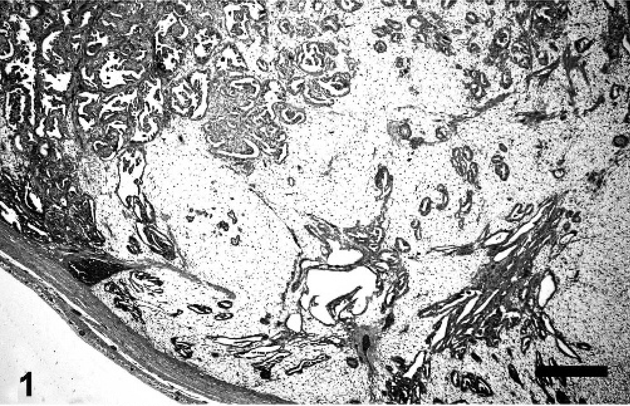
Uterus; malignant mixed müllerian tumor, rabbit. Neoplastic glands invade the myometrium. HE. Bar = 80 µm.
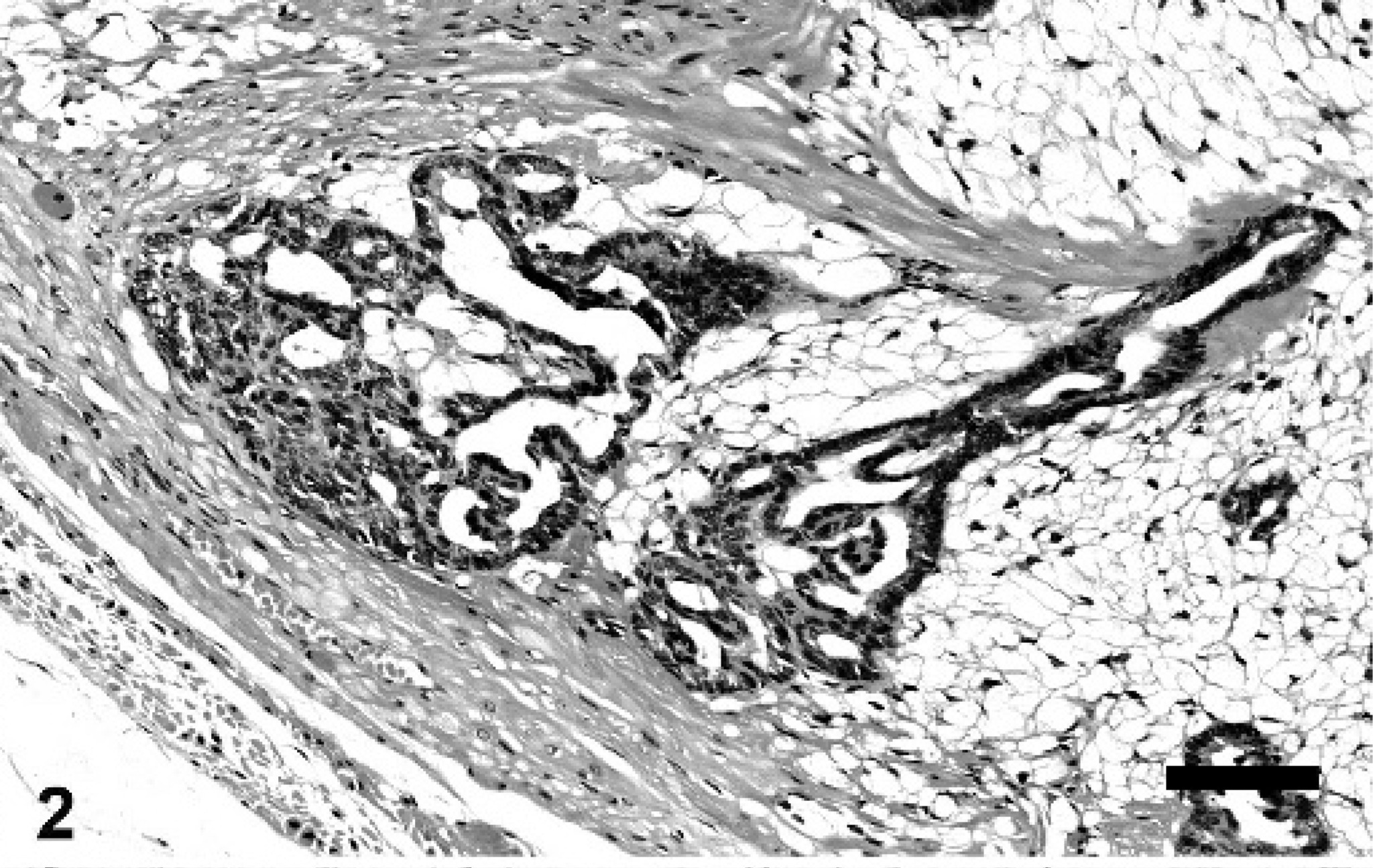
Uterus; malignant mixed müllerian tumor, rabbit. Decidual/stromal cells contain large amounts of intracytoplasmic glycogen. PAS and PAS with diastase digestion. Bar = 40 µm.
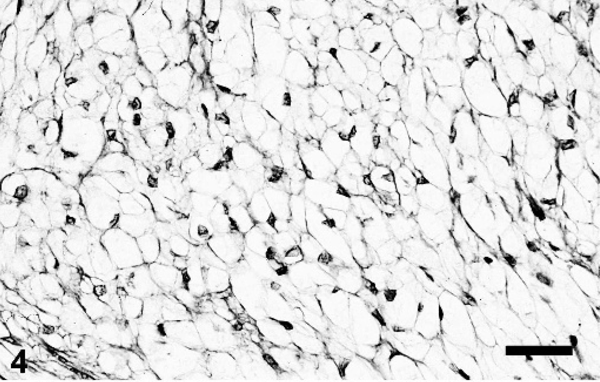
Uterus; malignant mixed müllerian tumor, rabbit. Decidual/stromal cells contain large amounts of intracytoplasmic glycogen. PAS and PAS with diastase digestion. Bar = 40 µm.
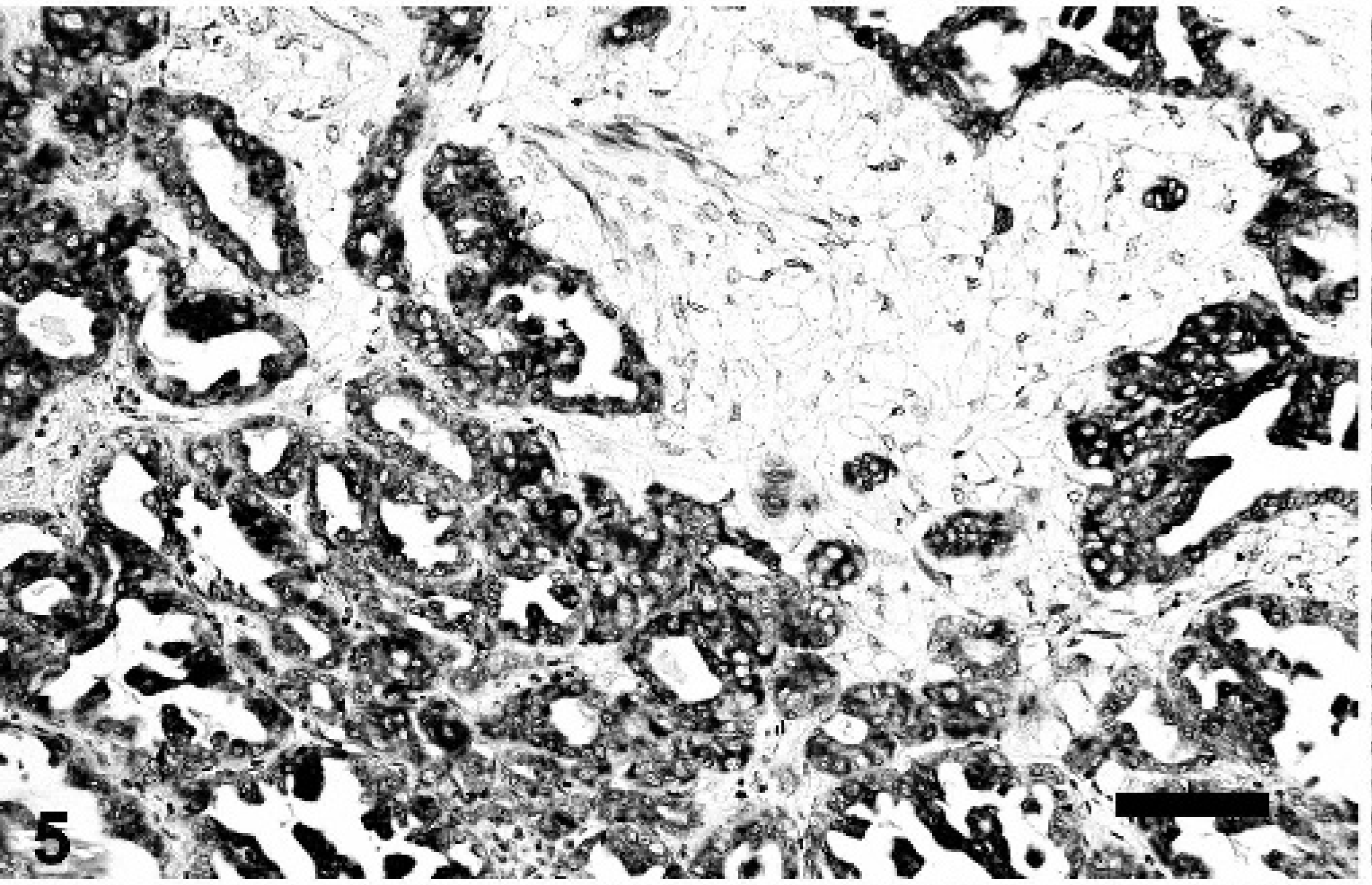
Uterus; malignant mixed müllerian tumor, rabbit. Epithelial components are stained with cytokeratin (AE1/3). Immunohistochemistry, DAB; counterstained with hematoxylin. Bar = 40 µm.
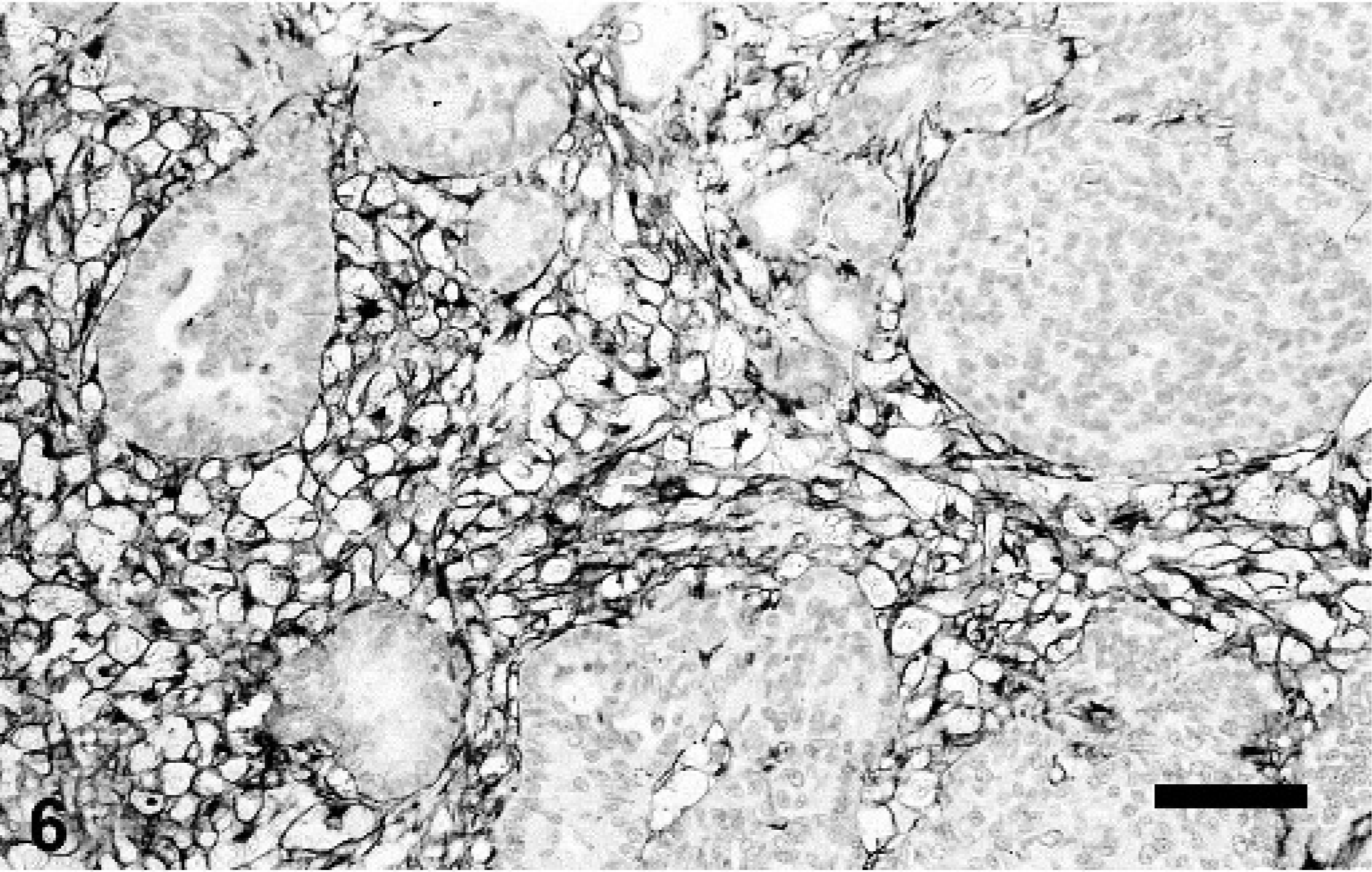
Uterus; malignant mixed müllerian tumor, rabbit. Decidual/stromal cells stain positive with vimentin (v9), and epithelial components are not stained. Immunohistochemistry, DAB; counterstained with hematoxylin. Bar = 40 µm.
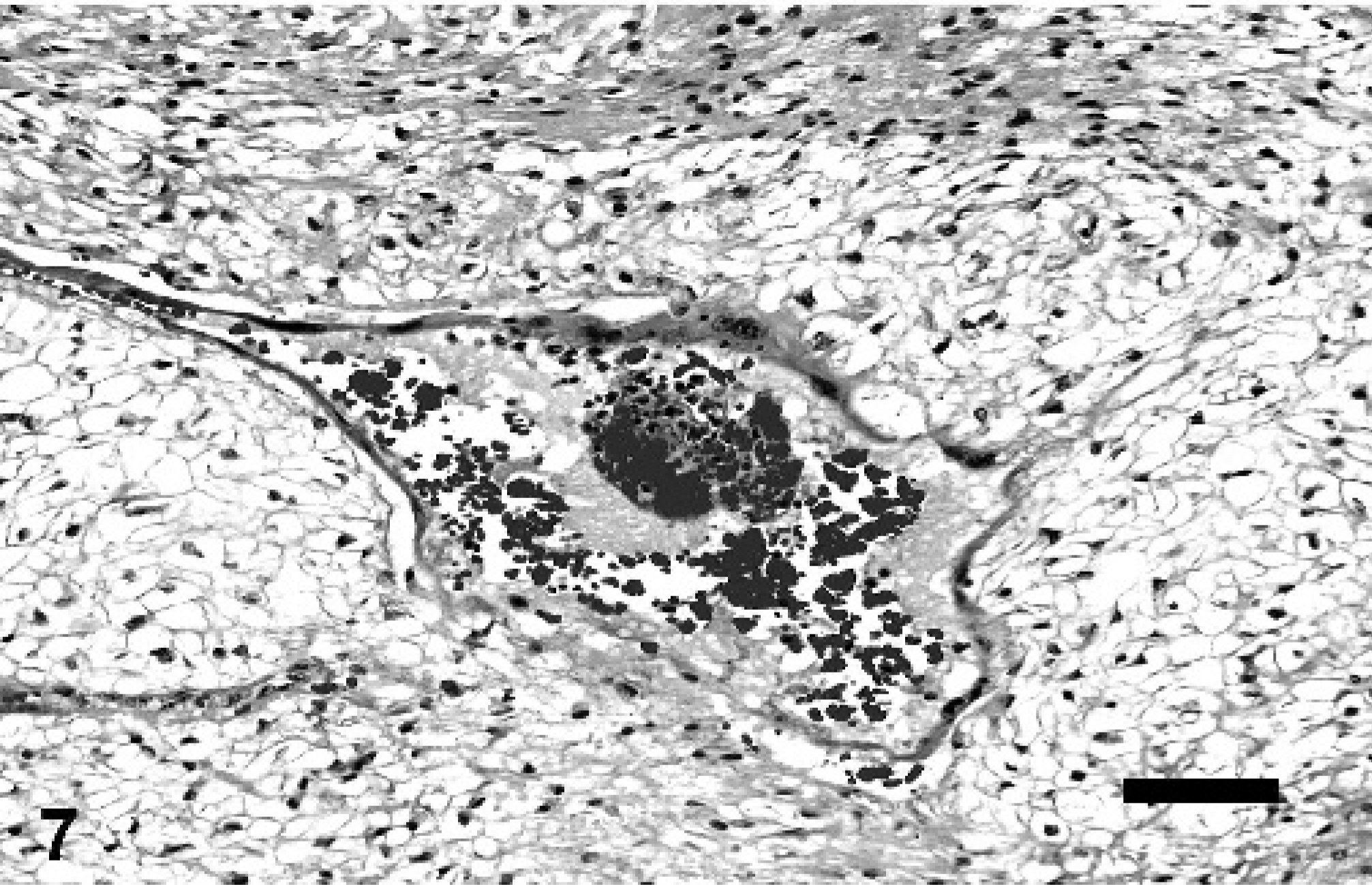
Uterus; malignant mixed müllerian tumor, rabbit. Vascular-like structures that contain blood cells in the lumen are lined by hypertrophic endothelial cells. HE. Bar = 90 µm.
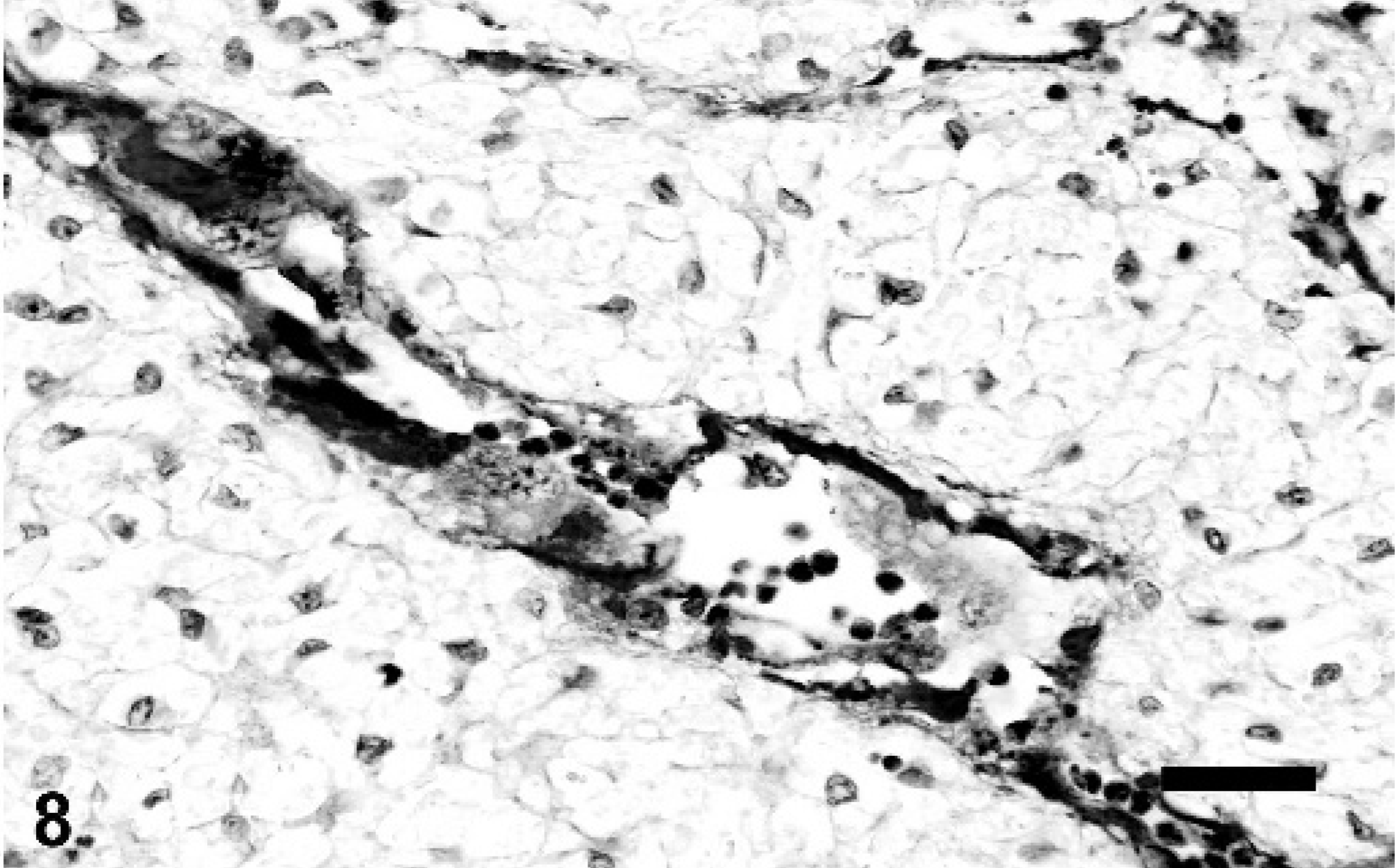
Uterus; malignant mixed müllerian tumor, rabbit. Hypertrophic endothelial cells stain positive with CD31. Immunohistochemical analysis, AEC; counterstained with hematoxylin. Bar = 45 µm.
Unfortunately, because this was a “rescue” rabbit, previous reproductive history and follow-up information were not available. Because the ovaries were not submitted for histologic examination, we were unable to determine whether there were any underlying ovarian abnormalities that could have induced an abnormally high concentration of progesterone, which may have been responsible for the decidualization of the neoplastic endometrial stromal cells.
In conclusion, we have described a unique uterine tumor in a rabbit with characteristic histopathologic, histochemical, and immunohistochemical features.
Footnotes
Acknowledgements
The assistance of Geza Acs, MD is greatly appreciated.