
Abstract
A 3-year-old female rabbit (Oryctolagus cuniculi) presented with apathy and indisposition for 2–3 days. Palpation revealed a mass in the caudal abdomen, namely, in the wall of the uterus. Ovariohysterectomy was performed, and the tissues were submitted for histopathologic examination. The mass consisted of 3 different (trophoblastic, syncytiotrophoblastic, and cytotrophoblastic) neoplastic cell types originating from the uterus. Immunohistochemistry was positive for cytokeratin in all 3 neoplastic cell types, and the syncytiotrophoblasts were positive also for human chorionic gonadotropin. Together these features allow the diagnosis choriocarcinoma. This report documents the first case of a spontaneous choriocarcinoma in a rabbit.
Choriocarcinoma is a highly malignant neoplasm of trophoblastic cells arising in placental tissue, ovary, testis, or sequestered remains of totipotential cells in the mediastinum or abdomen. 2 It is a rapidly proliferating, invasive, widely metastasizing tumor of low incidence in humans and animals. There are 3 reports of choriocarcinoma in rhesus monkeys and 1 of a cynomolgus monkey. 3,6,9,12 Furthermore, several cases in rodents have been described. 1,7,10 Experimentally induced chorionic tumors in 2 of 15 pregnant rabbits were reported in 1967. 5 In addition to rodents, choriocarcinoma has also been observed in an armadillo. 8 Here we document the first known case of a rabbit with choriocarcinoma that metastasized into the abdomen and lung possibly after removal of the original uterine neoplasm by ovariohysterectomy.
A 3-year-old female rabbit with a history of apathy and indisposition lasting for 2–3 days was presented. On physical examination, a large solid mass was palpated in the caudal abdomen. Ultrasonography demonstrated a mass in the uterus wall. The hematocrit values were low, there were monocytosis and lymphopenia, and glucose and cholesterol levels were increased. Surgical exploration revealed a firm nodulated, approximately 8 × 10 cm white mass, weighing 300 g, with multifocal extensive hemorrhage and cystic softening in the right uterine horn. The cut surface was white with scattered red to brown foci. Another white firm mass of 1-cm diameter was situated at the cranial end of the left uterine horn. Both ovaries were unaffected. The remaining abdominal organs were normal.
The rabbit recovered from surgery within a few days, but nearly 3 weeks later it presented again in bad general condition. This time, a mass was palpated in the cranial abdomen. The animal died before ultrasonography could be performed. At necropsy, multiple firm, disseminated white to red nodules were apparent in the mesentery and the lung (Fig. 1).
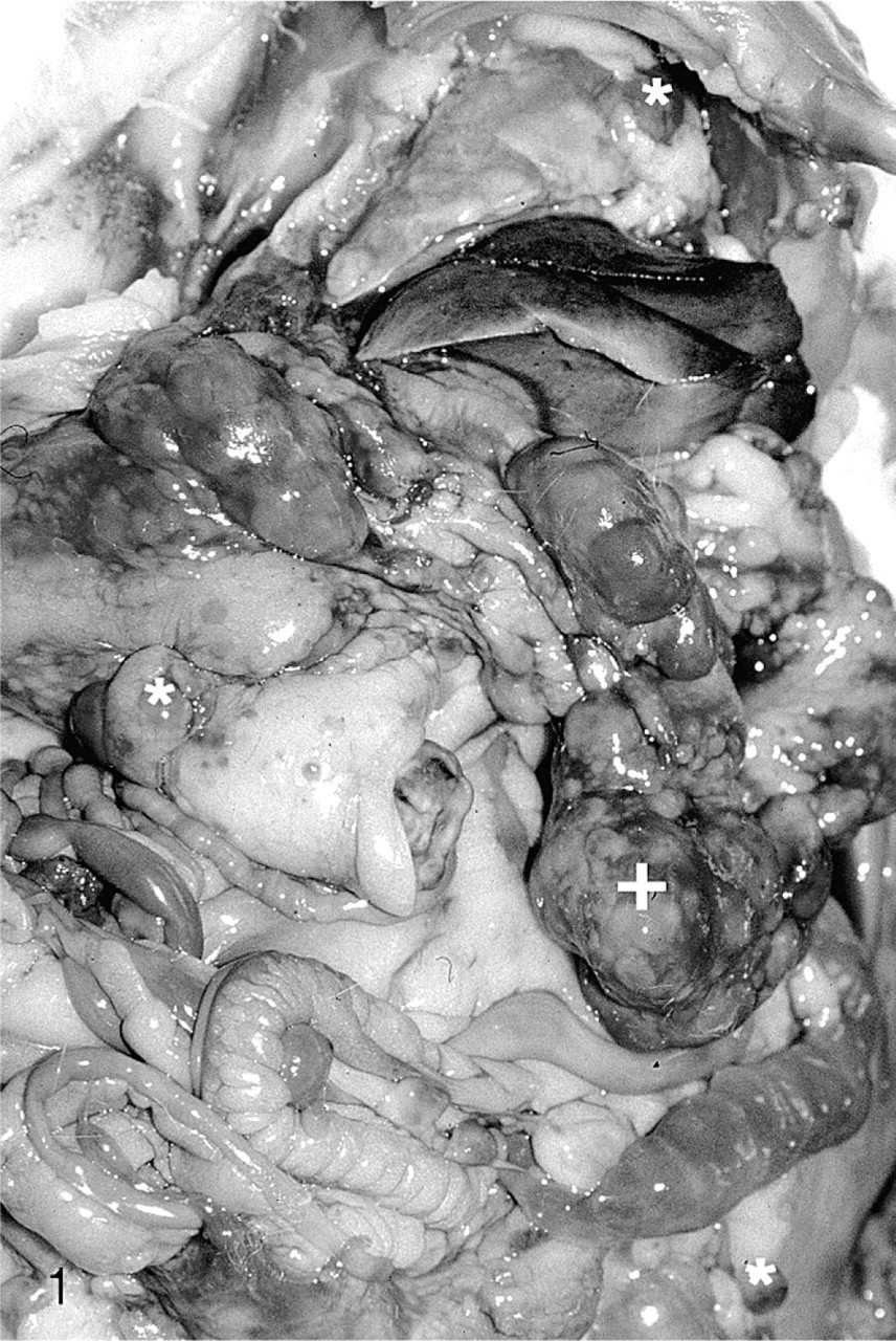
Abdominal and thoracal cavity; rabbit. Palpable mass (+) in the mesentery close to the site at which an ovariohysterectomy had been performed 3 weeks previously. There were additional metastases in the mesentery and the lung (∗).
Tissue samples from the uterus and the metastases from the lung and mesentery were fixed in 4% buffered formalin, embedded in paraffin, sectioned at 3–4 μm, and stained with HE for histopathologic evaluation. Immunohistochemistry was performed on sections of the uterine mass and the metastases using an avidin-biotin peroxidase complex method for human chorionic gonadotropin and a labeled streptavidin-biotin method for cytokeratin (DAKO EnVision, DAKO Corp., Carpinteria, CA).
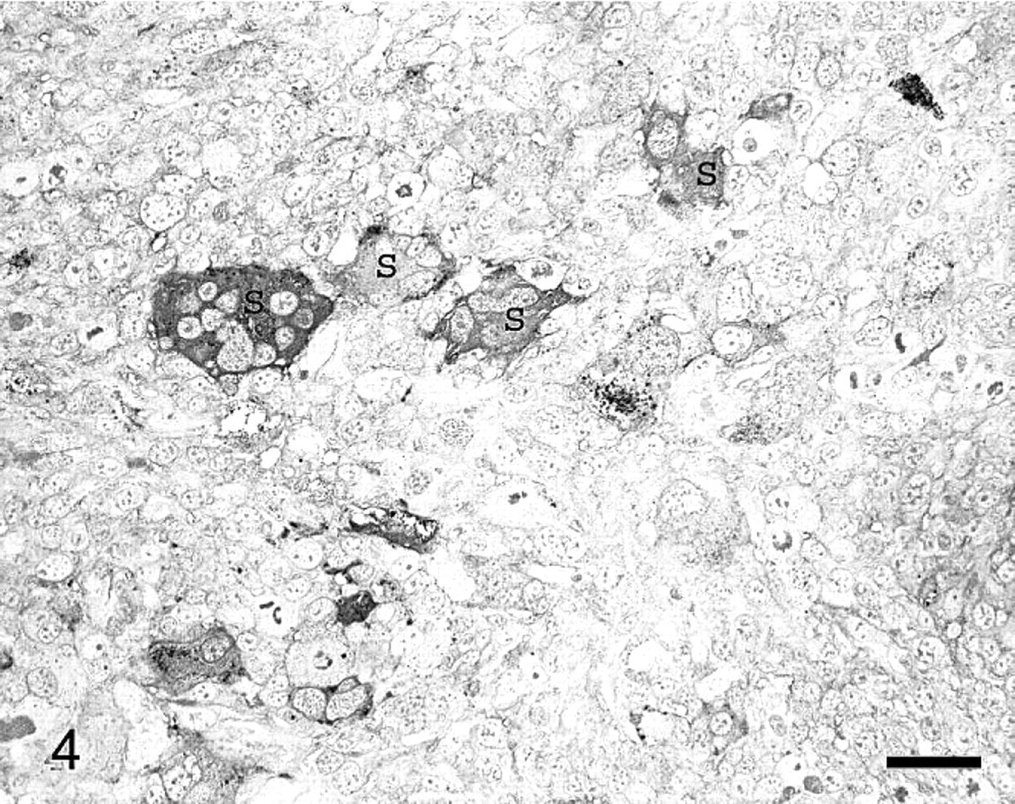
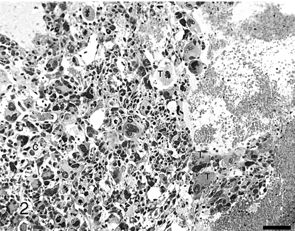
The mass in the left uterine horn was well demarcated and encapsulated by a thin fibrous layer. The tissue was composed of tubuli of variable diameter lined by predominantly homogenous cuboidal to high prismoidal cells. The nuclei were oval with only slight polymorphism and rare mitotic figures. The tubular lumina were partly filled with deeply staining homogenous eosinophilic fluid. An adenoma of the uterus was diagnosed. The original nodule in the right uterine horn and the metastases in the lung and mesentery were composed mainly of a highly pleomorphic cell population accompanied by multifocal necrosis and hemorrhage. A large number of trophoblastic cells were present. Both the cells and the nuclei were particularly variable in size and shape and mitotic, and atypical mitotic figures were observed regularly. The cytoplasm was amphophilic, was sometimes slightly granulated, or contained vacuoles (Fig. 2). Numerous multinucleated cells were identified as syncytiotrophoblastic cells. Only a few round cytotrophoblastic cells with central hyperchromatic nuclei and clear to amphophilic cytoplasma were present. There was multifocal to coalescing necrosis in all tissue sections. Hemorrhages lined by endothelial cells were visible in the periphery. Many apoptotic cells were scattered throughout the proliferations. The neoplasia showed aggressive infiltrative growth. Immunohistochemistry revealed that syncytiotrophoblasts were positive for human chorionic gonadotropin (Fig. 3) and some of the neoplastic cells were positive for cytokeratin (Fig. 4). These features were compatible with choriocarcinoma.
Uterus, choriocarcinoma; rabbit. Several neoplastic cells were immunoreactive for cytokeratin, and the strongest positivity was seen in syncytiotrophoblasts (S). Streptavidin-biotin method. Bar = 50 μm.
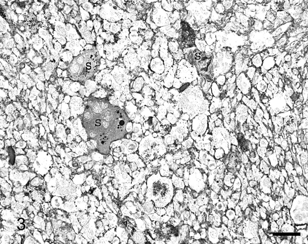
Uterus, choriocarcinoma; rabbit. Some of the syncytiotrophoblasts (S) are positive for human chorionic gonadotropin. Avidin-biotin-peroxidase complex method. Bar = 50 μm.
Uterus, choriocarcinoma; rabbit. Trophoblastics (T), syncytiotrophoblasts (S), and cytotrophoblasts (C). HE. Bar = 100 μm.
Choriocarcinomas may arise during the course of gestation or may be of germ cell origin. This rabbit had been kept with a castrated male in a private household, and to our knowledge a gestation had never occurred. An ovarian origin cannot be completely ruled out because the ovaries, having appeared normal at necropsy, had not been examined histologically. It is feasible but unlikely that a nodule of ovarian tissues could have been overseen in the large quantities of abdominal fat.
The immunohistochemical reaction patterns were similar to those described in the literature, in which syncytiotrophoblastic cells were positive for human chorionic gonadotropin 1,3,4,9,10,12 and some of the neoplastic cells were positive for cytokeratin. 4 In humans, the most common origin of choriocarcinoma is the placenta during normal or abnormal pregnancy. 2 Choriocarcinomas are rapidly growing, invasive neoplasms, often with distant metastasis typically to the lung, which is the most frequent extragenital metastatic site of choriocarcinoma in women. This was observed also in rhesus monkeys. 2,3,6 Endometrial hyperplasia and uterine adenocarcinoma are the most frequent uterine disorders in rabbits. They are related to aging, rabbits being most susceptible at the age of 4 to 5 years. 11
The histomorphologic appearance as well as the immunohistochemical staining patterns for cytokeratin and human chorionic gonadotropin confirmed the presence of a uterine choriocarcinoma with multiple pulmonary and mesenteric metastases in this rabbit and represents an unusual and rare disease entity. This is the first report on spontaneous choriocarcinoma in a rabbit.