
Abstract
In humans and animals, ossifying fibroma is a benign neoplasm that most frequently affects the mandible, often resulting in cosmetic deformities and malocclusion. It is considered rare in animals and most frequently affects young horses. A surgical biopsy of a solitary mass located beneath the gingiva in the right maxillary region, which had overgrown teeth and expanded the adjacent hard palate from a 6-year-old miniature Rex rabbit was submitted for light microscopic examination. The submitted incisional biopsy specimen was pale pink, firm, and nodular. Histopathologically, the neoplasm was composed of fibroblastic cells separated by abundant collagen. The neoplastic cells were interwoven with osteoblasts surrounding islands of mineralized, bony matrix containing few, widely spaced, often empty, lacunae. Minimal inflammation was present. Based on the histopathologic features, the tumor was diagnosed as an ossifying fibroma. To our knowledge, this is the first report of an ossifying fibroma in a rabbit.
Ossifying fibroma is a benign fibro-osseous neoplasm of the jaw in humans and animals that most frequently affects the mandible, causing distortion of normal bone contour, displacement and loss of teeth, and difficult mastication. 5, 8 Lesions that extend into the nasal cavities can obstruct airflow. In animals, ossifying fibroma is considered rare and is reported most frequently in young horses, in which it is classified as equine juvenile mandibular ossifying fibroma. 6 The tumor has also been reported in cats, 9 dogs, 3 a llama, 4 a greater kudu, 1 and a sheep. 7 In humans, ossifying fibromas typically affect the mandibles of young females. but a subset, juvenile ossifying fibroma, involves the maxilla and paranasal sinuses more frequently and is considered more aggressive. 8 Radiographically, solitary, well-demarcated, mixed or moderately radiolucent lesions expand bone. 5, 8 Histologically, ossifying fibroma is composed of fibroblastic spindle cells that resemble fibroblasts and undergo differentiation to osteoblasts that surround and form spicules of woven or lamellar bone or cementum-like material. 2, 5, 8
A surgical biopsy from a 6-year-old miniature Rex rabbit with a recent history of licking, opening the mouth wide, and inappetence was submitted for light microscopic examination. The biopsy was obtained from an approximately 1 × 2 × 2 centimeter, solitary oral mass located in the right maxillary region, which had overgrown teeth and expanded the adjacent hard palate (Fig. 1). The submitted incisional biopsy specimen was pale pink, firm, and nodular. Histopathologically, expanding the subgingival connective tissue and extending to submitted margins was an unencapsulated neoplasm composed of fibroblastic cells separated by abundant collagen (Fig. 2). The neoplastic cells were interwoven with osteoblasts surrounding islands of mineralized, bony matrix containing few, widely spaced, often empty, lacunae (Fig. 3). Neoplastic cells were characterized as having indistinct cell borders, scant eosinophilic fibrillar cytoplasm, and elongate nuclei. No mitotic figures were noted. Rarely, bony islands were lined by osteoclasts (Fig. 4). Minimal inflammation composed primarily of heterophils and eosinophils was present.
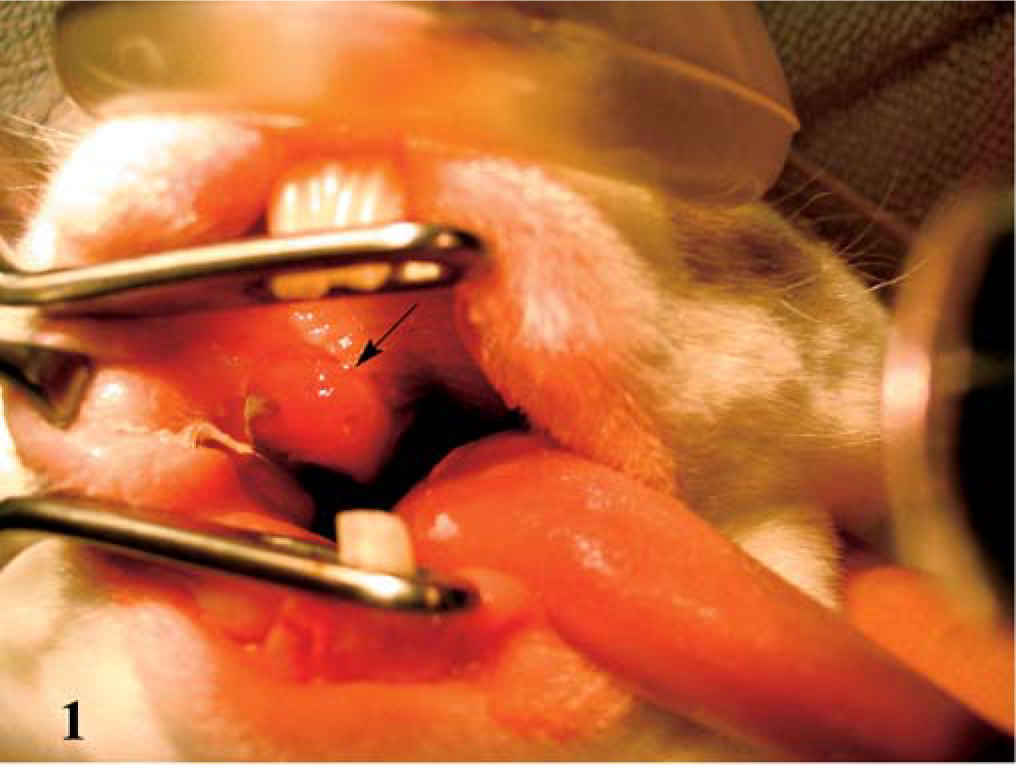
Oral cavity; rabbit. A solitary mass in the right maxillary region and expanding the adjacent hard palate (arrow).
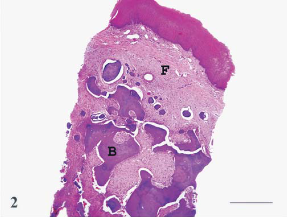
Maxilla; rabbit. An expansile, unencapsulated subgingival neoplasm composed of fibroblastic cells (F) separated by abundant collagen and islands of mineralized, bony matrix (B). Hematoxylin and eosin. Bar = 500 µm.
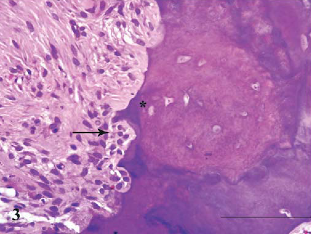
Maxilla; rabbit. The neoplastic cells blend with the osteoblasts (arrow) surrounding islands of mineralized, bony matrix that contain few, widely spaced, often empty, lacunae (asterisk). Hematoxylin and eosin. Bar = 100 µm.
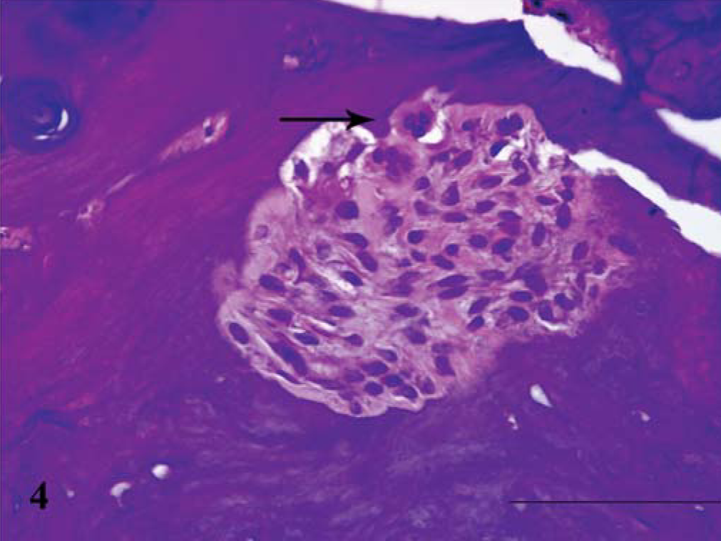
Maxilla; rabbit. Rare osteoclasts (arrow) line the mineralized, bony matrix. Hematoxylin and eosin. Bar = 100 µm.
Differential diagnosis included fibrous dysplasia, odontogenic dysplasia of lagomorphs, fibromatous epulis of periodontal ligament origin, and cementifying fibroma.
The presence of osteoblasts surrounding the bony spicules helps differentiate this case from fibrous dysplasia, a nonneoplastic fibro-osseous condition of the jaw in man and animals. 2, 5, 8 The lack of odontogenic elements, including odontogenic epithelium and enamel, dentinal, and periodontal ligament-type matrix, makes odontogenic dysplasia and fibromatous epulis of periodontal ligament origin less likely. 2 While there is some suggestion of cemental differentiation (scattered basophilic tide lines) within this neoplasm, the features are not as distinct and sharply defined as that which occurs in cementifying fibroma. 2
Ossifying fibroma is a benign, solitary, expansile tumor of the mandible and maxilla in man and animals. 5, 8 The histopathologic features of the neoplasm in this case are consistent with those described for ossifying fibroma. To our knowledge, this is the first report of this tumor in a rabbit.
Footnotes
Acknowledgements
We are grateful to Dr. Kevin Brumfield and Dr. Mark Smith for their photographic support. We would also like to thank Dr. Kevin Torske, Department of Oral Pathology, and Dr. Francis Gannon, Department of Orthopedic Pathology, Armed Forces Institute of Pathology, for their consultative assistance.
K. A. Whitten is a Major and D. A. Belote and M. G. Mense are Lieutenant Colonels in the US Army. The opinions or assertions contained herein are the private views of the authors and are not to be construed as official or as reflecting the views of the Department of the Army or the Department of Defense.