
Abstract
Ossifying fibroma (OF) is a slow-growing, expansive, and benign fibro-osseous neoplasm that is rare in cattle. It mainly affects the craniofacial bones, especially the mandible. Here, we report 2 cases of mandibular OF in Nelore and mixed-breed steers with enlarged masses in the rostral portion of the mandible. Radiographic analysis of case 1 revealed an oval, lobed mass with the radiopacity of bone tissue that displaced the incisors laterodorsally. Histologically, both masses were composed of a proliferation of spindloid-to-stellate cells, supported by a dense fibrovascular stroma, with bony trabeculae surrounded by stroma and covered by a single layer of osteoblasts. Cellular pleomorphism was low, anisocytosis and anisokaryosis were negligible, and mitotic figures were not observed. The clinical, radiologic, gross, and histologic changes are compatible with OF. The primary differential diagnoses of OF are fibrous dysplasia and osteoma; their differentiation can be difficult. Fibroma, low-grade osteosarcoma, and multilobular sarcoma of bone are also differential diagnoses, with striking features that facilitate their exclusion.
Ossifying fibromas (OFs) are benign mesenchymal neoplasms with variable amounts of bone tissue in fibrous tissue stroma. Most are slow-growing and expansive but can be invasive; OFs occur mainly in the craniofacial bones, especially the jaw, 4 with reports of occurrence in humans, 8 horses, 10 dogs, 9 cats, 17 sheep, 14 rabbits, 19 antelopes, 20 and non-human primates. 15 However, reports of OFs in cattle are scarce. 2
Morphologically, OF is a single, firm mass, sometimes ulcerated,1,11,16 generally displacing or deforming the bone, leading to dental malocclusion, making it challenging to grasp food; therefore, even benign behavior can have clinical effects. 4 Here we report 2 cases of OF in beef cattle in the state of Mato Grosso (MT), Brazil.
Cases 1 and 2 had enlarged masses in the rostral portion of the jaw, without historical timelines on growth. Case 1, a ~24-mo-old Nelore steer, from a beef farm in the municipality of Cuiabá, MT was transported to the Universidade Federal de Mato Grosso Veterinary Hospital’s Large Animal Surgical Center (HOVET/UFMT; Cuiabá, Brazil) for clinical evaluation. Euthanasia was carried out due to the severity of the lesion, and the carcass was forwarded immediately for postmortem examination to the UFMT Veterinary Pathology Laboratory (LPV/UFMT). The Imaging Diagnostic Sector of HOVET/UFMT performed a postmortem radiographic examination of the head.
Case 2 was a 2-y-old mixed-breed steer from the municipality of Pontes e Lacerda, MT. The steer was sent to slaughter, where the veterinary service official sequestered the head during the sanitary inspection and sectioned the rostral region of the mandible before forwarding it to the LPV/UFMT for histologic evaluation. Samples of the masses from these 2 animals were preserved in 10% neutral-buffered formalin and processed routinely for histologic examination.
In case 1, the patient was in poor body condition. A nodular 20 × 15 × 15-cm mass covered by mucosa with extensive areas of ulceration projected from the rostral portion of the oral cavity, involving the incisor teeth, mainly on the left side of mandible (Fig. 1). Given the difficulty in prehending food, the animal was euthanized, and an autopsy was performed immediately. In the postmortem radiographic examination, the mass was oval, lobed, and contiguous with the rostral portion of the left ramus of the mandible; the incisor teeth were displaced laterodorsally. The mass had the radiopacity of bone, with a discretely radiated appearance and well-defined, predominantly smooth contours, without evidence of cavitations or osteolysis in the rostral portions of the mandible (Fig. 2). Upon sectioning, the mass macroscopically had a hardened central area (mineralization), with radial projections towards the periphery, surrounded by firm, white material, occasionally with a mucinous appearance (Fig. 3). In sections in the median plane, it was possible to observe infiltration and deformation of the incisor and molar dental alveoli.
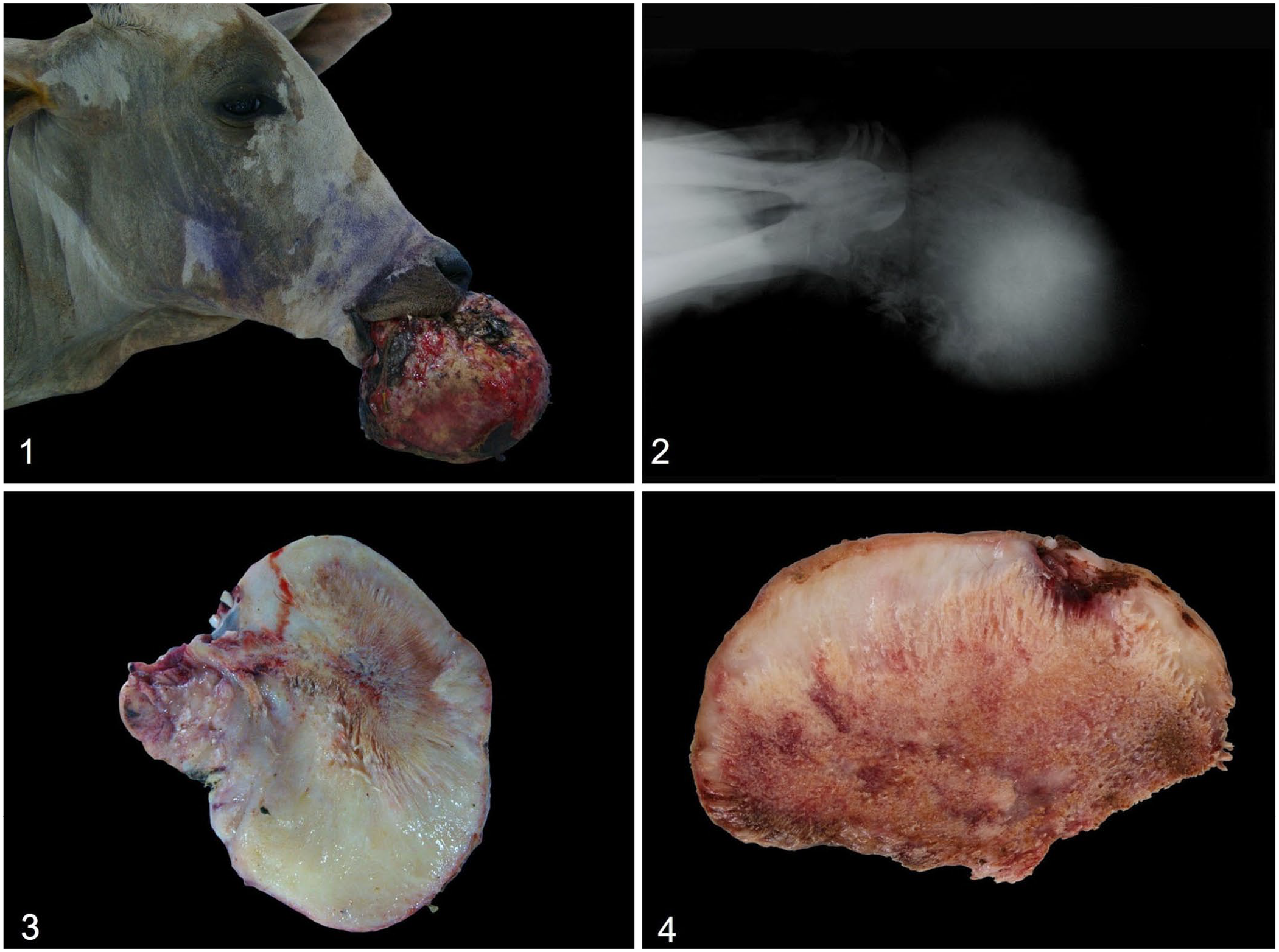
Mandibular ossifying fibroma (OF) in 2 cattle.
Macroscopically, the samples from case 2 had a crusty, brown surface, bordering soft-to-firm white tissue that extended to the base of the mass, where the tissue was hard and brown-red (Fig. 4). The samples from both cases and the jaw from case 1 were fixed in 10% neutral-buffered formalin. The mineralized tissues were demineralized in 10% nitric acid solution. The samples were processed routinely, and 4-µm sections were stained with H&E.
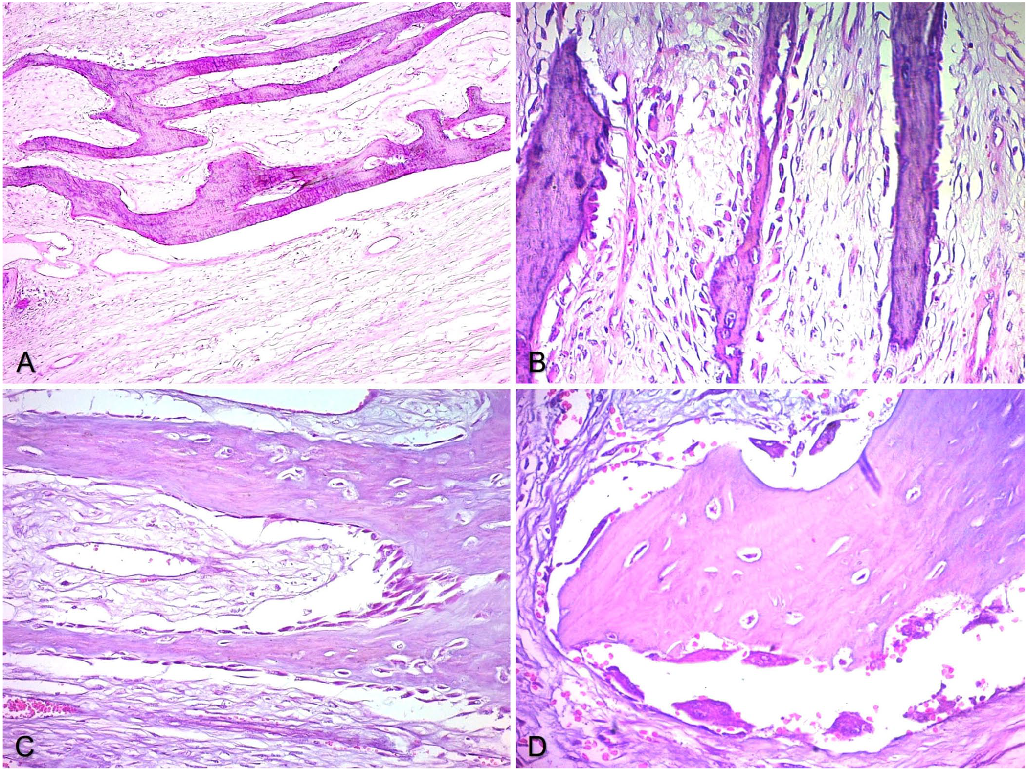
In both cases, the histologic findings were similar. Masses of spindloid-to-stellate mesenchymal cells with low pleomorphism were distributed in multidirectional bundles. The tumor cells had large, pale, basophilic, and sometimes vacuolated cytoplasm and a hyperchromatic basophilic nucleus with barely evident nucleoli. Anisocytosis and anisokaryosis were insignificant. No mitotic figures were observed. Intermingled with the fibrovascular matrix were significant amount of myxoid matrix and multidirectional bone trabeculae (Fig. 5A), sometimes mineralized, regularly spaced (Fig. 5B), and covered by a single layer of typical osteoblasts (Fig. 5C), with rare osteoclasts in resorption (Howship) lacunae (Fig. 5D). In places of higher cell density, there were small sites of necrosis with neutrophil infiltrates.
Mandibular ossifying fibroma in case 1.
The clinical and lesional findings of mandibular changes in our 2 cases are compatible with OF. OF and fibrous dysplasia (FD) comprise a complex group of benign lesions called proliferative fibro-osseous lesions. 12 Osteoma (OM) is a benign neoplasm in which mature bone tissue proliferates. 16 These lesions commonly arise in the bones of the skull, especially those that develop through intramembranous ossification. 1 Although it is not a true neoplasm, FD is a tumor-like lesion that should be considered a differential diagnosis in cases of expansile lesions in the mandible, as in our cases. Differentiation of the 3 tumors can be complex; however, histologic characteristics can allow identification of the main pattern.1,12,16
OF is more often recognized in horses <1-y-old, occurring in the rostral region of the mandible 10 ; although reports of this neoplasm in cattle are rare, it is typical for the rostral region of the mandible to be the leading site of occurrence, 2 a situation observed in both of our cases. The histogenesis of this neoplasm is possibly related to the periodontal ligament. 13 OFs are well-defined, firm-to-hard osseous masses, distorting the anatomy of the involved bone. Microscopically, a proliferation of fibrovascular stroma produced by fibroblasts and by bone trabeculae is typically covered by a single layer of osteoblasts, which may be arranged perpendicular to the mesenchymal surface layer or irregularly shaped; this proliferation replaces typical bone architecture. The intertrabecular spaces are filled with fibrous connective tissue, and there are areas of bone resorption by osteoclasts. No features of malignancy, or of a cartilage or periosteal envelope, are observed. 1 These macroscopic and microscopic findings were present consistently in both of our cases.
OM is characterized by the proliferation of compact and trabecular bone with reduced fibrovascular stroma, which may be related to the endosteum (central) or periosteum (peripheral). The architecture comprises trabecular and, caudally, lamellar bone, often exhibiting ordered zones between these 2 bones and a reduced and sparse connective matrix. Furthermore, OMs have a covering of peripheral connective tissue typical of periosteum,9,12 unlike in our case, in which the proliferation mainly had a fibrovascular character.
FD is a rare fibro-osseous change very similar to OM and OF; in humans, it is associated with a somatic mutation in gene GNAS encoding the Gsα subunit of the G protein, which increases cAMP, interfering with osteoblastic differentiation.5,6 Proliferating mesenchymal cells produce trabecular bone in a disorganized and disoriented manner, without an apparent transition from mesenchymal cells to osteoblasts and may be surrounded by a thin layer of dense bone produced by the periosteum.10,11 The differentiation of these 3 tumors is often confusing and complex; some authors propose that FD is the initial stage of OF, and OM is its final stage.11,16
On radiographic evaluation, OF can have 2 different radiologic patterns, ranging from single or multiple radiolucent cysts to mixed or radiopaque areas, which usually are well-defined,3,7 with their radiopacity linked to the type of tissue mineralization. In case 1, the well-circumscribed, oval-shaped proliferated tissue with a radiolucent cyst was compatible with the known radiographic patterns for OF. Furthermore, the expansion of bone tissue and the displacement of teeth and dental alveoli are typical of OF. 4
In humans, OF occurs in middle-aged adults, with the mandibular bone as its leading site. 7 Reported as a slow-growing neoplasm that does not recur after surgical excision, there are descriptions of conditions with more aggressive behavior with a tendency to recur. These cases occur in children and are called juvenile OF. 18 As in veterinary medicine, the diagnosis of OF in humans is complex due to the significant similarity with other fibro-osseous lesions, 8 such as dysplasia, florid cemento-osseous dysplasia, and focal cemento-osseous dysplasia, making it necessary to associate histopathology with radiography and clinical evolution for accurate differentiation. 18
Cemento-OF (COF) is a variation of OF that should be considered in the differential diagnosis. In COF, cemental spheroids and isolated cementum islands are formed. The cementum is more basophilic than osteoid matrix and is organized in circular or irregular clusters. 11 Other neoplasms such as fibroma, low-grade osteosarcoma, and multilobular sarcoma of the bone are differential diagnoses that must also be ruled out. The presence of bone tissue is one of the keys to excluding fibroma. Osteolysis occurs in the most aggressive form of low-grade osteosarcoma and can be observed radiographically, helping in differentiation. Multilobular sarcoma of bone is classically formed by countless nodules with a trilaminar histologic appearance, facilitating its exclusion.1,11,16
Footnotes
Declaration of conflicting interests
The authors declared no potential conflicts of interest with respect to the research, authorship, and/or publication of this article.
Funding
We are grateful to the Brazilian Federal Agency for the Support and Evaluation of Graduate Education (CAPES) for financial support through a Masters’ scholarship and to the Research Support Foundation of the State of Mato Grosso (FAPEMAT) for financial support through scientific initiation scholarship.