
Abstract
The gross and histopathologic lesions of meningoencephalitis tuberculosa in a 4-year-old Holstein cow showing clinical signs compatible with bovine spongiform encephalopathy are described in this report. Grossly, numerous gray to yellow, firm and caseous nodules were seen on the ventral surfaces of the brain and in the lateral and fourth ventricles. Histopathologically, foci of caseation and dystrophic mineralization were surrounded by multinucleated giant cells, epitheloid macrophages, plasma cells, lymphocytes and fibrous proliferation. Ziehl-Neelsen stains of the lesions revealed masses of slender acid-fast bacilli in the necrotic centers of lesions and within surrounding giant cells.
Tuberculosis is typically a chronic disease caused by bacteria of the genus Mycobacterium. 7 Various forms of the disease have many features in common, but the exact pattern differs according to the species of Mycobacterium and the species of animal affected. Bovine tuberculosis has been one of the most important diseases of cattle. 5 The usual routes of Mycobacterium infection are respiratory and alimentary. In adult cattle, primary infection is usually in the lungs and is caused by inhalation of infected droplet nuclei. 1, 8 Less common routes of infection are cutaneous, congenital, and genital. Tuberculosis in the central nervous system (CNS) is rare in cattle. 2 In a large abattoir survey in Turkey, 4 bovine tuberculosis was detected most often in lungs, spleen, liver, uterus, and testes, but was not reported in the CNS. Tuberculosis in the CNS begins mainly as a meningitis, and is more common in the cerebral than in the spinal meninges. Involvement of cranial meninges is almost always hematogenous, with the initial lesions occurring usually in the basilar meninges and extending from there via the arachnoid spaces to the choroid plexus and ventricles. To a limited extent, infection can spread via the Virchow-Robin spaces into the brain parenchyma itself. Involvement of the spinal meninges may spread from a vertebral osteomyelitis, or by a hematogenous route. The meningeal lesions are similar to those of serous membranes but are generally more exudative and necrotizing. Miliary or conglomerate tubercles are uncommon in the CNS. 1
The case material was a cranium taken from a 4-year-old Holstein dairy cow. The cerebral hemispheres, cerebellum, and brain stem were collected and fixed in neutral buffered formalin solution. After routine histopathology processing, 5–6 micron paraffin sections were prepared and all sections were stained with hematoxylin-eosin (HE) and the Ziehl-Neelsen method for acid-fast bacteria. 3
The submitting veterinarian had treated this cow with fluid electrolytes, antibiotics, vitamins, and rumenotorics because of anorexia, decreased milk production, and poor condition. Initial observations included mastication, incoordination, and a stumbling gait. By 12 days after the onset of clinical findings, severe neurologic findings included apathy, nervousness, and hyperexcitability. Because bovine spongiform encephalopathy (BSE) was a possible cause, the animal was slaughtered and the cranium was brought to our laboratory (Konya Veterinary Control and Research Institute) for histopathologic examination.
Grossly, many blood vessels of cerebrum and cerebellum were hyperemic. The leptomeninges were markedly thickened, mainly over the cerebellum, the basal surfaces of the brain stem and the optic chiasm. There was an accumulation of fibrin-like and opaque exudate in affected areas (Fig. 1), and numerous nodules from 2 to 15 mm in diameter were found on the basal surfaces of the brain. These nodules were grayish to yellowish in color, firm and caseous, and covered the brain stem, including the optic chiasm, cerebral crus, medulla oblongata, and pons (Fig. 1). Only a few granulomas could be seen on the meninges of the cervical medulla spinalis. When the both hemispheres were separated, several nodules were observed on the surfaces of lateral ventricles (Fig. 2) and a few nodules were seen within the fourth ventricle. All these nodules were gray to yellowish in color and caseous. When the brain stem, cerebellum, and cervical medulla spinalis were longitudinally sectioned, it was confirmed that these caseous nodules were located only within the meninges and on ventricular surfaces. There were no visible macroscopic nodules on sagittal sections of the cerebral hemispheres, brain stem, and cervical medulla spinalis. Gross lesions were not detected in the oral cavity or in the retropharyngeal or submandibular lymph nodes, and gross lesions were not reported in other organs by the submitting veterinarian.
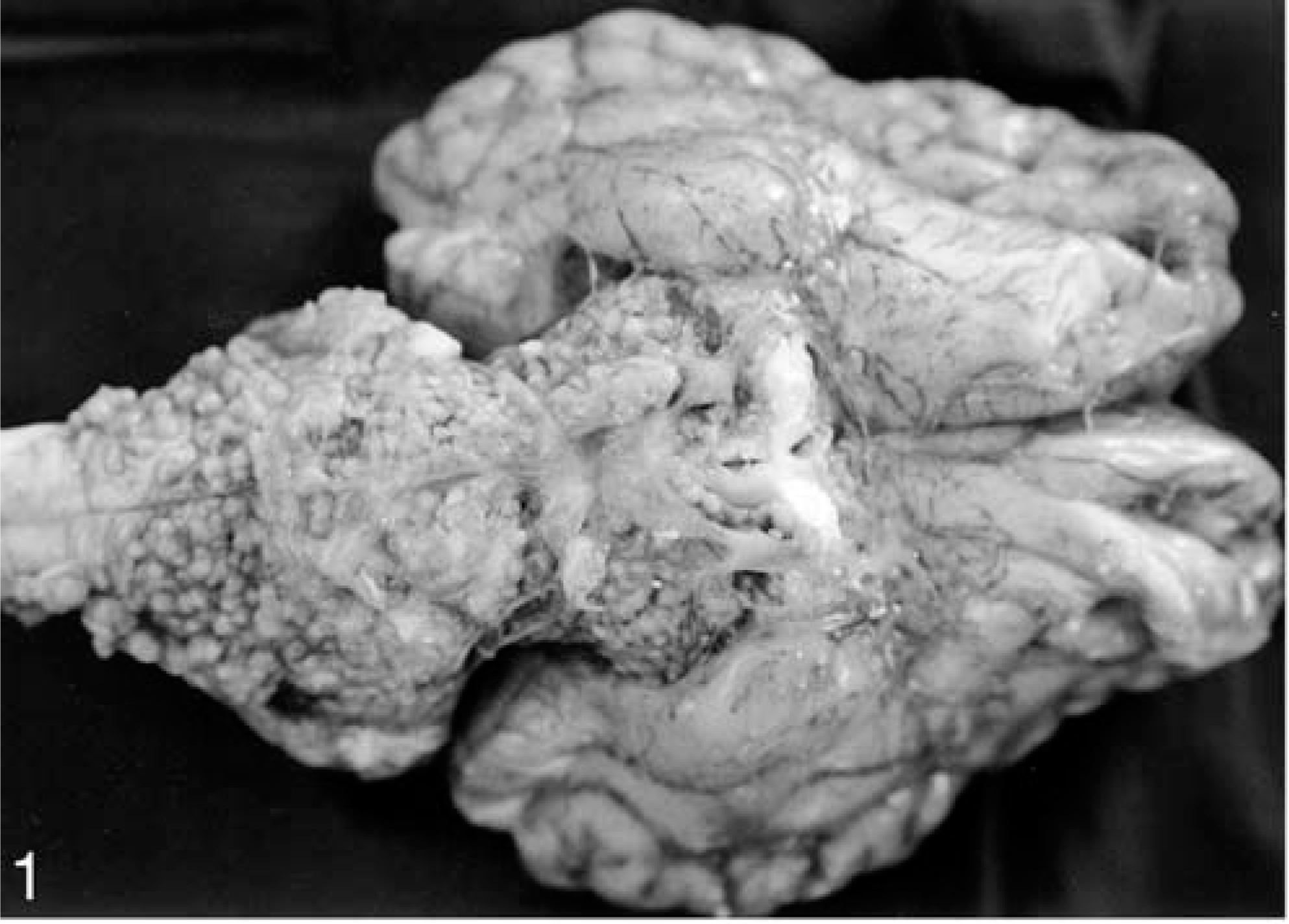
Base of brain; Holstein cow. Numerous grayish to yellowish, firm nodules, opacity of leptomeninges, and accumulation of fibrin-like exudate.
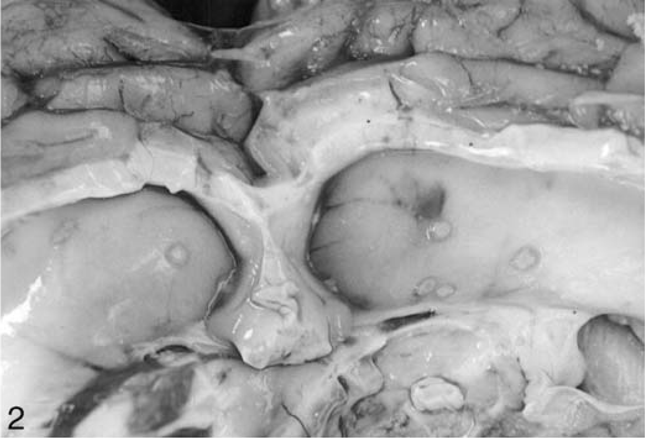
Lateral ventricles; Holstein cow. Several tuberculous nodules on the right and left lateral ventricles.
Meningeal capillaries were generally hyperemic, and prominent granulomas were detected in the meninges of cerebellum, medulla oblongata, pons, and optic chiasm. Many meninges contained central areas of caseification and dystrophic mineralization. These extensive necrotic areas were surrounded by multinucleated giant cells, epitheloid macrophages, plasma cells, lymphocytes, and fibrous proliferation (Fig. 3). The granulomatous lesions compressed adjacent tissues. In deeper areas of the cerebellum and brain stem, there were perivascular lymphoid infiltrates and diffuse gliosis. Ziehl-Neelsen stains of the lesions revealed masses of slender acid-fast bacilli in the necrotic centers of lesions and within surrounding giant cells (Fig. 4). There were no microscopic findings of lesions typically associated with BSE, such as neuronal vacuolation or spongiosis.
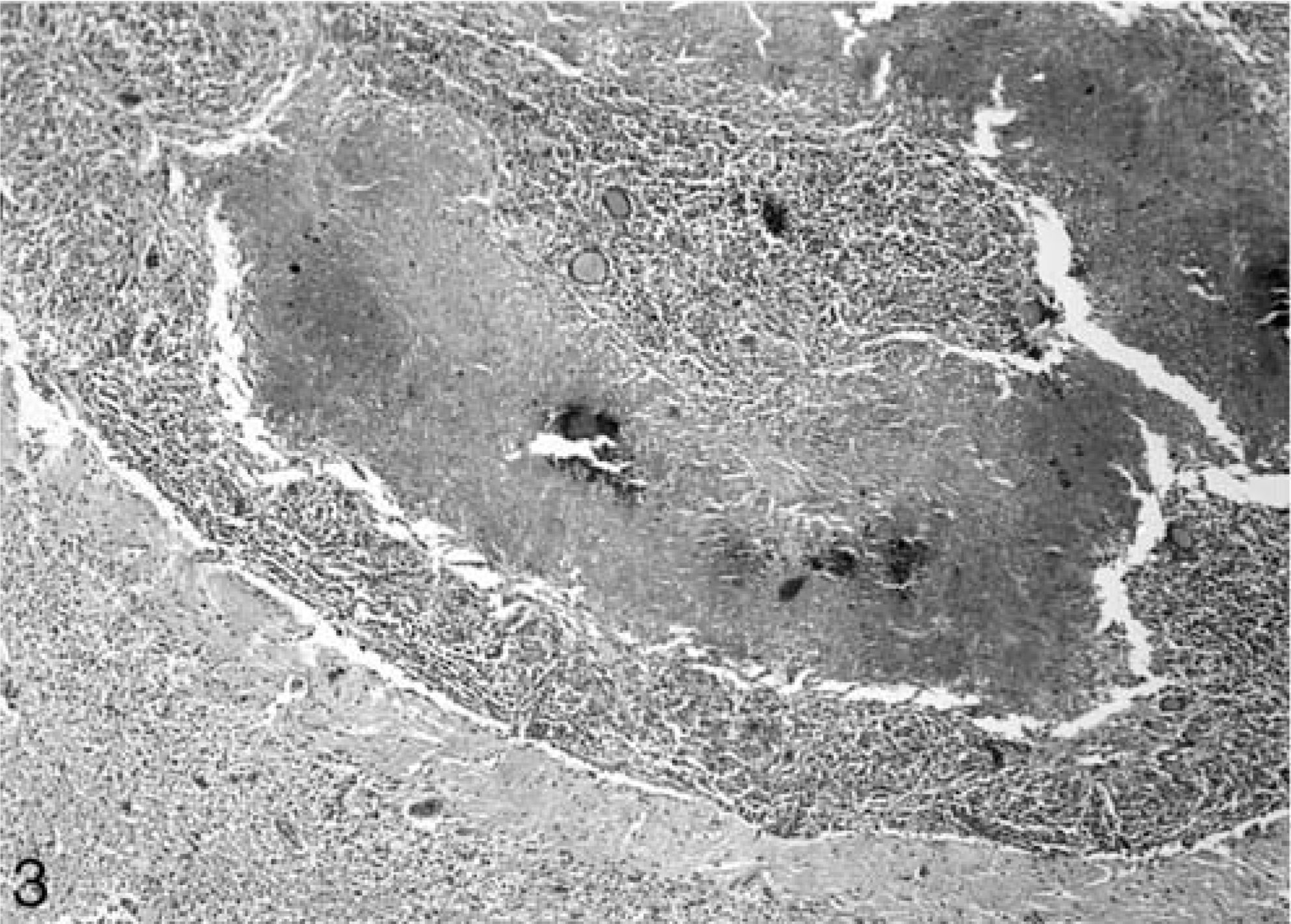
Cerebellar meninges; Holstein cow. Central area of caseation necrosis and dystophic mineralization surrounded by multinucleated giant cells, epitheloid macrophages, plasma cells, lymphocytes and fibrous proliferations. HE.
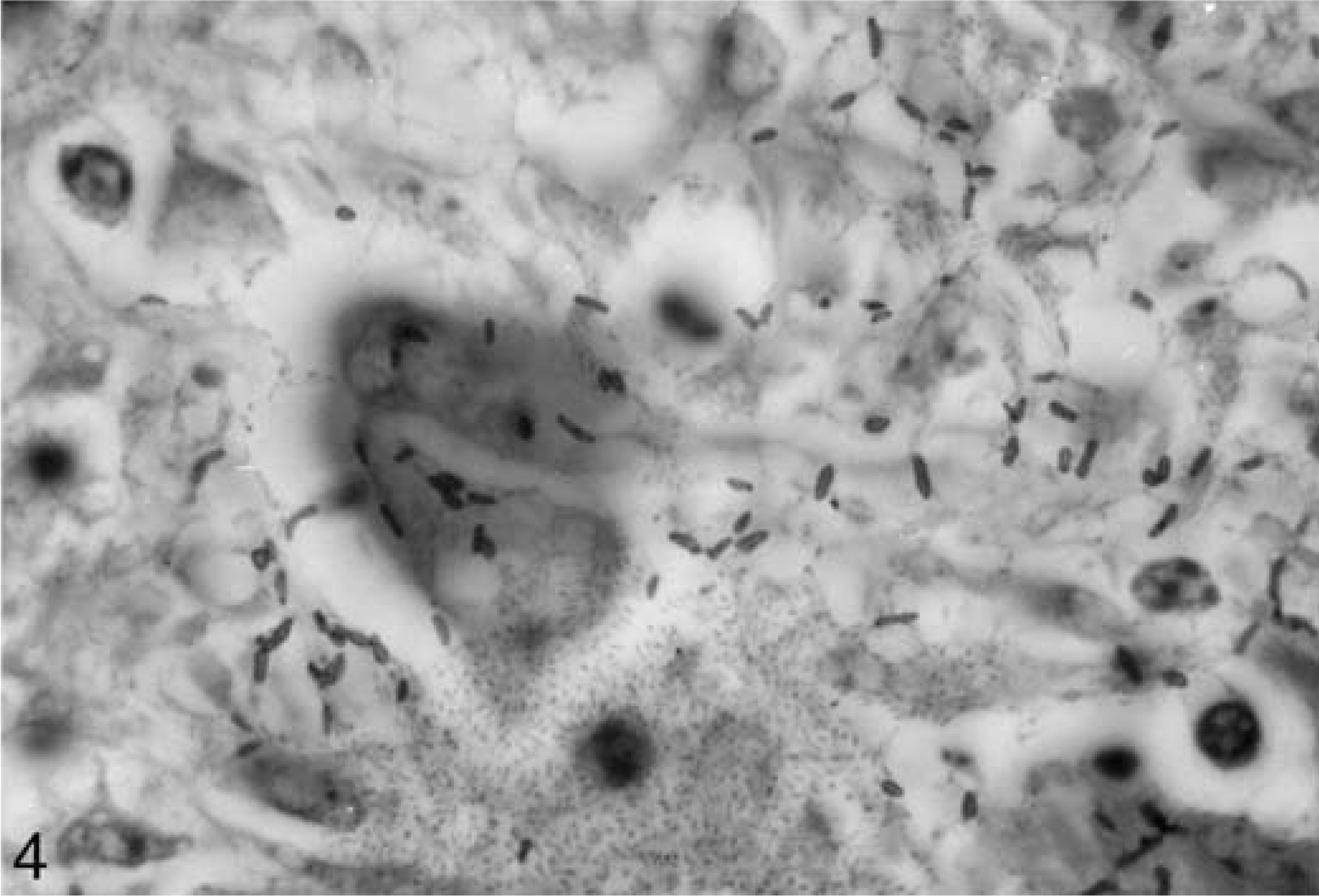
Cerebellar meninges; Holstein cow. Masses of slender acid-fast bacilli in the necrotic centres of lesions and in surroinding giant cells. Ziehl-Neelsen.
Bovine tuberculosis is still one of the most important diseases of cattle in Turkey and many other parts of the world. 4, 5, 7 However, tuberculosis lesions are infrequently encountered in the CNS. 2 The reported clinical findings in this case, such as anorexia, incoordination, apathy, stumble, mastication, and behavioral abnormalities were similar to those described in a previous report 6 and the tuberculous lesions in the CNS were also present predominately in the meninges of the base of the brain. 1, 6 This suggests that the infection reaches the brain by a hematogenous route. The few tubercles seen within ventriculi suggest that circulating organisms were retained in the choroid plexus and meningeal vessels before reaching the cerebrospinal fluid and the ventricular system. The histopathologic appearance of the productive tubercles detected in this case, with caseous necrosis, dystrophic mineralization, epitheloid and Langhans giant cells, lymphocytes, plasma cells, and fibrous proliferation were similar to previous reports of tuberulosis in the CNS and other organs. 2, 4, 6
In conclusion, tuberculosis of the CNS should be included as a differential diagnosis in cattle demonstrating severe neurologic symptoms.