
Abstract
To determine whether cyclooxygenase-2 (COX-2) is expressed in canine hemangiosarcoma (HsA), histiocytic sarcoma (Hs), and grade-II mast cell tumor (MCT), we performed immunohistochemistry using COX-2 antibodies in the aforementioned tumors. Twenty cases of each tumor type were selected initially from the Laboratory of Pathology archives of cases submitted through the Matthew J. Ryan Veterinary Hospital of the University of Pennsylvania. Immunohistochemistry was performed, using a polyclonal antiprostaglandin endoperoxide synthase immunoglobulin G COX-2 antibody. Sections from the kidneys of young dogs, in which the macula densa stains positive for COX-2, served as positive controls. Slides were reviewed by a single pathologist (M. H. Goldschmidt) and graded for COX-2 expression according to previously established scales.18 Descriptive data is given for each tumor type. COX-2 expression was identified in 0 of 19 HSA, 1 of 20 HS, and 1 of 17 grade-II MCT. Although COX-2 has been shown to be overexpressed in selected human sarcomas and hematopoeitic tumors, these results indicate that canine HSA, HS, and MCT do not express COX-2 in any appreciable fashion.
Cyclooxygenase-2 (COX-2) is a member of the family of cyclooxygenase enzymes that catalyze the conversion of arachidonic acid to prostaglandin G2 (PGG2; Product # PG 27B, Oxford Biomedical Research, Oxford, MI). PGG2 is further metabolized to a variety of other prostaglandins (PG) and thromboxanes (TXA). 24 PG and TXA are expressed ubiquitously in many tissues and are essential molecules in the maintenance of cellular homeostasis. COX-2 is an inducible enzyme and is expressed in response to numerous cellular conditions, inflammatory stimuli, cytokines, and cancer. 24 Specifically, COX-2 is induced readily by proinflammatory mediators such as vascular endothelial growth factor (VEGF) and basic fibroblast growth factor (bFGF) as well as oncogenes (ras and scr) and specific cellular conditions such as hypoxia. 7, 25 Cells that participate in inflammation such as macrophages and synoviocytes have been shown to express COX-2 and produce PGs. 11
The role of COX-2 in neoplasia has been investigated extensively. 7, 8, 11, 24 Specific mechanisms whereby COX-2 contributes to the development of malignancies include the inhibition of cellular apoptosis, promotion of tumor angiogenesis, increased tumor cell motility invasiveness, immunomodulatory activity, and the conversion of procarcinogens to carcinogens. 8 COX-2 has been shown to upregulate the expression of proangiogenic molecules such as VEGF, bFGF, and platelet-derived growth factor as well as matrix metalloproteinases 2 and 9, which are essential in tumor invasiveness. 3
Expression of COX-2 has been identified as a prognostic factor in several human neoplasms. 2, 6, 9, 13 A link exists between COX-2 expression in familial adenomatous polyposis and the development of colonic adenocarcinoma, which has lead to the use of inhibitors of COX-2 as chemopreventative agents. 23, 26 Tumor cells and their associated neovasculature have also been shown to express COX-2. 8 Levels of COX-2 expression have been correlated with prognosis in human breast carcinoma, 2 malignant mesothelioma, 6 squamous cell carcinoma, 13 and chronic myelogenous leukemia. 9 Overexpression of COX-2 has been identified previously in a variety of canine neoplasms, including intestinal carcinoma, 16 transitional cell carcinoma of the bladder (TCC), 17 squamous cell carcinoma, 18 mammary carcinoma, 5 and renal cell carcinoma. 12 These findings prompted the investigation of the use of nonsteroidal anti-inflammatory drugs (NSAIDs) therapeutically. NSAIDs have been shown to be as efficacious as conventional chemotherapy in dogs with TCC and oral squamous cell carcinoma. 14, 21
To date, there have been no reported studies examining COX-2 expression in canine sarcomas and round cell malignancies such as mast cell tumors (MCT). This study examines COX-2 expression in three separate canine tumors that have not been examined previously. COX-2 is overexpressed in a variety of human sarcomas 4 and gastric mucosal–associated lymphoid tissue lymphoma, 15 which supports this effort. The aim of this study was to determine whether COX-2 is expressed in canine hemangiosarcoma (HSA), histiocytic sarcoma (HS), and MCT.
Cases of HSA, MCT (grade II), and HS were examined by immunohistochemistry to determine levels of COX-2 expression. Cases were deemed eligible for selection if they were treated at Matthew J. Ryan Veterinary Hospital of the University of Pennsylvania (MJR-VHUP) and had complete follow-up information available. Medical records were reviewed until 20 eligible cases were collected for each tumor type. Biopsy samples were collected from the Laboratory of Pathology archives of cases submitted through the MJR-VHUP. All cases were reviewed by a single veterinary pathologist (Dr. M. H. Goldschmidt) in a blinded fashion and verified as the correct tumor type. Control tissue consisted of sections from the normal kidney of young dogs that stain the cells of the macula densa positive for COX-2. Case and control samples were processed in a timely and appropriate manner.
The immunohistochemical evaluation was performed using the DAKO EnVision Plus technique and the DAKO Autostainer (DAKO Corporation, Carpinteria, CA) to standardize the procedure and reduce the chances of technical error. Routinely processed, paraffin-embedded tissues were cut at 4 μm and mounted on ProbeOn Plusb microscope slides. The tissues were heated to 60 C and deparaffinized using clearant and ethyl alcohol followed by rinsing in deionized water and Tris-buffered saline. No antigen retrieval procedure was required. The primary antibody used was COX-2 AB, a polyclonal anti-PG endoperoxide synthase immunoglobulin G (Oxford Biomedical Research, Oxfor, MI). The primary antibody was applied at room temperature for 30 minutes at a 1 : 250 dilution. The secondary antibody is also applied at room temperature for 30 minutes. This detection uses the Envision+ (polyclonal) system (DAKO Corporation, Carpinteria, CA), which incorporates the secondary antibody with the enzyme detection in one step at a ready to use dilution. The normal avidin–biotin complex method was replaced by a dextran polymer conjugated with horseradish peroxidase and goat anti-mouse secondary antibodies (DAKO Corporation, Carpinteria, CA). Diaminobenzedene was used as the chromogen. Three cases initially reported as grade-II MCT and one case initially reported as HSA were stained but later deemed ineligible for inclusion because of either incorrect diagnosis or an inadequate amount of tissue for staining. Control samples were run on each batch of 40 slides. The control samples, which were kidneys from young dogs, all had evidence of positive COX-2 staining in the macula densa.
Nineteen of the 20 HSA cases were included in the final analysis. All samples were from splenic biopsies. One case was deemed ineligible after COX-2 staining was performed on the tissue because of inadequate tumor in the sample. Five of the 19 cases, which were used in an initial pilot study to validate methodology, were collected from outside submissions, and therefore, descriptive data were not available for these cases. Eleven different breeds were represented in the 14 cases that were taken from the VHUP clinical archives. German Shepherds (n = 2) and Cocker Spaniels (n = 2) were represented more than once. Age at the time of diagnosis ranged from 5.8 to 11.8 years (median, 10.2 years). Of the 14 cases with adequate descriptive data available, eight were female (all spayed) and six were male (2 male castrated (MC), 4 male intact (MI)). None of the 19 HSA samples stained positive for COX-2.
A total of 20 cases of HS were eligible for inclusion. The majority (n = 9) of biopsies were taken from the spleen. All dogs had disseminated disease at the time of diagnosis, and consequently, it was impossible to determine the site of the primary tumor. Eight different breeds of dog were represented, with Golden Retrievers (n = 6) and Rottweilers (n = 5) the most common. A total of four different breeds were represented more than once. Age at the time of diagnosis ranged from 2.8 to 14.8 years (median, 8.6 years). Fourteen males (5 MC, 9 MI) and six females (5 female spayed (FS), 1 female intact (FI)) were present in the study population. One of the 20 cases stained positive for COX-2 expression. The degree of positive staining is reported in Table 1.
Results of immunohistochemical evaluation.∗
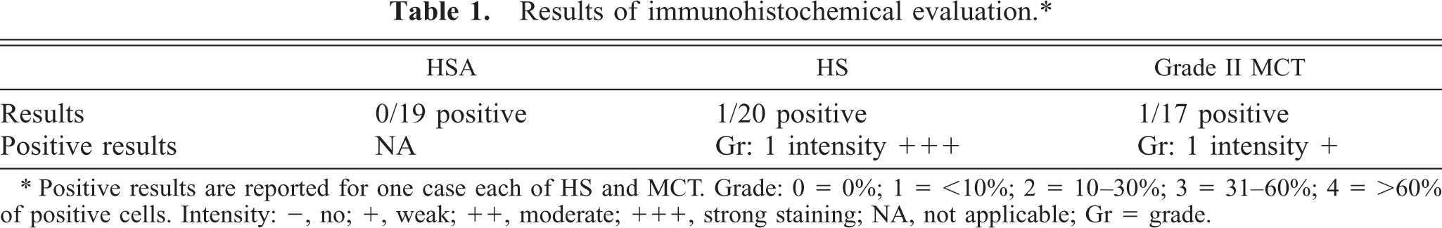
∗ Positive results are reported for one case each of HS and MCT. Grade: 0 = 0%; 1 = <10%; 2 = 10–30%; 3 = 31–60%; 4 = >60% of positive cells. Intensity: −, no; +, weak; ++, moderate; +++, strong staining; NA, not applicable; Gr = grade.
Seventeen of the 20 cases of Grade II MCT were deemed eligible for inclusion. Three cases were excluded after their biopsy samples were cut and stained because of either incorrect specimen labeling or absence of tumor in the submitted samples. Eleven different breeds of dog were represented, with mixed breed dogs (n = 4), Labrador Retrievers (n = 3), and Golden Retrievers (n = 2) represented more than once. Age at the time of diagnosis ranged from 5.8 to 10.7 years (median 8.2 years). Ten females (7 FS, 3 FI) and seven males (4 MC, 3 MI) were present in the study population. The site of the primary tumor was highly variable; however, all were cutaneous MCT. One of the 17 samples stained positive for COX-2 expression. The degree of positivity is reported in Table 1.
Canine HSA is a highly malignant neoplasm of endothelial cells. Using conventional combination chemotherapy protocols, mean survival times exceeding 6 months have not been achieved in dogs with stage II or higher disease. 10, 19 HS is an aggressive tumor of histiocytic cells that is usually widely disseminated at the time of diagnosis. Responses to conventional chemotherapy are poor. MCTs are the most common cutaneous tumor of the dog. In grade I or II, stage-I disease, complete surgical excision is potentially curative. 22 However, treatment options for disseminated disease are limited. In this study, we evaluated samples from spontaneously occurring canine HSA, HS, and MCT for COX-2 expression.
COX-2 has been demonstrated to be overexpressed in human sarcomas 4 and round cell tumors. 15 In addition, over-expression of COX-2 has been shown to be of prognostic significance in human mesothelioma, 6 squamous cell carcinoma, 13 chronic myelogenous leukemia, 9 and breast carcinoma. 2 The tumors examined in this study had not been examined previously for COX-2 expression. Two of the three tumors studied (HS and HSA) have a poor long-term prognosis with conventional therapies 1, 27 ; hence, novel therapeutic options such as the inhibition of COX-2 might prove to be valuable additional alternative therapies. Mast cells were evaluated in this study because previous in vitro experiments have demonstrated rapid and transient upregulation of COX-2 in activated mast cells, 20 thereby providing the rationale to look for COX-2 expression in MCT.
None of the tumor types evaluated in this study had any significant degree of COX-2 expression. These findings may suggest a lack of efficacy of these cancers to COX-2–selective inhibitors. However, studies in human colon cancer cell lines have demonstrated a response to COX inhibitors (acetylsalicylic acid) in COX-2–negative tumors. 29 The responses in these COX-2–negative tumors may be because of either inhibition of COX-2 in the associated neovasculature or through COX-2–independent mechanisms. Furthermore, other pathways involved in tumor growth and progression, such as the upregulation of the antiapoptotic protein Bcl-2, may be altered in the presence of a COX inhibitor. 28
Future studies to evaluate COX-2 expression in tumor neovasculature and the effects of COX-2 inhibition on other pathways involved in tumor growth are warranted. However, unless such studies demonstrate an important role for COX-2 in canine HSA, HS, and MCT, routine treatment of these tumors with COX-2 inhibitors is not supported by these findings.
Footnotes
Acknowledgements
We thank the Commonwealth of Pennsylvania for their contributions. This study was funded by the Departmental Research Resources Committee of the MJR-VHUP.