
Abstract
Two Vietnamese pot-bellied pigs (Sus scrofa) were euthanatized after they developed abdominal distension. Necropsy of both pigs revealed large myometrial neoplasms and cystic endometrial hyperplasia. Multiple discrete smaller myometrial neoplasms were also observed in one pig; however, distant metastases were not observed in either animal. The tumors were diagnosed as leiomyomas on the basis of histologic examination and immunohistochemistry. This is the first detailed report of uterine leiomyomas in swine, and it is suggested that this diagnosis may become more common as more aging pigs are examined.
Keywords
Leiomyomas are the most common uterine tumors in women; approximately 25% of all women will develop such a tumor during their active reproductive life.7 The prevalence of uterine tumors in swine has been estimated from slaughterhouse surveys to be between 5.4 × 10⁻⁵%2 and 0.8%,1 depending on the age of the population studied. Approximately half the tumors were described as benign smooth-muscle tumors in both surveys.1,2
The popularity of Vietnamese pot-bellied pigs (Sus scrofa) as pets peaked between 1991 and 1993, resulting in a rapid increase in pig numbers.17 As a result of this sudden increase, an aging pet pig population is now developing diseases that are rarely seen in swine raised for meat production. This report describes two pot-bellied sows that developed large uterine leiomyomas. This is the first detailed report of such tumors in swine, and it is believed that this diagnosis may become more common as more of these older pot-bellied pigs are examined.
Pig No. 1 initially developed abdominal distension and tenesmus. This pig was 10 years old and was from a herd of six pigs that included three other females and two castrated males. She had farrowed twice before the age of 2 years and was reported to have had regular estrus cycles approximately every 22 days since. This pig subsequently developed anorexia and was euthanatized. Pig No. 2 was 8 years of age when she developed abdominal distension and a serosanguineous vaginal discharge. Pig No. 2 was reported to be nulliparous, cycling regularly, and was the only pig on the property. A large abdominal mass was detected by ultrasound examination. The pig was euthanatized after an ultrasound-guided fine needle aspirate suggested a diagnosis of leiomyosarcoma.
Necropsy of both pigs revealed large quantities of subcutaneous and abdominal fat and hoof-wall overgrowth. Examination of the left uterine horn of pig No. 1 revealed three discrete nodular white masses. These masses had a firm, fibrous texture. The largest mass measured 30 cm in diameter and weighed approximately 4 kg. This tumor contained multiple small areas of hemorrhage and necrosis. The other two masses were smaller, with diameters of 9 and 5 cm. All three masses were restricted to the wall of the uterus, did not extend into the lumen, and were covered by uterine serosa. Examination of the right uterine horn of pig No. 2 revealed a single 35-cm-diameter mass, which grossly resembled the large mass observed in pig No. 1. No distant metastases were observed in either pig. Myriad 5- to 15-mm-diameter endometrial cysts were present diffusely throughout the body and uterine horns of both pigs. Examination of the left kidney from pig No. 1 revealed dilation of the renal pelvis and the proximal 5 cm of the ureter. Tissue samples were fixed in 10% buffered formalin and routinely processed for histologic examination.
Histologic examination of the uterine tumors from both pigs revealed expansile, encapsulated, nodular masses within the myometrium. The masses consisted of densely packed cells arranged in broadly woven fascicles and bundles (Fig. 1). The cells were supported by a scant fibrous stroma with numerous capillaries. The neoplastic cells were moderately monomorphic large spindle-shaped cells with ill-defined cell borders. They generally contained a large, centrally placed cigar-shaped vesicular nucleus with large quantities of eosinophilic fibrillar cytoplasm (Fig. 2). Numerous foci of necrosis, up to 3 cm in diameter, were present within both tumors. These foci contained amorphous eosinophilic material surrounded by a thin zone of degenerate smooth-muscle cells with nuclear pyknosis and faded eosinophilic hyaline cytoplasm. The necrotic smooth-muscle cells were not associated with a significant inflammatory reaction. Approximately two mitotic figures were visible per 10 high-powered fields within tumors from both animals.
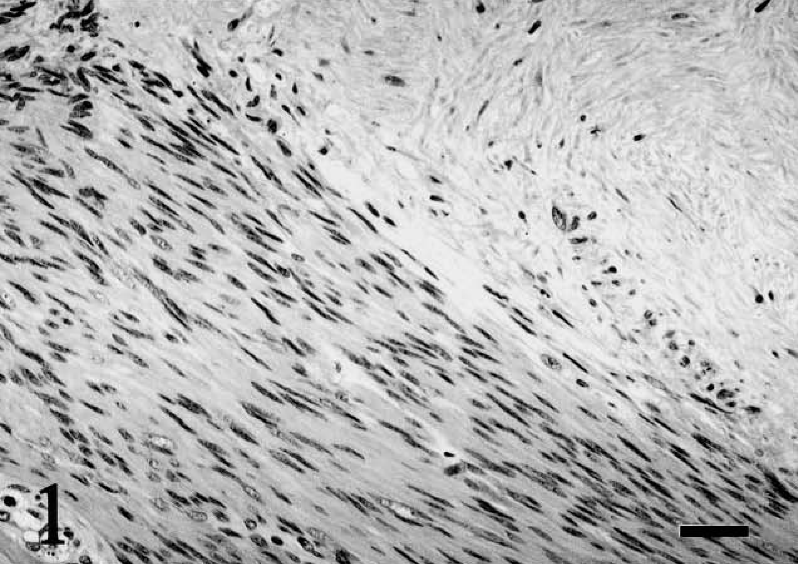
Uterus; pot-bellied pig. Pig No. 1. Uterine leiomyoma composed of bundles of spindle-shaped cells with an area of gradual hyaline fading of cells characterizing infarct-type necrosis. HE. Bar = 30 μm.
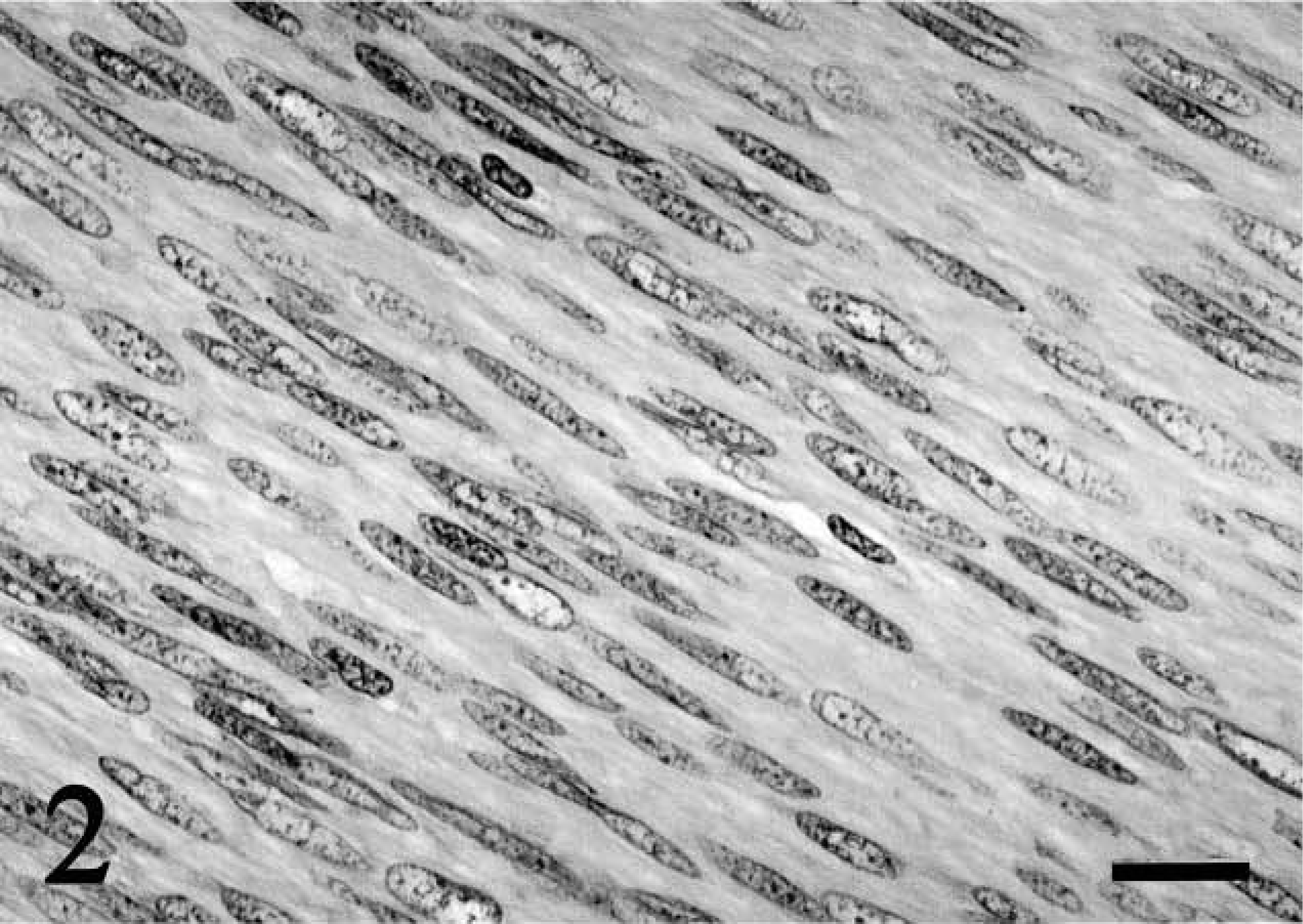
Uterus; pot-bellied pig. Pig No. 1. Uterine leiomyoma composed of elongated cells with characteristic large vesicular cigar-shaped nuclei. HE. Bar = 20 μm.
Neoplastic cells stained red with Masson's trichrome stain. Immunohistochemical staining of tumors from both animals revealed cytoplasmic staining with smooth-muscle actin (Fig. 3) (Dako Corporation, Carpinteria, CA), desmin (Biogenex, San Ramon, CA), and S-100 (Biogenex). Neoplastic cells did not react with cytokeratin (Biogenex) antibodies. The ABC system (avidin–biotin peroxidase complex, Vector Laboratories, Burlingame, CA) was used to visualize all immune reactions.
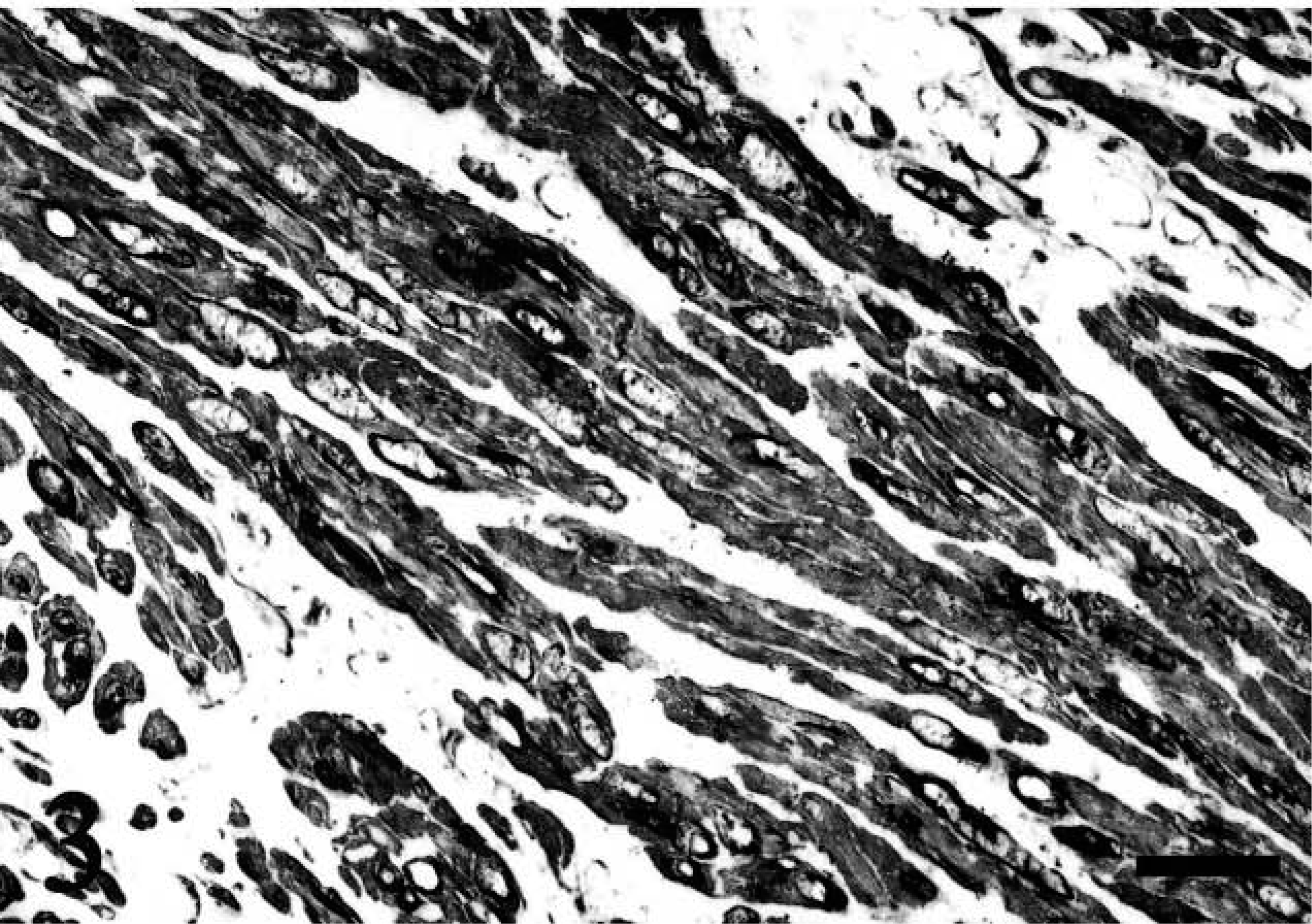
Uterus; pot-bellied pig. Pig No. 1. Neoplastic cells have strong cytoplasmic smooth muscle actin immunoreactivity. Avidin–biotin–peroxidase complex system and Gill's hematoxylin. Bar = 20 μm.
Examination of endometrium from both pigs revealed hyperplasia and cystic dilation of endometrial glands. The glands measured up to 15 mm in diameter, contained pale eosinophilic amorphous material, and were lined by attenuated columnar epithelium.
Ovaries from pig No. 1 contained three 2- to 3-mm postovulatory follicles that were lined by large eosinophilic luteal cells and multiple areas of hemorrhage. Two regressing corpora lutea less than 1 mm in diameter and multiple atresic follicles were also visible. The ovaries from pig No. 2 contained three tertiary follicles that measured up to 3 mm in diameter. A partially regressed corpus luteum that measured approximately 2 mm in diameter was also visible in one of the sections.
Moderate dilation of the left renal pelvis with flattening of the renal papillae and dilation of the proximal ureter was visible on examination of sections from pig No. 1. This hydronephrosis and hydroureter was considered most likely to have been caused by compression of the ureter by the large space-occupying tumor.
The microscopic appearance of these tumors is similar to that reported in human uterine leiomyomas.6 Two patterns of necrosis have been characterized in human leiomyomas to enhance differentiation between benign and malignant tumors. The first, as observed in this case, consists of neoplastic cells that gradually become increasingly hyaline with nuclear karyolysis and is designated “infarct-type” necrosis (Fig. 1).3,12 Necrosis of this type is commonly observed in human uterine leiomyomas. The second pattern is referred to as “coagulative tumor cell” necrosis and occurs in 80% of human uterine leiomyosarcoma but rarely in leiomyomas. This is characterized by a sudden transition from viable neoplastic cells to necrotic cell debris. Islands of viable cells surrounding blood vessels within areas of necrosis are observed within coagulative tumor cell necrosis, but not infarct-type necrosis.3,12
In pigs, cystic endometrial hyperplasia has been observed to result from hyperestrogenism induced by experimental administration of zearalenone.5 Chronic estrogen stimulation in women has been associated with both uterine leiomyomas and endometrial hyperplasia.7 It is possible that repeated estrus cycling in these nonbreeding pigs may have led to hyperestrogenism, which subsequently promoted endometrial hyperplasia and tumor development. Alternatively, endometrial and myometrial estrogen receptor activity is up-regulated in estrus but down-regulated during gestation.14 Repeated estrus cycling could therefore have resulted in the uterine lesions because of increased estrogen receptor activity without increased serum estrogen concentrations.
Uterine leiomyomas are reported regularly only in dogs, among domestic species. Although only representing 0.08% of the total neoplasms,8 uterine leiomyomas constitute 10% of the tumors of the tubular reproductive tract in this species.4 Other regularly reported uterine tumors in the domestic species include uterine lymphosarcoma and endometrial adenocarcinoma, both of which are most commonly observed in cows,13 and endometrial adenocarcinoma in rabbits.11
The prevalence of uterine tumors in swine has been investigated in two slaughterhouse studies. Comparison of these two reports suggests that the rate of uterine tumors increases with age. Only two uterine tumors (an adenomyoma and a leiomyoma) were identified within 3,700,000 pigs approximately 6 months old (5.4 × 10⁻⁵%).2 However, when a population of pigs with a mean age of 4 years was examined, the prevalence increased to 11 tumors in 1445 animals (0.8%).1 Of these 11 tumors, six were classified as leiomyomas, whereas the remainder included three fibromas, a cystadenoma, and a fibroleiomyoma.1
Vietnamese pot-bellied pigs were first imported into the USA in 1986. The popularity of these pigs as pets peaked between 1991 and 1993, and a rapid increase in pig numbers occurred between 1991 and 1995.17 Currently, in the USA, there are 35,000 registered pot-bellied pigs and an estimated 200,000 unregistered pigs (J. Blaney, personal communication). Because of the increase in popularity of these pigs in the early to mid 1990s, many of these pigs are from 5 to 10 years of age.
In contrast to the high prevalence of uterine leiomyomas in women, few animals naturally develop these tumors frequently enough to be used as animal models. A transgenic mouse model was recently described in which a uterine leiomyoma prevalence rate of 100% was reported.16 Prevalence rates in an inbred Ecker rat model9 and an inbred colony of guinea pigs10 are considerably lower at 30% and 8%, respectively. However, uterine and abdominal leiomyomas can be reliably induced by estradiol-17β implants in ovariectomized guinea pigs.15 Cystic endometrial hyperplasia was not reported in any of these rodent models. The prevalence of uterine leiomyomas in pot-bellied pigs is probably also too low for these animals to provide a model of human disease. However, it is interesting to note that uterine tumors in these pigs appear to mimic their human counterparts with regard to predisposing factors, histologic appearance, and behavior.
In conclusion, this report describes two large uterine leiomyomas in pot-bellied pigs. As more aging pigs are examined, increasing rates of these benign tumors can be anticipated, and the presence of such a tumor should be considered if a large abdominal mass is observed. Identification is beneficial because the benign behavior of these tumors allows the possibility of surgical cure.