
Abstract
Thirty-two 4-month-old to 19-year-old female miniature pet pigs were spayed. Uterine lesions were present in all except 8 pigs. The 24 remaining pigs had diffuse cystic endometrial hyperplasia, of which 14 had smooth muscle tumors, including leiomyomas and leiomyosarcomas, in the uterus or broad ligament. Nodular endometrial lesions—including adenocarcinomas, adenomas, and/or adenomyosis—were present in 10 pigs, 3 of which had concurrent smooth muscle tumors. Pyometra was present in 3 pigs. In uterine sections with cystic endometrial hyperplasia, adenomyosis, or adenomas, approximately 70% of epithelial nuclei expressed estrogen receptor and progesterone receptor immunohistochemically; in adenocarcinomas, expression was 20%. Regardless of malignancy, more than 50% of nuclei in smooth muscle tumors expressed estrogen receptor and progesterone receptor. Aging was associated with the development of uterine lesions in miniature pet pigs.
Keywords
Miniature pigs are a subspecies of the domestic pig (Sus scrofa) but, unlike domestic pigs, are often raised as pets. Because the females are usually nulliparous and live longer than domestic pigs, they may be predisposed to development of reproductive tract lesions. The increased numbers of Vietnamese potbellied pigs kept as pets in the United States in the early to mid-1990s correspond with the initial reports of uterine lesions from 2002 to 2004. 11,12 An inbred population may have provided much of the base stock for the entire Vietnamese potbellied pig population in the United States, 14 but the role of inbreeding in the development of uterine lesions is unknown. Uterine smooth muscle tumors in potbellied pigs usually occur as single or multiple benign tumors, mainly in the uterine horns. 11,12 Cystic endometrial hyperplasia (CEH) and endometrial carcinoma have also been reported in Vietnamese potbellied pigs. 4,6,11,12 The purpose of this study was to survey randomly selected female miniature pet pigs of different ages for reproductive tract lesions. Expression of estrogen and progesterone receptors was evaluated immunohistochemically in hyperplastic, benign, and malignant uterine lesions.
Local pig sanctuaries were contacted in search of intact female miniature pet pigs. During 2007–2008, 32 female miniature pet pigs from local rescues were subjected to ovariohysterectomy at the Veterinary Teaching Hospital, University of Tennessee College of Veterinary Medicine; the reproductive tracts were submitted for gross and histologic examination, and all gross lesions in the uteri and broad ligaments were recorded. Ovaries were grossly evaluated for the presence of follicles, corpora lutea (CL), cysts, and paraovarian cysts. Ovarian cysts were defined as fluid-filled cysts ≥ 11 mm in diameter. Follicles were identified as fluid-filled cysts < 11 mm in diameter. 8 CL were defined as round yellow-to-pink solid ovarian nodules.
Representative sections of ovaries, uteri, mucosal masses, and tumors from the uterine wall and broad ligament were evaluated histologically. Tissues were fixed for 24 hours in neutral-buffered 10% formalin, trimmed, embedded in paraffin, sectioned at 5-μm thickness, routinely processed, and stained with hematoxylin and eosin.
Immunohistochemistry was performed on all uterine sections for estrogen receptor (ER) and progesterone receptor (PR) and on smooth muscle tumor sections for smooth muscle actin (SMA). Primary antisera were mouse monoclonal antibodies against ER-α (Clone CC4-5, Santa Cruz Biotechnology, Santa Cruz, CA), PR (Clone PR10A9, Immunotech-Beckman Coulter, Fullerton, CA), and SMA (Clone 1A4, Dako, Carpinteria, CA). Plasma estrogen and progesterone concentration was determined by radioimmunoassay before ovariohysterectomy.
Smooth muscle tumors were classified as leiomyoma or leiomyosarcoma. Criteria for distinguishing between benign and malignant smooth muscle tumors were mitotic index, degree of pleomorphism, presence of necrosis and hemorrhage, and peripheral invasion. A mitotic index of ≥ 10 mitotic figures per 10 high-power fields (400×) was considered diagnostic of malignancy. A mitotic index of 6 to 9 with increased pleomorphism or peripheral invasion was also considered diagnostic for leiomyosarcoma. Leiomyomas had a mitotic index of 0 to 2. Nodular endometrial lesions were classified as adenomyosis, adenoma, or adenocarcinoma. Adenomyosis was defined as the presence of well-differentiated endometrial glands within the tunica muscularis. Adenomas formed discrete nodules with low pleomorphism and few mitotic figures without invasion of surrounding tissue, whereas adenocarcinomas had higher pleomorphism and mitotic index with peripheral invasion and scirrhous response.
For statistical analysis, the fit of continuous data to a normal distribution was tested by the method of Shapiro–Wilk. P values ≤ .05 were considered significant.
Results
Eight pigs had no gross or histologic uterine abnormalities. PR and ER expression in pigs without uterine lesions was mild to intense, diffuse, and observed in stromal, epithelial (surface and glandular), and myometrial cells.
CEH (Fig. 1) was the most prevalent gross and histologic change in the 24 (75%) pigs with uterine lesions. ER and PR expression varied in distribution in sections of CEH, but all sections had strong expression of both receptors in approximately 70% of the cells.
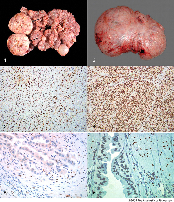
Fourteen pigs (44%) had concurrent CEH and smooth muscle tumors. Smooth muscle tumors were single (7 pigs) or multiple (7 pigs), and varied from < 1 cm in diameter to as large as 35 × 30 × 40 cm (Fig. 2); the largest mass weighed 24.4 kg. These tumors were firm, smooth, and gray to white with cystic degeneration in gray-to-yellow necrotic foci evident on cross-section. Smooth muscle tumors were detected in the serosa and myometrium of the uterine horns and in the broad ligament. Sections of 19 representative smooth muscle tumors from 14 pigs were classified as 12 leiomyomas and 7 leiomyosarcomas. All tumors were strongly positive for SMA; all expressed ER and PR in > 50% of nuclei (Figs. 3, 4).
Multiple endometrial masses were present in 10 pigs (31%), 3 of which had concurrent smooth muscle tumors. Twenty-three endometrial masses from 10 affected pigs were classified as follows: 12 adenomas, 9 adenomyosis, and 2 adenocarcinomas. In adenomyosis and adenomas, approximately 70% of epithelial nuclei expressed ER and PR; in adenocarcinomas, the expression was only 20% (Figs. 5, 6). Six adenomas had multifocal squamous differentiation with no ER or PR expression.
Pyometra and endometritis were present in 3 pigs (9%); lesions varied from segmental (1 pig) to diffuse involvement of both horns (2 pigs). Additional gross lesions included uterine segmental aplasia of 1 uterine horn (1 pig) and cysts within the oviduct wall (5 pigs). Histologically, these oviduct cysts were most consistent with paramesonephric duct cysts.
Gross ovarian changes in the 24 pigs with uterine lesions included multiple ovarian follicles, CL, single or multiple ovarian cysts (5 pigs), and a paraovarian cyst (1 pig). According to the ovarian findings, 2 affected pigs were not cycling normally at ovariohysterectomy. One 19-year-old pig had inactive ovaries; the other (8 years old) had bilateral polycystic ovaries. Pigs with no uterine lesions had gross ovarian follicles and/or CL. Macroscopic ovarian cysts were not present in pigs without uterine lesions.
A single histologic section of one ovary of each pig was evaluated. Histological findings included follicles (primordial, primary, secondary, and tertiary), regressing follicles, CL, corpora albicans, and follicular and luteinized cysts. Ovarian cysts were present in 5 pigs with uterine lesions. One 8-year-old pig with a uterine smooth muscle tumor had bilateral polycystic ovaries without evidence of follicular development or CL. In the other 4 pigs (including 3 with uterine smooth muscle tumors and 1 with multiple endometrial adenomas), ovarian cysts were multiple (2 pigs) or single (2 pigs), and concurrent follicular development and presence of CL were observed.
Plasma progesterone concentrations were evaluated in 31 pigs; values ranged from 0.03 to 43.3 ng/ml. Total plasma estrogen concentration was evaluated in 23 pigs and ranged from 49.0 to 464.4 pg/ml. The available plasma volume was not enough to perform both analyses in all pigs.
Age data were available for 27 of the 32 pigs and so ranged from 4 months to 19 years. Specifically, age data were available for 4 of the 8 pigs without uterine lesions; the ages of the other 4 pigs, though unknown, were indicated by the caretakers as being less than 5 years. Pigs with uterine lesions (9.9 ± 4.1 years) tended to be older than pigs without uterine lesions (2.6 ± 3.0 years; P = .003). Table 1 summarizes age and pathological findings.
Comparison of uterine lesions and ages of 32 female miniature pet pigs.
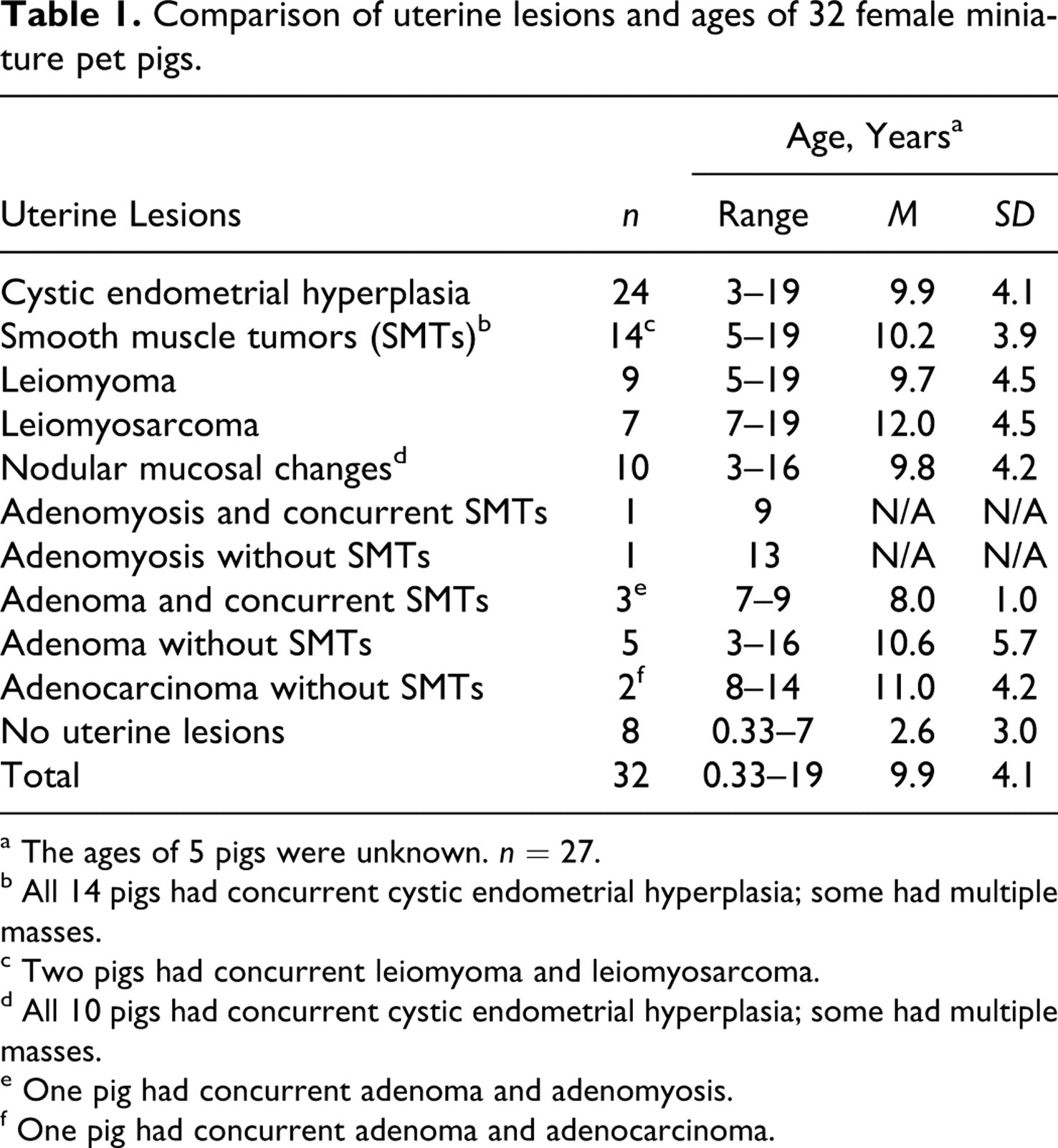
a The ages of 5 pigs were unknown. n = 27.
b All 14 pigs had concurrent cystic endometrial hyperplasia; some had multiple masses.
c Two pigs had concurrent leiomyoma and leiomyosarcoma.
d All 10 pigs had concurrent cystic endometrial hyperplasia; some had multiple masses.
e One pig had concurrent adenoma and adenomyosis.
f One pig had concurrent adenoma and adenocarcinoma.
Discussion
The results of this study indicate a high prevalence of hyperplastic and neoplastic uterine lesions in miniature pet pigs. Of the 32 pigs, 24 (75.0%) had CEH; neoplastic lesions were seen in 20 pigs (62.5%). In contrast, the prevalence of reproductive tract tumors in domestic pigs is considered low. In a large slaughterhouse study of 1,445 adult sow reproductive tracts, 11 (0.76%) had uterine tumors, which were classified as 6 leiomyomas, 3 fibromas, 1 fibroleiomyoma, and 1 cystadenoma. 1 One cervical tumor was also identified. Smooth muscle tumors and endometrial adenocarcinomas have all been described in female Vietnamese potbellied pigs, 4,6,11,12 but most reports were of isolated cases. 4,6,12 Only 1 study involving large numbers of pigs has been published. 11
Aging is a known factor associated with the development of neoplasia in humans and domestic animals. 9 Smooth muscle tumors were identified in animals 5 years old or older, similar to results of a previous study in Vietnamese pot bellied pigs. 11 In the large slaughterhouse study, the age of domestic pigs was not available, but the mean parity of studied sows was 6.8 and the mean parity of sows that developed tumors was 11.6, suggesting that domestic sows that developed tumors were older.
Twenty-four pigs in this study had CEH. In domestic pigs, estrogen stimulation may play a role in the development of cystic uterine changes. Hyperestrogenic syndrome associated with ingestion of subterranean clover was suspected, although not confirmed, as the cause of a high incidence of cystic ovaries and CEH in pigs in Tasmania. 16 Cystic dilation of endometrial glands and metaplasia of endometrium were observed histologically with experimental administration of zearalenone in swine. 2 Squamous metaplasia was observed in a uterine adenocarcinoma in a potbellied pig 6 and in a portion of the adenomas in the present study.
Endogenous hormones can play a role in the development of tumors in the organs of the female and male reproductive tracts, 9 and strong evidence suggests that action of hormones can increase the risk of mammary carcinomas in dogs as well. 9 Little is known about the pathogenesis of uterine neoplasms in domestic pigs, but ER and PR were recently identified in a cervical leiomyoma in a 6-month-old domestic pig. 15 In the present study, nuclear expression of ER and PR was intense and diffuse in cells of smooth muscle tumors and in benign epithelial hyperplastic and neoplastic lesions in female miniature pet pigs. The intensity and distribution of ER and PR expression were fairly similar between these neoplasms and uterine sections of pigs without uterine lesions. Progesterone and estrogen may contribute to the development of smooth muscle tumors in other species, including humans and dogs. 3,10,17 Uterine smooth muscle tumors in potbellied pigs may be a valuable animal model for studying human fibroids. 11 ER and PR expression has been observed in epithelial and spindle cell neoplasms of the reproductive tract of women, 3,5,17 and ER and PR receptors are both expressed in women with uterine leiomyomas. 3,17 Although data are conflicting, many studies have revealed greater concentrations of ER and PR in leiomyomas when compared with those of normal myometrium in women. 3,13 Other studies in humans have demonstrated that leiomyosarcomas have lower ER and PR expression than that of leiomyomas. 17 Loss of ER and PR expression is associated with more aggressive biological characteristics in endometrial proliferations in women. 5
Additional studies are necessary to elucidate the role of estrogen and progesterone in the development of uterine lesions in miniature pet pigs. None of the pigs in this study had a history of pregnancy, but breeding history before rescue was largely unknown. Signs of estrus were not seen at any time by the animal caretakers, but silent estrus may have occurred. In the present study, most pigs had follicles and corpora lutea, consistent with ovarian cycling. Estrogenic stimulation is increased by repeated estrous cycling in unbred animals. Studies have shown an inverse relationship between parity and the risk of fibroids in women; a progressive decline in risk relative to increase in the number of births has also been reported. 3
A feature of hormonal carcinogenesis in humans—and one that may also be important in animals—is its association with an inherited genetic predisposition to tumor formation. 9 Genetic changes and inbreeding can also be considered as predisposing factors for the uterine changes in these miniature pet pigs. CEH was found in a high proportion of an inbred strain of miniature swine with increased homozygosity at loci associated with the major histocompatibility complex of swine lymphocyte antigen complex. 7 The genetic pool for miniature pet pigs in the United States may be similarly restricted.
In conclusion, female miniature pet pigs have a tendency to develop hyperplastic and neoplastic uterine lesions, with a strong association between increased age and development of neoplastic lesions. ER and PR were consistently expressed in hyperplastic and neoplastic uterine lesions. Other factors in the pathogenesis and development of uterine lesions in miniature pet pigs need further study. Earlier ovariohysterectomy should decrease the prevalence of uterine lesions in pet female miniature pigs.
Footnotes
Acknowledgements
We would like to thank Ms Ladonna Mrkonjich for technical assistance with immunohistochemical section preparation, Ms Deborah Haines for photographic assistance, Ms Misty Bailey for assistance with text editing and formatting, Ms DeAnne Gibbs for assistance with hormone determinations, and Ms Peggy Couey from the Shepherd’s Green Sanctuary, Cookeville, Tennessee, for the submission of numerous potbellied pigs that participated in this study.
The authors declared that they had no conflicts of interest with respect to their authorship or the publication of this article.
The authors declared that they received no financial support for their research and/or authorship of this article.