
Abstract
A 4-year-old native-breed cow had a mass with wide areas of ulceration and hemorrhage at the base of the tail at the same level as the vulva. The tumor was 19 X 13 X 11 cm, appeared red-brown, and was firm to hard, with gritty areas apparent on cut surface. Histologically, the tumor mass was composed of multilayered epithelial cells forming glandular structures with occasional apical blebs and rare solidly packed cells in nests. The stroma included fibrous connective tissue, scattered or periglandular sheets of spindle-shaped cells resembling myoepithelium, several cartilaginous formations, and numerous irregular islands of mineralized osteoid, well-formed bone trabeculae lined by osteoblasts, and many osteoclast-like multinucleated giant cells among or near the neoplastic epithelium. Immunohistochemically, the neoplastic epithelium was positive for pan-cytokeratin (AE1/AE2) and cytokeratin 19 but was negative for cytokeratin 18. Spindle-shaped cells were stained with alpha smooth muscle actin (αSMA) and to a lesser extent vimentin antibodies. The cells of osteogenic lineage and spindle cells closely associated with the osteoid showed strong immunostaining for vimentin but not for αSMA. Immunostaining for neuron-specific enolase and S100 protein was not observed in any component of the tumor mass. These findings suggested that the origin of bone formation was undifferentiated mesenchymal cells with osteogenic potential.
Apocrine sweat gland tumors occur most frequently in dogs, occasionally in cats, and rarely in other domestic animals.9,10 Two equine cases have been reported, in a mare3 and an aged pony.1 The tumor is equally rare in the cattle. To our knowledge, only two cases of bovine mixed apocrine sweat gland adenocarcinoma have been documented,6,12 and no immunohistochemical data are available. Here, we describe various morphologic and immunophenotypical features of this rare neoplasm, mixed apocrine sweat gland adenocarcinoma, in the tail base of a cow in Van, Turkey.
A 4-year-old native-breed cow was presented with a 4-month history of a slowly progressive growth at the base of the tail. The owner first noticed a localized subcutaneous swelling on the ventral surface of the base of the tail at the same level as the vulva and reported that the swelling, with an ulcerated and hemorrhagic area ventrally, continued to enlarge. The cow was referred to the Faculty of Veterinary Medicine, Yuzuncu Yil University, because the mass bled continuously for 7 days. The mass was 19 cm long, 13 cm in diameter dorsoventrally, and 11 cm in diameter transversally. It was located below the second and fifth coccygeal vertebrae (C2–C5) and was firmly adherent to the deep tissues. The tail with its attached tumoral mass was amputated at the C1–C2 articulation. On cross section, the tumor was red to brown, was subdivided into lobules by thin white septa, and contained extensive hemorrhagic and necrotic areas. The mass contained dense white and gritty areas (Fig. 1). Specimens from a number of sites throughout the tumor mass were fixed in 10% neutral buffered formalin and embedded in paraffin, and sections were cut at 5 μm and stained with hematoxylin and eosin (HE), periodic acid–Schiff (PAS) with and without diastase, Alcian blue–PAS, Masson's trichorome, and Perls's iron. Some parts of the tumoral mass were processed after being decalcified with 5% nitric acid solution. Immunohistochemistry was used to further define the mixed composition of the tumor and the relationship between the osteoid tissue and the proliferating myoepithelium. The selected sections were immunostained for reactivity to commercially available antibodies using the avidin–biotin–peroxidase complex (ABC) method (Table 1).
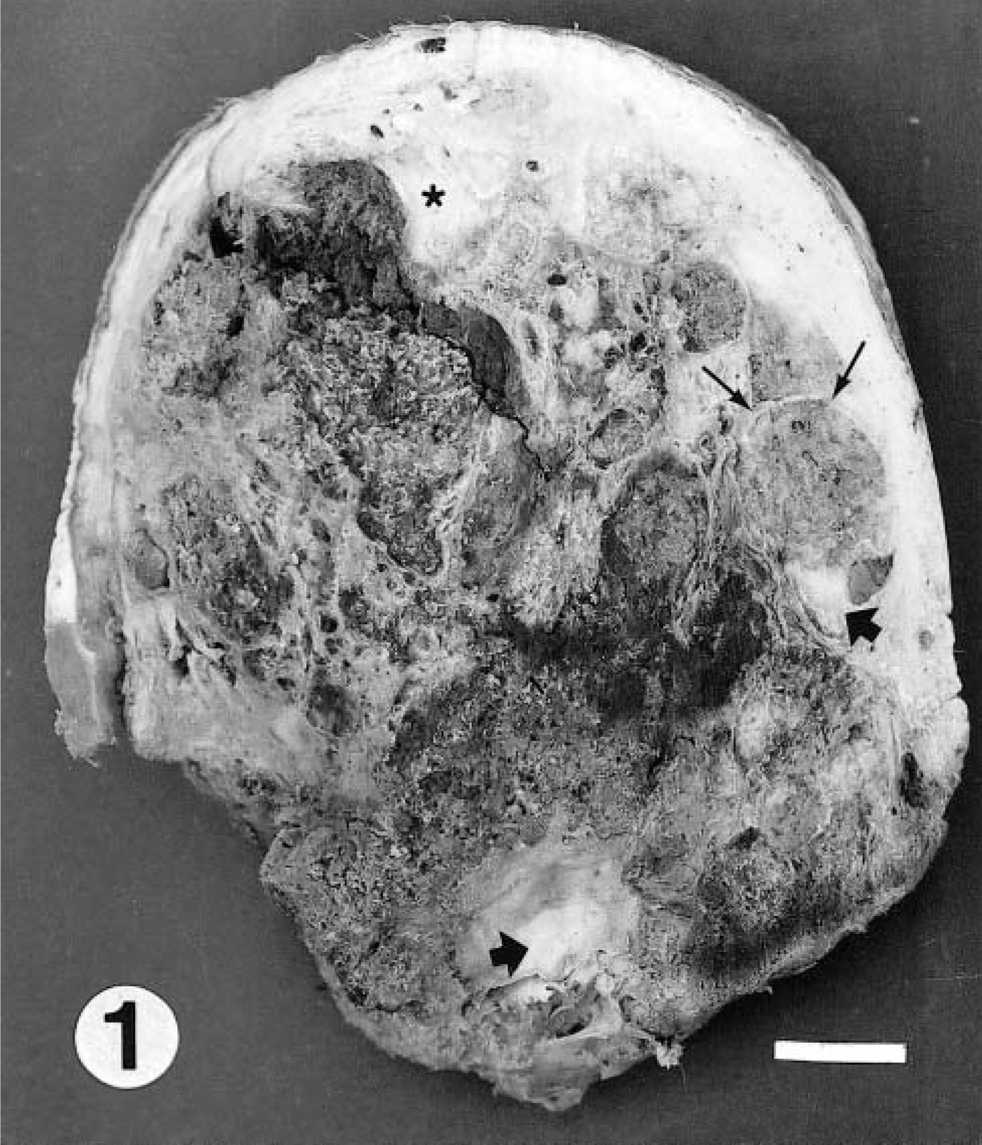
Mixed apocrine sweat gland tumor; cow. Cut surface of the tumor is subdivided into lobules by thin white septa (thin arrows), including newly formed bone tissues (thick arrows) and ventrally ulcerated and hemorrhagic areas. Star indicates the coccygeal vertebra. Bar = 1.5 cm.
Some properties of antibodies and antigen retrieval techniques employed to evaluate a mixed apocrine sweat gland tumor on the tail of a cow.
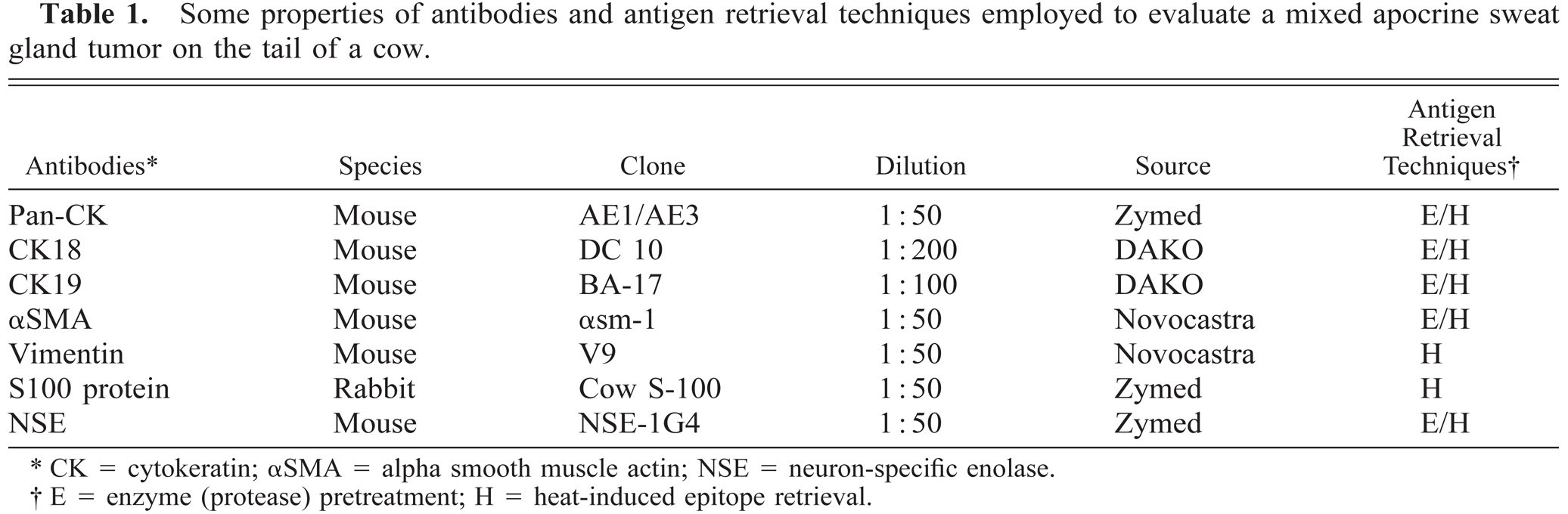
CK = cytokeratin; αSMA = alpha smooth muscle actin; NSE = neuron-specific enolase.
E = enzyme (protease) pretreatment; H = heat-induced epitope retrieval.
Microscopic examination of the tumor mass revealed frequently multilayered neoplastic epithelium, which contained pseudoadenomatous and papillary projections, arranged as glandular structures with irregular lumina of various sizes (Fig. 2) and rare ductlike structures or cells solidly packed in nests. These cells were cuboidal to columnar in shape and had eosinophilic cytoplasm. Vesicular nuclei were round to plump or oval and often had distinct nucleoli. Mitotic figures were infrequent. The luminal surface of well-formed glandular structures was occasionally blebbed (decapitation secretion) (Fig. 3). The luminal basophilic secretory material stained positively with PAS and was resistant to diastase predigestion. The neoplastic epithelium also had PAS-positive, diastase-resistant cytoplasmic granules. Intracellular iron-positive granules were detected by Perls's iron stain. The stromal elements were composed of coarse fibrous connective tissue, solidly proliferated or periglandular sheets of spindle-shaped cells admixed with occasionally irregular islands of cartilaginous formation, and abundant osteoid, which was arranged as anastomosing trabeculae lined by osteoblasts and many osteoclast-like multinucleated giant cells among or near the neoplastic epithelium (Fig. 2). Some trabeculae were partially mineralized. Spindle-shaped cells had pale cytoplasm and oval nuclei with distinct nucleoli and occasionally showed mitotic figures. In some areas, many osteoclast-like giant cells intermingled with spindle cells among or near the neoplastic epithelial cells, which formed ductlike structures or cells solidly packed in nests and were not associated with the osteoid or trabeculae (Fig. 4). The slitlike spaces between epithelium and stroma were regarded as artifacts. The tumor mass had extensive necrotic and hemorrhagic areas admixed with numerous hemosiderin-laden macrophages. Mixed leucocyte infiltrations were present within the stroma, often in association with the ulcerated areas. Immunohistochemically, most of the luminal epithelial cells stained positively for pan-cytokeratin (AE1/AE3) (Fig. 5) and cytokeratin (CK) 19 and negatively for CK18 and the other antibodies used. Spindle-shaped cells resembling myoepithelium, which formed a thick layer underlying epithelial cells and were scattered or solidly packed into the stroma and smooth muscle of the blood vessels, had a strong immunopositive reaction against alpha smooth muscle actin (αSMA) antibody (Fig. 6a), whereas these cells were negative with anti CK antibodies. In some regions of the tumor, positive staining for αSMA showed a discontinuous layer or disappearance of the myoepithelial cells surrounding epithelial structures (Fig. 6b). The cells of osteogenic lineage and spindle-shaped cells in close association with the osteoid gave strong immunostaining for vimentin (Fig. 7) but were negative for αSMA. In some areas, some spindle-shaped cells also showed a positive reaction to vimentin antibody. Immunoreactivity to neuron-specific enolase (NSE) and S100 protein was not detected in any component of the tumor mass.
Mixed apocrine sweat gland tumor; cow. The stroma near the neoplastic epithelium displays osteoid trabeculae (o) with multinucleated giant cells (thick arrows). The slitlike spaces are artifacts (thin arrow). HE. Bar = 80 μm.
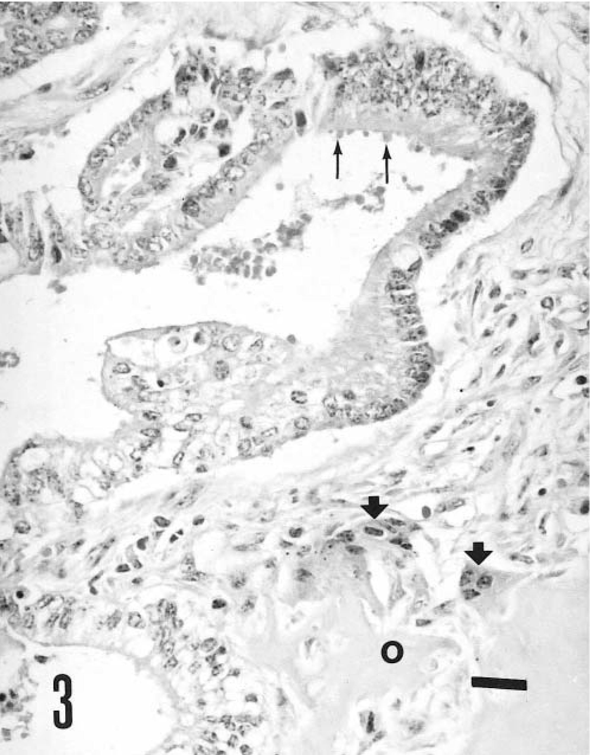
Mixed apocrine sweat gland tumor; cow. Note apical blebs (thin arrows) on the luminal surface of neoplastic glandular epithelium near the osteoid formation (o), with osteoclast-like giant cells (thick arrows). HE. Bar = 40 μm.
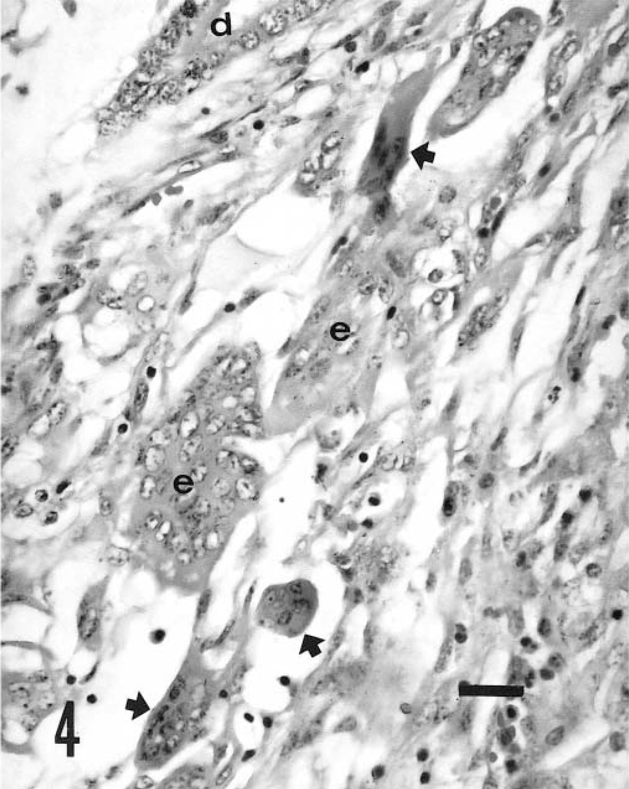
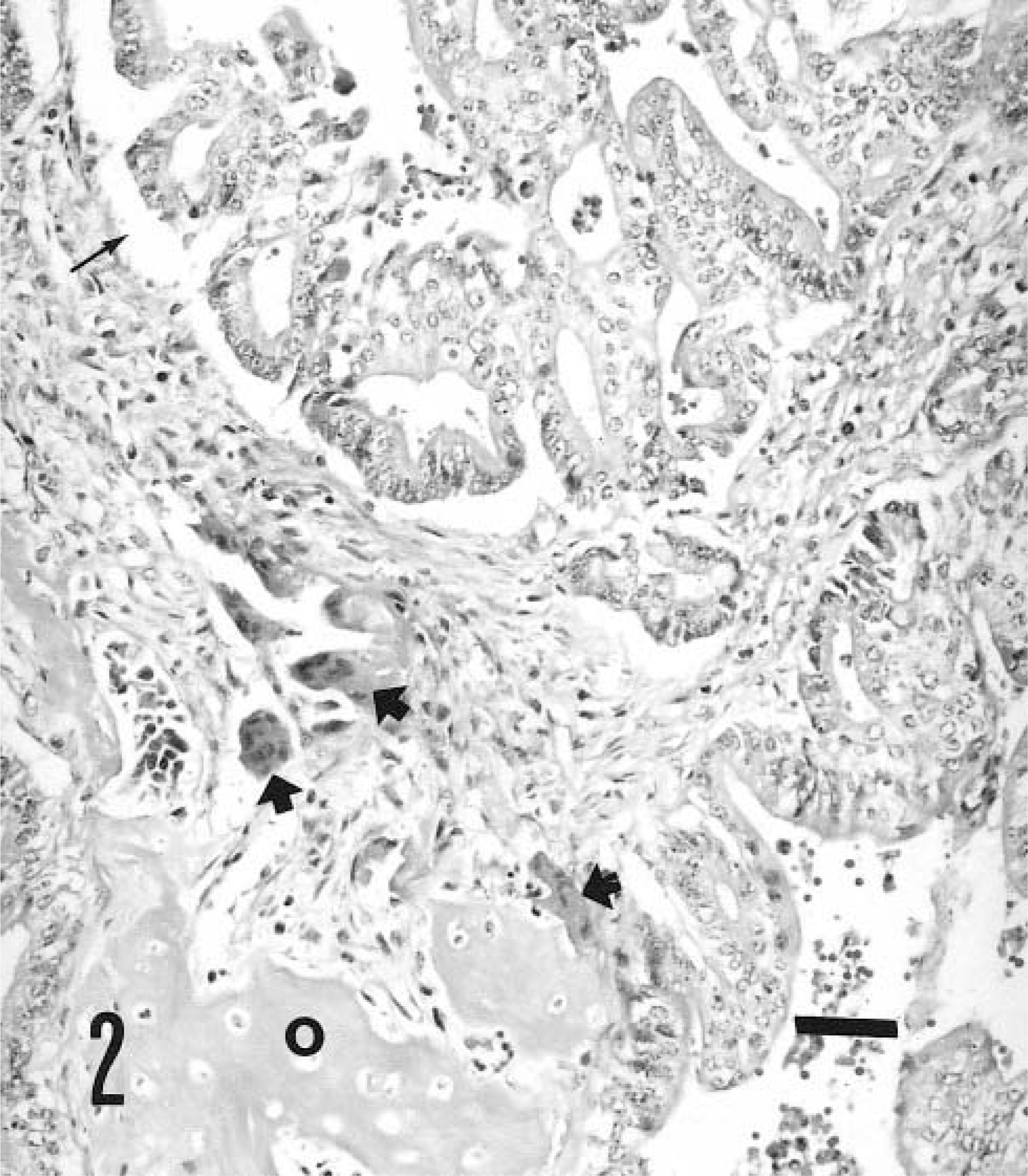
Mixed apocrine sweat gland tumor; cow. Osteoclast-like multinucleated giant cells (arrows) are not associated with the bone tissue but are intermingled with proliferating spindle-shaped cells (myoepithelium) among and/or near the ductlike structures (d) or epithelial nests (e). HE. Bar = 40 μm.
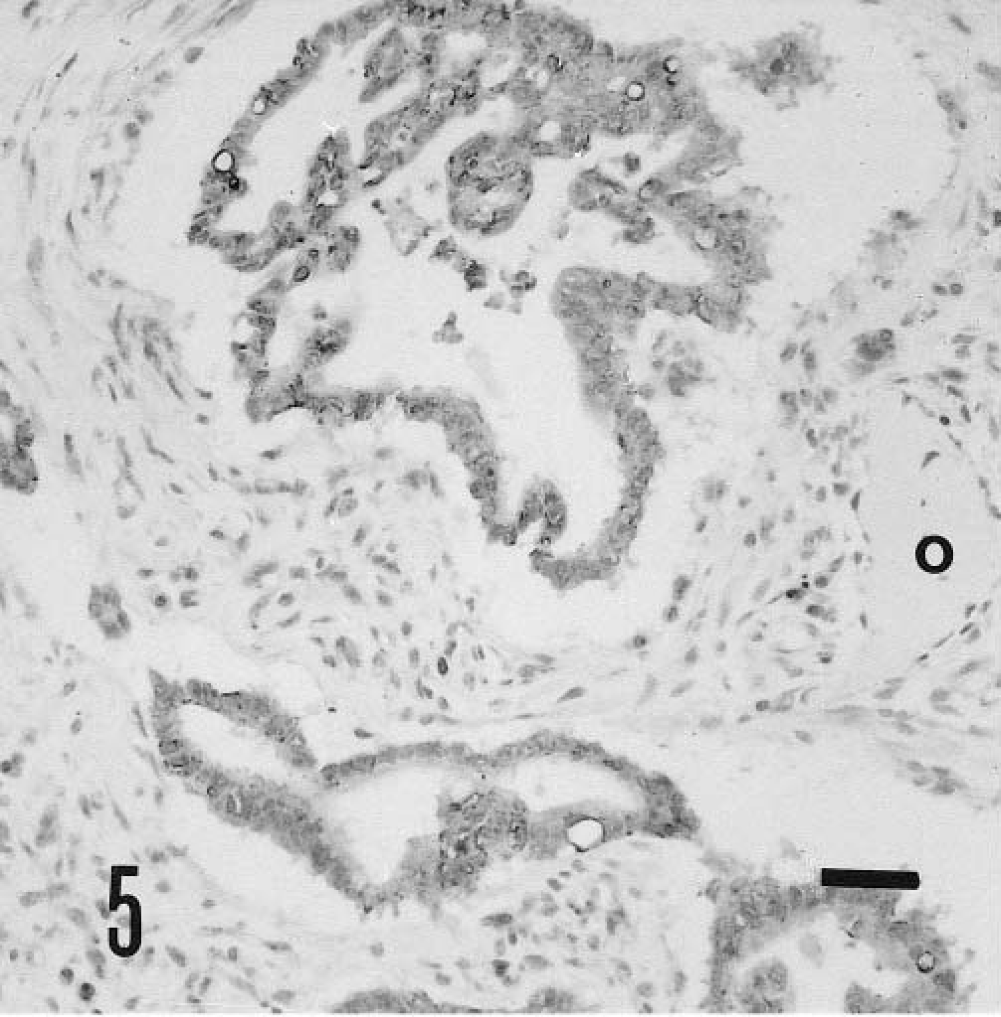
Mixed apocrine sweat gland tumor; cow. Immunostaining for AE1/AE3 in the luminal cells. Note lack of immunostaining in the osteoid formation (o) and the stroma. ABC method, Mayer's hematoxylin counterstain. Bar = 80 μm.
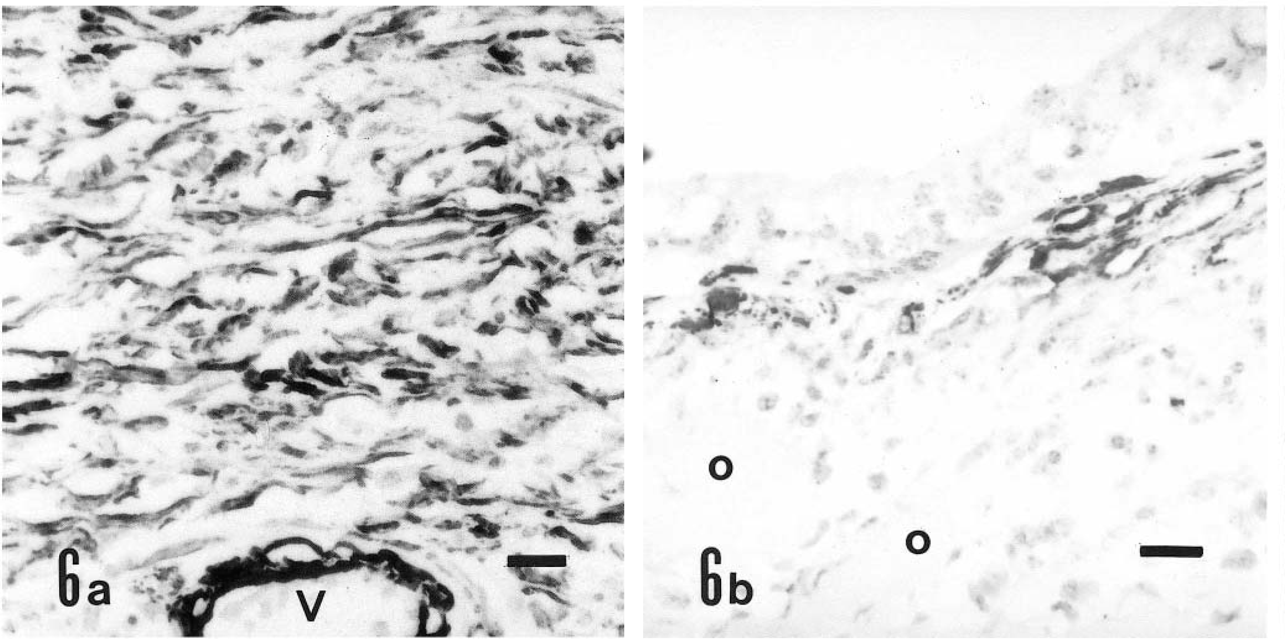
Mixed apocrine sweat gland tumor; cow. Positive immunoreactivity for αSMA in proliferating spindle-shaped cells and smooth muscle of a blood vessel (V) in the stroma (
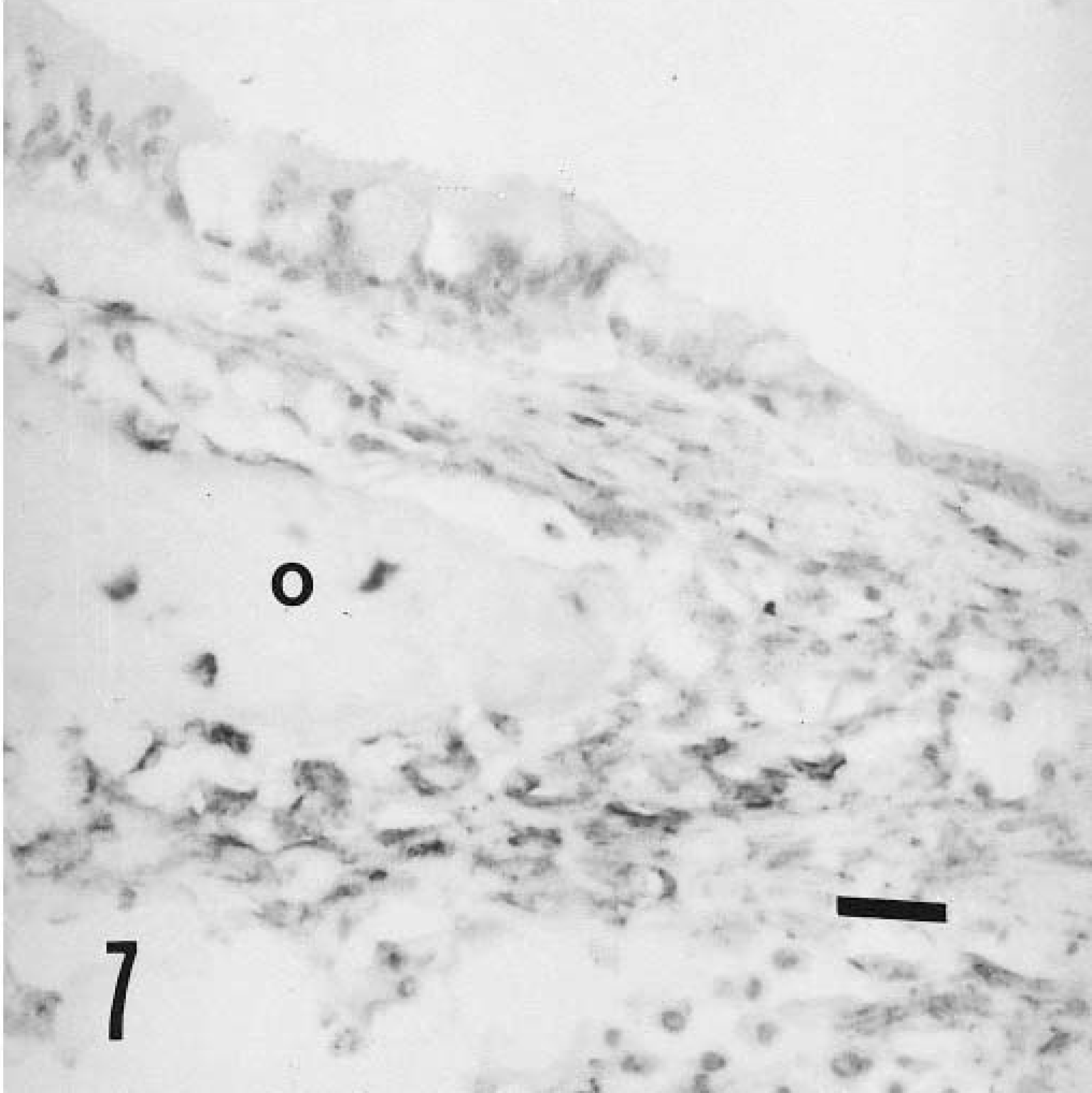
Mixed apocrine sweat gland tumor; cow. Positive immunostaining in cells of the osteoid lineage for vimentin and to lesser extent in basally located spindle cells (o = osteoid formation). ABC method, Mayer's hematoxylin counterstain. Bar = 40 μm.
This tumor was considered to be of apocrine origin based on the intracellular PAS-positive, diastase-resistant granules within the apical cytoplasm of many of the epithelial cells, the intracellular iron-positive granules, and the occasional apical secretory blebs (decapitation secretion) typical of normal apocrine glands.1,9,12 Moreover, the presence of osteoid formations among or near neoplastic epithelial cells warranted a diagnosis of malignant mixed tumor of an apocrine sweat gland.
Two bovine cases of this neoplasm have been described, one at the base of the tail of a mature Holstein bull in Canada6 and the other on the ventral surface of the tail, about 20 cm from its base, in a pregnant Friesian cow in the UK.12 Because this tumor occurred in only two cows and limited clinical data were included in these reports, no conclusions concerning age, breed, sex, or site predilections can be made. However, the skin on the ventral surface of the tail base seems predisposed to sweat gland tumors, as seen in all three cows.
CKs 8, 18, and 19 are reliable general basic markers for gland differentiation in cutaneous epithelial tumors. In canine sweat gland tumors, positive immunostaining in the secretory and luminal cells for AE1/AE3, CAM5.2 (CK8 and CK18), and CK19 and positive staining of the myoepithelium for LP34 (CK6 and CK18) by means of the complementary reactions have been demonstrated.16 In the present case, neoplastic epithelium was positive for AE1/AE3 and CK19 but negative for CK18. αSMA has been used for demonstrating evidence of myoepithelial differentiation or myoepithelial cells within human sweat gland tumors.4,15 αSMA-positive cells correspond to myoepithelial cells in bovine udder8 and S100 protein–positive cells in dogs.5 Anti-CK14 antibody is also a useful marker of the myoepithelium in the bovine udder; however, it is less spesific than anti-αSMA antibody.8 In the present case, CK and S100 protein antibodies did not stain the myoepithelium, whereas myoepithelium was strongly positive for αSMA and to some extent vimentin. Mixed sweat gland tumors may be analogous to the mixed mammary tumor of the dog,9 and heterotopic (metaplastic) bone formation has been reported in mixed mammary tumors7 and salivary gland tumors;11 however, there has been controversy about the origin of cartilage and bone in these tumors. In connection with the origin of the mesenchymal component from mixed mammary and salivary gland tumors, the roles of myoepithelial cells or undifferentiated mesenchymal cells have been considered.7,10,11 Moreover, bone has arisen by endochondral ossification of cartilage formed by myoepithelial cells in mixed mammary tumors.10 In the present case, the absence of αSMA immunostaining in cells located within or near the osteoid supports the hypothesis of undifferentiated stem cell origin in contrast to myoepithelial origin of the bone formation. However, with regard to myoepithelial origin of osteoid formation, immunostaining properties in the transformed cells may indicate an alteration in the content of some cytoplasmic filaments during the metaplasia of myoepithelium. The identification of origin of osteoid formation by specific antigen expression may be misleading, because the phenotypic expression in neoplasia can be variable and does not necessarily reflect its origin. The occurrence of ectopic ossification demonstrates the existence of osteogenic precursors at extraskeletal sites, and metaplastic changes arise from a resident population of undifferentiated mesenchymal cells with osteogenic potential, termed inducible osteogenic precursor cells.2 An osteoinductive stimulus produced by the neoplastic cells that acts on undifferentiated pluripotential stem cells can cause their metaplastic transformation into osteoblasts or chondroblasts, resulting in a heterogenous histologic appearance. Some cytokines, such as those of the transforming growth factor β superfamily and related bone morphogenetic proteins, are particularly important in osteoinduction, stimulating differentiation of primitive mesenchymal cells into bone-forming tissues.9,14 In the majority of human sweat gland neoplasms, vimentin-positive cells represent remnants of myoepithelium, as indicated by their expression of αSMA. Moreover, in rare cases, vimentin expression in anaplastic tumor cells may reflect reduced cell-to-cell contact.4 Scattered myoepithelial cells throughout the stroma and disrupted or discontinuous myoepithelial layers may be interpreted as a loss of the barrier to invasion in sweat gland carcinomas.15 The possibility of widespread metastasis of sweat gland adenocarcinomas should also be considered.10 In the present study, although local invasion was observed, there was no evidence of distant metastasis, in accordance with previous reported cases,6,12 and the cow remained in good health for at least 5 months after tumor excision. Sometimes, myoepithelial cells may not represent a neoplastic component of sweat gland carcinomas; rather, they can be considered residual cells undergoing displacement of destruction by neoplastic cells.15 In the tumor in this cow, the presence of myoepithelial cells throughout the stroma and even disrupted or discontinuous myoepithelial layers indicates that the myoepithelium was an important stromal component but, in light of the immunohistochemical findings, was not responsible for osteogenesis. The limited number of NSE-positive tumor cells in canine apocrine adenocarcinoma is compatible with immunostaining of parathyroid hormone-related protein (PTHrP) and is important in the pathogenesis of malignancy-associated hypercalcemia.13 In the present case, no immunoreactivity to NSE was observed in this tumor, and immunostaining for PTHrP was not employed.
Additional bovine cases and further investigations, including immunohistochemical studies with more specific antibodies raised against bovine tissues, are required to clarify the factors and mechanisms involved in the biological behavior of this tumor.
Footnotes
Acknowledgements
We thank O. Faruk Ercin from Vet-Pa Veterinary Clinic for contributing this case.