
Abstract
Liposarcomas are rare neoplasms in domestic animals, but have been reported to occur in many species. in humans, liposarcoma is one of the most common malignant mesenchymal tumors. classification of liposarcomas in humans has been well established and categorization by type can be of prognostic value; no such unique classification scheme has been established for liposarcomas in animals. Liposarcoma of the head and neck in humans are uncommon, and are rarely reported in the nasal cavity, sinuses, and nasopharynx. To our knowledge, a liposarcoma has never been reported in the nasal cavity of a domestic animal. In this report we describe a liposarcoma that developed in the nasal cavity of a cow, with local invasion into the oral cavity.
Liposarcomas are rare neoplasms in domestic animals, but have been reported to occur in many species. 6 In humans, liposarcoma is one of the most common malignant mesenchymal tumors and accounts for 20% of all sarcomas. 2 Common sites of occurrence in humans include the thighs, buttocks, and retroperitoneum. 4 Liposarcomas of the head and neck in humans represent approximately 1% of all head and neck tumors, and are rarely reported in the nasal cavity, sinuses, and nasopharynx. 5, 9 Among domestic animals, liposarcoma has been most frequently described in dogs and typically arises within subcutaneous tissues in this species. 1 Sporadic cases have been reported in other domestic animals, including 2 cases affecting cattle: a 14-month-old Holando-Argentino cow with numerous intra-abdominal masses attached to serosal and peritoneal surfaces, 13 and an adult cow (age and breed unknown) with multiple masses within lymphatic vessels of the head, neck, and thorax, and on abdominal parietal surfaces. 12 To our knowledge, a liposarcoma has never been reported in the nasal cavity of a domestic animal. In this report we describe a liposarcoma that developed in the nasal cavity of a cow, with local invasion into the oral cavity.
A 7-year-old Angus cow presented to the Texas A & M University Veterinary Teaching Hospital for evaluation of a facial swelling over the left maxilla of 2–3 weeks duration. Several days before admission, the cow was reported to experience difficulty breathing. The owner had treated the cow with long-acting oxytetracycline systemically (13 mg/kg; 6 mg/lb SC q 48 hours) and locally at the lesion with no improvement in clinical symptoms or reduction in size of the mass. The cow had retained a good appetite and did not exhibit any apparent difficulty in mastication.
Upon presentation, the cow was febrile with a temperature of 103.3°F (39.6°C) and was dyspneic with intermittent open-mouthed breathing. Minimal airflow was noted from the right nare and airflow was absent from the left nare. A small amount of blood was present in the external opening of the left nare. A large, firm mass was present on the face over the left maxilla. Oral palpation revealed a firm mass surrounding the upper left molars and premolars. During palpation, 1 molar and premolar were easily removed by hand from the left upper dental arcade. A foul odor was appreciable within the oral cavity. The submandibular lymph nodes appeared to be moderately enlarged. Rectal examination revealed a nongravid uterus with an adhesion of the left horn that prevented complete retraction of the uterus. Given the perceived extent of the mass, pregnancy status, value of the animal, expense of proposed therapy, and poor prognosis for recovery, the owner elected humane euthanasia and the cow was submitted for necropsy.
Postmortem examination revealed a soft, multilobulated, yellow to tan to white mass filling the left nasal cavity (Fig. 1). The mass encompassed the nasal turbinates and displaced the nasal septum to the right. The left maxilla overlying the mass was thickened and elevated. Multifocal, variably sized pockets containing yellow purulent material were within the mass. The hard palate on the left side was thin and mildly friable. Within the oral cavity immediately ventral to the nasal mass was a fleshy, dark red to black, fissured, focally extensive mass surrounding the remaining molars of the left upper dental arcade. The molars were loosely held within the gingiva. Dissection into the tissue around the molars revealed yellow, purulent material and feed material surrounding the tooth roots.
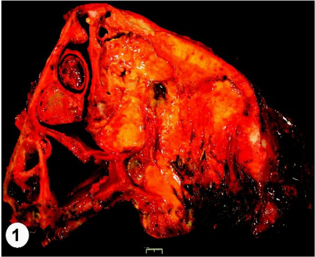
Nasal cavity, liposarcoma; cow. Transverse section of the nasal cavity showing expansile mass with septal deviation.
Radiographs of the skull taken during necropsy revealed an expansile lesion of the maxillary sinus with osteolytic and osteoblastic activity in the maxilla. An air–fluid line in the maxillary sinus and a soft tissue swelling overlying the maxilla were present. The alveolar pockets surrounding the remaining teeth in the upper left dental arcade were expanded (Fig. 2).
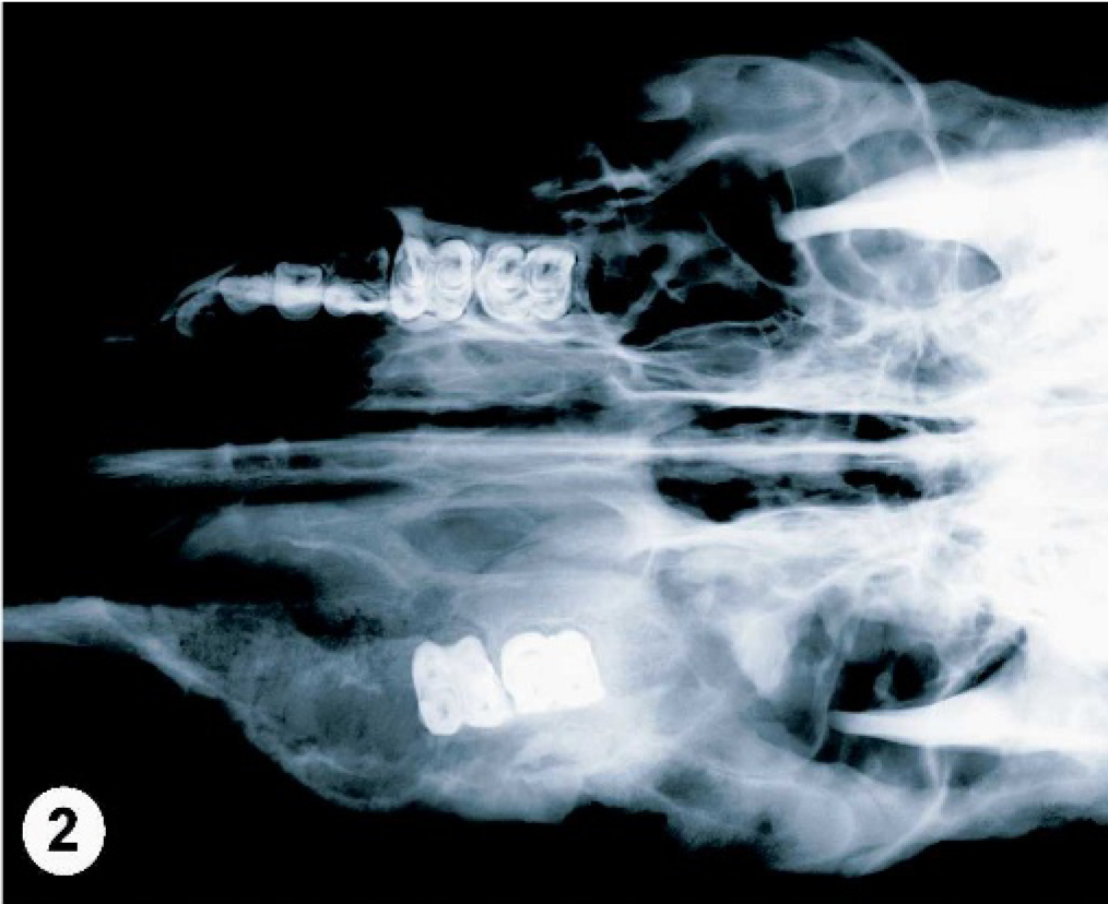
Head; cow. Dorsoventral radiograph of the head showing unilateral expansile soft tissue mass within the region of the left maxilla.
Tissue from the mass was fixed in 10% formalin and processed for routine histopathology. Histologically, the tumor consisted of densely cellular lobules separated by a variably dense fibrovascular stroma (Fig. 3). In the nasal cavity, lobules of neoplastic cells were expanding the submucosa and encompassing remnants of the nasal turbinates. Neoplastic cells were spindle-shaped to stellate to polygonal, with marked cellular atypia and frequent mitoses. Cells exhibited large, round to oval to irregular nuclei with coarsely clumped to vesicular chromatin and 1–2 prominent nucleoli. Many cells exhibited scant to moderate amounts of pale eosinophilic cytoplasm containing numerous clear, round, variably sized, well-defined vacuoles (Fig. 4). Lesser numbers of neoplastic cells exhibited a single, large, clear, intracytoplasmic vacuole that displaced the nucleus to the periphery. Multifocal abscesses were present within the nasal mass and contained numerous mixed bacterial colonies and mineralized material. Neoplastic cells infiltrated into the hard palate and multifocal regions of fibrosis and woven bone formation were present in these areas. The oral mucosa was markedly expanded by granulation tissue that surrounded infiltrative lobules of neoplastic cells. The oral mucosa was ulcerated and hemorrhagic and the underlying granulation tissue contained a marked infiltrate of degenerate neutrophils and mixed bacterial colonies. The submandibular and retropharyngeal lymph nodes were moderately hyperplastic and edematous, but were not infiltrated by neoplastic cells.
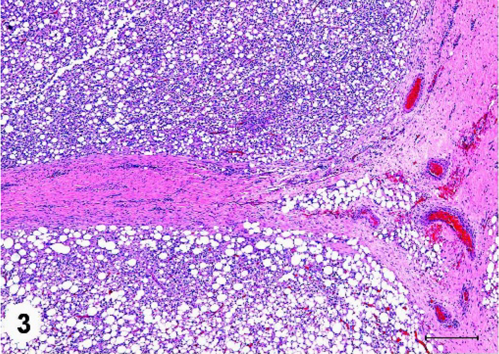
Nasal cavity, liposarcoma; cow. Neoplastic cells are arranged in densely cellular lobules within a fibrovascular stroma. HE. Bar = 300 μm.
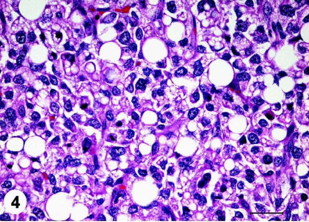
Nasal mass, liposarcoma; cow. Neoplastic cells exhibit marked cellular atypia and numerous clear, round, variably sized, well-defined, intracytoplasmic vacuoles. HE. Bar = 20 μm.←
Immunohistochemical staining on sections of the neoplasm was performed using antibodies for pancytokeratin (monoclonal mouse antibody, clone MNF116; DAKO, Carpinteria, CA) and vimentin (monoclonal mouse antibody, clone V9; DAKO). The tumor cells were strongly positive for vimentin and negative for cytokeratin; these findings are typical for tumors of mesenchymal origin. Oil-red-O (Fisherbiotech, Fisher Scientific International, Inc.) and osmium (Osmium [VIII] tetroxide, 99.9+%; ACROS Organics, Fisher Scientific International, Inc., Hampton, NH) stains on frozen sections were positive for intracytoplasmic lipid within the neoplastic cell population (Figs. 5, 6).
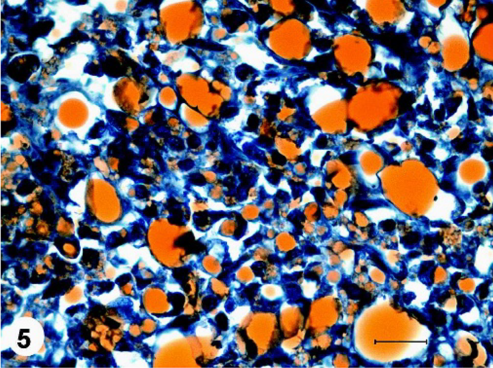
Nasal cavity, liposarcoma; cow. Neoplastic cells exhibit cytoplasmic staining for lipid within cytoplasmic vacuoles. Oil red O and counterstain with hematoxylin. Bar = 20 μm.
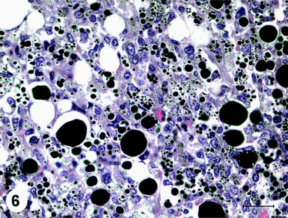
Nasal cavity, liposarcoma; cow. Neoplastic cells exhibit cytoplasmic staining for lipid within cytoplasmic vacuoles. Osmium (VIII) tetroxide and counterstain with hematoxylin. Bar = 20 μm.
In humans, classification of liposarcomas based on histologic pattern and cell morphology has been well established and categorization by type has been shown to be of prognostic value in predicting long-term survival rate and potential for metastasis. 2, 4, 11 The World Health Organization classification scheme for liposarcomas includes the following categories: well-differentiated (subdivided into adipocytic, sclerosing, inflammatory, and spindle cell); dedifferentiated; myxoid (including round cell variant); and pleomorphic. 4 The biologic behavior of these types is quite variable and ranges from no metastatic potential (well-differentiated liposarcoma) to a 30–50% rate of metastasis and 40–50% mortality rate (pleomorphic liposarcoma). 2, 4 The myxoid and round cell liposarcomas are considered to represent low- and high-grade variants, respectively, of the same subtype, with histologic progression from myxoid to round cell morphology associated with a significantly lower 5-year survival rate and poorer prognosis. 2, 4, 11, 14 Further characterization of liposarcomas in humans has included investigation of chromosomal patterns and morphology. Chromosomal morphologic abnormalities, including ring forms and giant marker chromosomes, chromosomal translocations and fusions, and overexpression of certain genes such as MDM2, TP53, and CDK4 have been described in human liposarcomas. 2, 4, 14 These cytogenetic changes have further supported the separation of liposarcomas into the above-mentioned categories, as certain cytogenetic features are consistently present in certain types of liposarcoma. 2, 4, 11, 14 Diagnosis of liposarcoma in humans is achieved through evaluation of the above-mentioned histologic, immunohistochemical, and cytogenetic features.
A unique classification scheme for liposarcoma in animals has yet to be established, likely reflecting the relatively low incidence of this tumor in domestic species. Generally, characterization of liposarcomas in animals has been adapted from the human literature, and includes well-differentiated, pleomorphic, and myxoid types. 6 Classification of a myxoid liposarcoma in a dog, based on histologic pattern, cytologic characteristics, and ultrastructural findings, has been previously reported. 8 Using the WHO classification scheme, the tumor described in this report is most appropriately categorized as a pleomorphic liposarcoma. In comparison, histopathologic characteristics of the tumor observed in the 14-month-old Holando-Argentino breed cow include pleomorphic, slightly vacuolated, elongated cells, an abundant mesenchymal stroma, and islands of chondrocytes, 13 features consistent with a dedifferentiated liposarcoma. In the bovine liposarcoma described by Piercy et al., 12 characteristics included large, pleomorphic cells containing lipid (interpreted as neoplastic lipoblasts), frequent multinucleation, and peripheral inflammation including many multinucleated giant cells; this tumor may be best classified as a pleomorphic liposarcoma.
A retrospective study of 56 liposarcomas in dogs evaluating biologic behavior and potential prognostic indicators found no significant association between survival time and histologic type. 1 In the study, the only factor significantly associated with survival time was the type of surgical excision performed. 1 Recurrence of liposarcoma following surgical removal is reportedly common, but metastasis is rarely documented. 6 Further studies evaluating the biological behavior of liposarcoma in domestic animals may allow development of prognostic criteria.
To date, there have been no studies evaluating the biologic behavior and histologic features of liposarcoma in the bovine species. The low number of reported cases of liposarcoma suggests that occurrence is rare in this species in comparison to some other domestic animals, and has thus far precluded specific characterization. The 2 previously reported cases in cattle described multiple liposarcomas within the abdominal or thoracic cavities. In comparison, the single liposarcoma reported in this case was restricted to the nasal and oral cavities, and did not exhibit gross or histologic evidence of metastasis. Additionally, the histologic features described in each of the previously reported cases of bovine liposarcoma differ both from one another and from the histologic features described in this case.
Most described cases of liposarcoma in animals are considered spontaneous, but some reports have suggested potential inciting agents. Liposarcoma associated with foreign bodies has been reported in 2 dogs. 7, 17 One dog developed a liposarcoma surrounding a microchip implanted at the site approximately 1.5 years previously 17 and liposarcoma surrounded a glass fragment in another dog with a history of trauma 10 years before the appearance of the mass. 7 Foreign-body carcinogenesis in animals has been described primarily in experimental studies involving rodents, including studies using microchip animal identification devices. 3 Review of the literature reveals isolated cases in humans describing development of sarcomas in association with foreign material. In this case, no foreign body was found during postmortem examination or observed in radiographs of the head.
Two reports of virus-associated liposarcoma have been described in domestic animals. One case involved a 4-month-old kitten infected with feline leukemia virus; ultrastructurally, retroviral particles were demonstrated within neoplastic cells. 16 However, subsequent attempts to induce development of liposarcoma in cats through transmission of virus-laden neoplastic cells were unsuccessful. 15 A hamster inoculated with bovine papilloma virus type 4 subsequently developed a liposarcoma at the site of injection, and multiple unintegrated copies of the BPV-4 genome were found within the DNA of this tumor. 10 No attempts to isolate a virus or ultrastructural examination of tissues were performed in the case reported here, so potential association with viral infection is unknown.
In conclusion, the gross, histologic, histochemical, and immunohistochemical findings in this case are consistent with an intranasal liposarcoma with local extension into the oral cavity. It is presumed that tumor infiltration around the molar and premolar tooth roots on the left dental arcade permitted entry of feed material and bacteria into the gingival mucosa, provoking granulation tissue formation in the oral submucosa and abscess formation within the nasal mass. To our knowledge, this is the first case report of a liposarcoma arising within the nasal cavity of a domestic animal and 1 of only 3 reports of liposarcoma in cattle. Unlike the previous reports of liposarcoma in cattle that describe numerous masses, this tumor was confined to the nasal cavity and exhibited locally invasive and destructive behavior.
Footnotes
Acknowledgements
We thank Dr. Michael Walker for his assistance in interpretation of radiographic findings and Dr. Neil Hooper for his assistance in evaluation at clinical presentation.