
Abstract
Immunohistochemistry, using a monoclonal antibody to Melan A and a polyclonal antibody to S100 protein, was applied to 48 formalin-fixed, paraffin-embedded specimens of feline melanoma. Forty-two cutaneous, three oral, one mucocutaneous, and two metastatic melanomas comprised the tumors. Thirty-two tumors (67%) were positive for Melan A and 42 (87.5%) were positive for S100. All but one of the tumors that were positive for Melan A were also positive for S100. S100 was detected in 11 of 16 tumors that were negative for Melan A. Seventy-five percent (9 of 12) of amelanotic melanomas were negative for Melan A. Normal adrenal cortex, the cerebellum, and the skin had cells that were positive for Melan A. Sebaceous adenoma was the only nonmelanocytic tumor examined that reacted with antibody to Melan A. Although less sensitive than S100 protein, Melan A is more specific for melanoma and is useful in differentiating feline cutaneous melanoma from the more common pigmented basal cell tumor.
The most common site of feline primary melanoma is the eye, followed by the skin and oral cavity. 5,13 Cutaneous melanoma is uncommon in the cat, accounting for 1–4% of all cutaneous tumors. 8,12,20 Diagnosis is based on microscopic examination of hematoxylin and eosin (HE)-stained slides. S100 protein is the most widely used immunohistochemical marker in human and animal melanomas and stains up to 100% of feline melanomas. 20 However, the specificity of this marker is low because it detects a variety of normal tissues and nonmelanocytic neoplasms. 19 Antibodies directed against specific melanocytic differentiation antigens, such as HMB-45 (against glycoprotein 100 of premelanosomes 1 through 3), NKI/C3 (against premelanosomal vesicles), HMSA-1 and HMSA-5 (against human melanosome-specific antigens), and Melan A 1,3,18,21 have been applied to canine melanomas but only Melan A, and less commonly HMSA-1 and HMSA-5, have been immunohistochemically detected. 2,16
The purpose of this study was to determine whether Melan A could be immunohistochemically detected in routinely processed feline melanomas and to determine the specificity of this marker to distinguish feline melanomas from nonmelanocytic tumors. A search of the databases of the University of Missouri Veterinary Medical Diagnostic Laboratory and the Michigan State University Animal Health Diagnostic Laboratory for feline melanomas yielded a total of 55 tumors. After reviewing HE slides, 48 tumors with enough available tissue to perform immunohistochemistry were selected for this study. All tissues had been fixed in formalin for an undetermined period of time. Forty-two tumors were from the skin, three were from the oral cavity, one was from the lung, one was from the liver, and one was from the lip. The pulmonary tumor was metastatic from the skin and the primary site of the hepatic tumor was undetermined. The primary tumor site of the metastatic pulmonary tumor was not available for immunohistochemistry. The predominant cell type was epithelioid in 14 (29.2%) tumors, spindle-cell type in 17 (35.4%), mixed (epithelioid and spindle-cell types) in 16 (33.3%), and signet-ring type in 1 (2.1%) (Fig. 1). Thirty-six tumors (75%) were melanotic, of which 25 were moderately to heavily pigmented. Immunohistochemical staining for Melan A with a monoclonal antibody (Dako, Carpinteria, CA) was done with a non–avidin–biotin detection method (EnVision+, Dako, Carpinteria, CA) and for S100 protein was done with polyclonal antibody (Novocastra Laboratories, Newcastle upon Tyne, UK), recognizing S100 A and S100 B, with a streptavidin–biotin–peroxidase method as previously described. 14,16 In heavily pigmented tumors, the diaminobenzidine (DAB) product was distinguished from melanin with Azure B counterstain. 10,16 To determine the specificity of Melan A for melanomas, an additional 126 formalin-fixed, paraffin-embedded feline normal tissues and feline nonmelanocytic tumors were immunostained for Melan A. Nonmelanocytic tumors included basal cell tumors, mast cell tumors, squamous cell carcinoma, and fibrosarcomas, which in our experience are the most common feline cutaneous tumors. 11
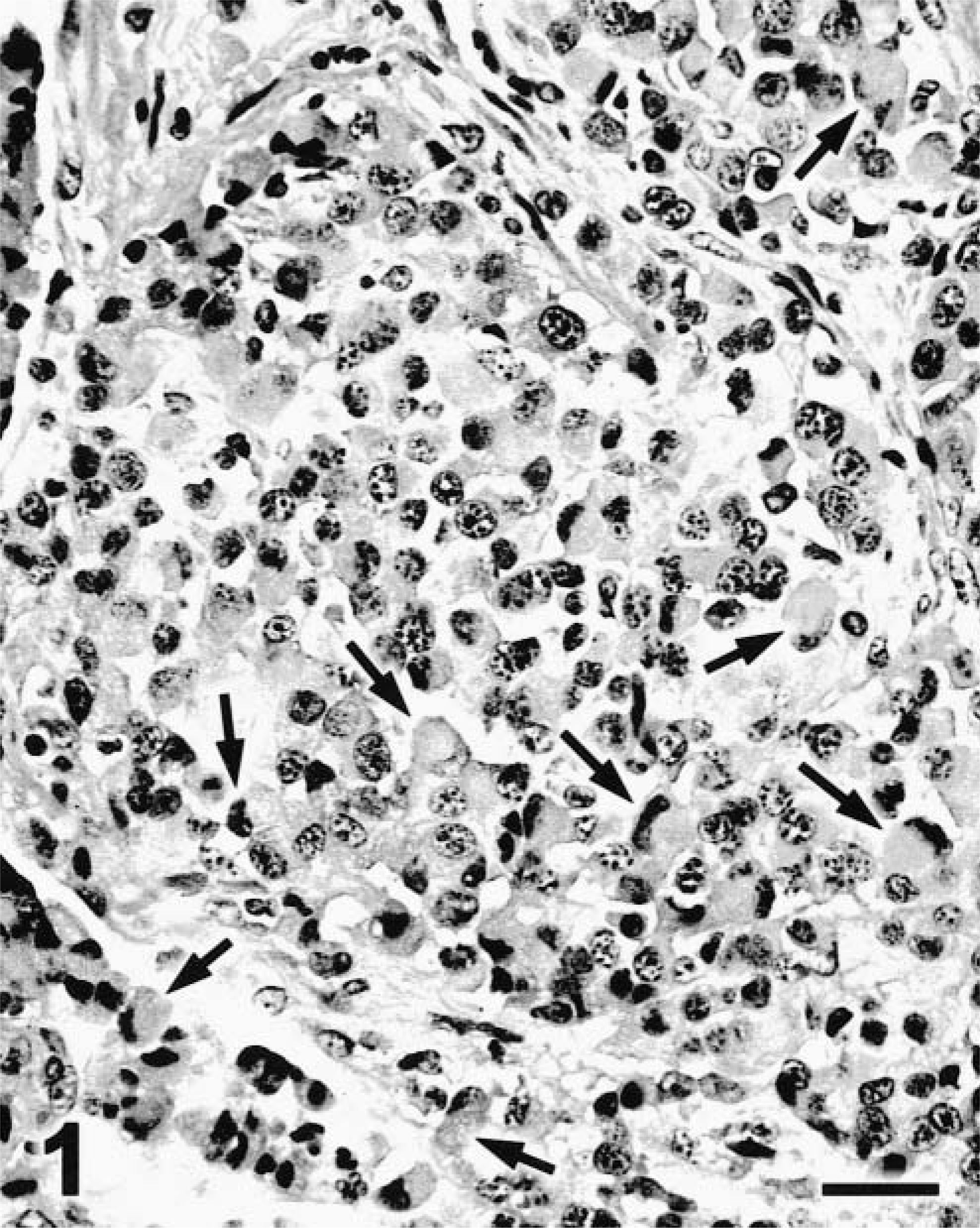
Lung; cat. Melanoma. Signet-ring cell type. Numerous cells have eccentric nuclei and abundant eosinophilic cytoplasm (arrows). HE stain. Bar = 27 µm.
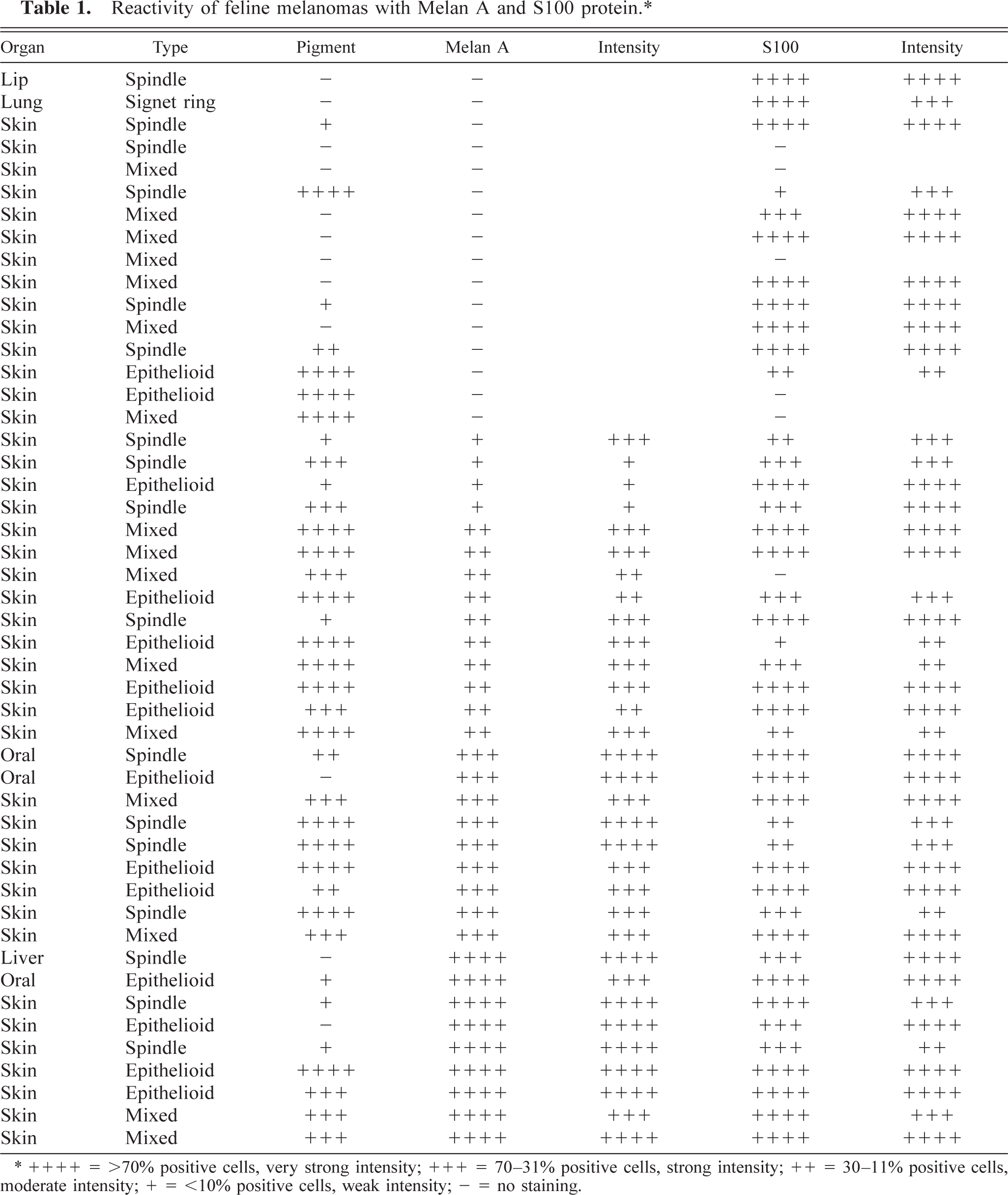
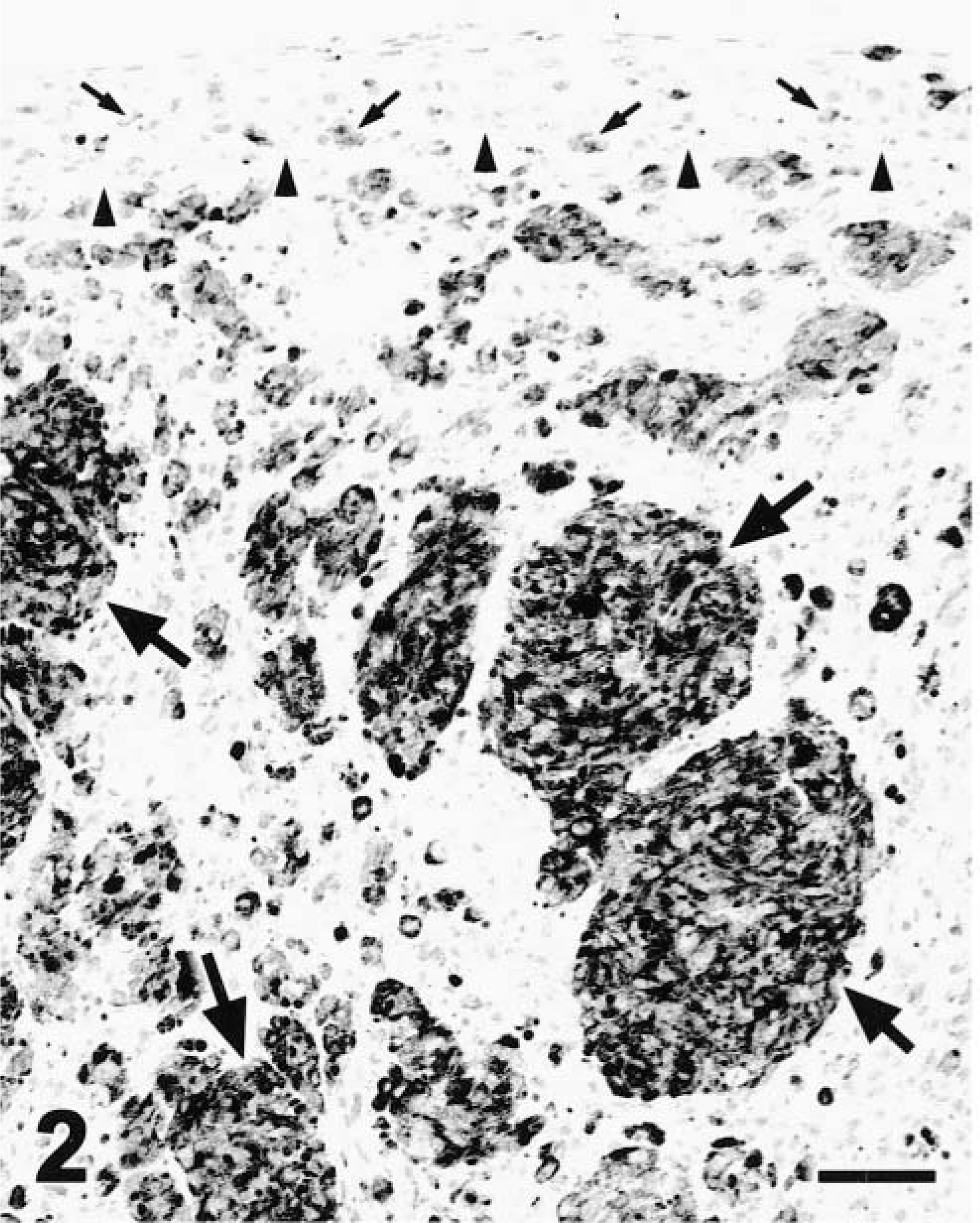
Staining with Melan A antibody was always diffusely cytoplasmic and never nuclear (Figs. 2, 3). Thirty-two melanomas (67%) were positive for Melan A, including 12 of 14 (86%) epithelioid tumors, 11 of 17 (65%) spindle-cell type tumors, 9 of 16 (56%) mixed cell tumors, and 0 of 1 (0%) signet-ring tumors (Table 1). Three of 32 (9.4%) tumors positive for Melan A were amelanotic and 9 of 12 (75%) amelanotic tumors were negative for Melan A. Intraepithelial melanocytes also were positive. The number of positive tumor cells was low (<10%) in 3 tumors, mild (11–30%) in 10 tumors, moderate (31–70%) in 10 tumors, and high (>70%) in 9 tumors. The intensity of staining was low in 6 tumors and moderate or high in 26 tumors. It was not uncommon for positive cells to be heavily laden with melanin. In those heavily pigmented tumors, counterstain with Azure B was necessary to detect the immunohistochemical reaction. With Azure B, melanin was dark green and the DAB product remained brown (Fig. 4). Those cells with both melanin and the DAB product had brown-green cytoplasm.
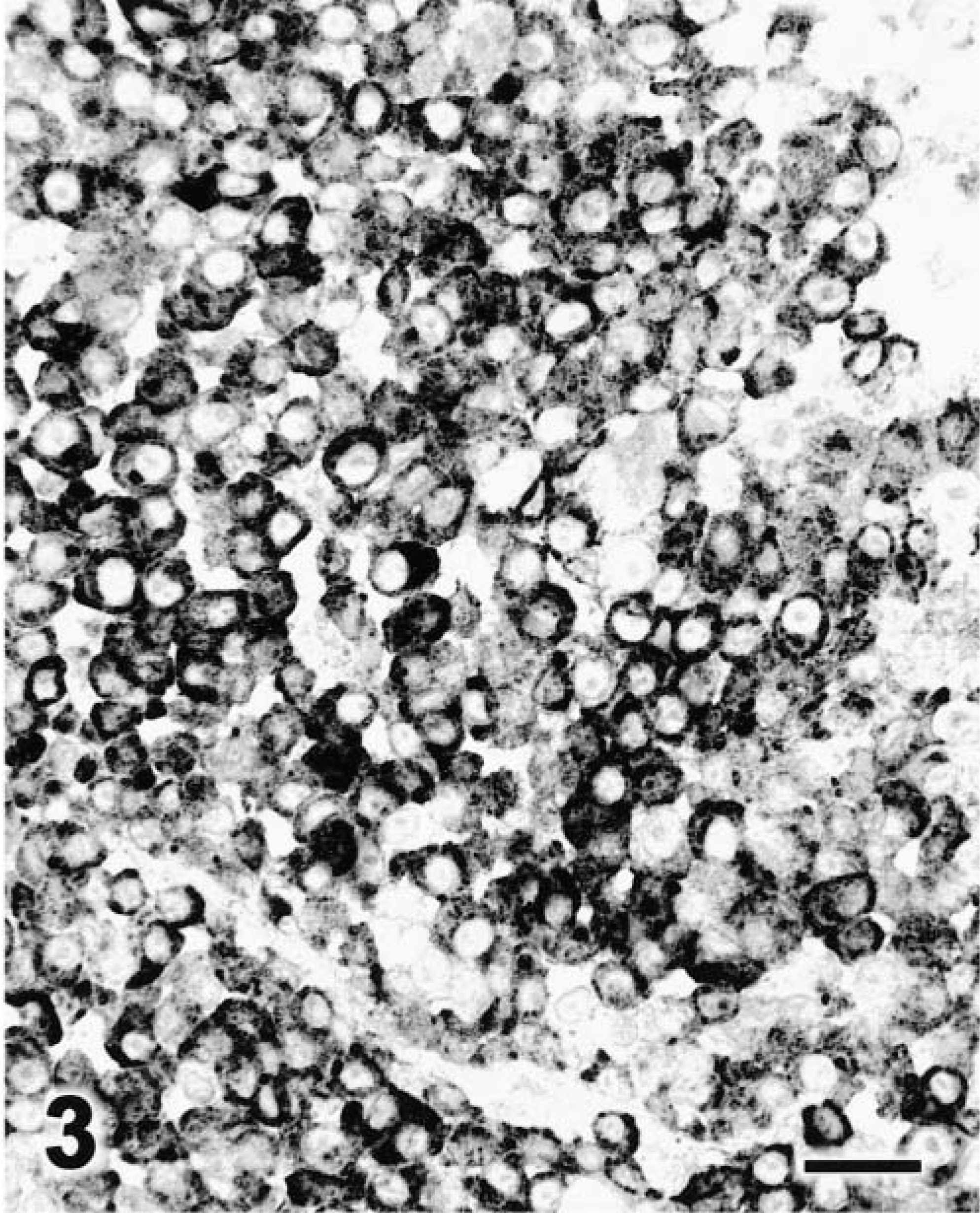
Reactivity of feline melanomas with Melan A and S100 protein.∗
∗ ++++ = >70% positive cells, very strong intensity; +++ = 70–31% positive cells, strong intensity; ++ = 30–11% positive cells, moderate intensity; + = <10% positive cells, weak intensity; = no staining.
Skin; cat. Melanoma. The epidermis (delimited by arrowheads) contains small groups of cells positive for Melan A (small arrows). The dermis also contains clusters of positive cells (large arrows). EnVision+-peroxidase stain with Mayer's hematoxylin counterstain. Bar = 54 µm.
Dermis; cat. Melanoma. Epithelioid cell type. Numerous cells have strong cytoplasmic staining for Melan A. EnVision+-peroxidase stain with Mayer's hematoxylin counterstain. Bar = 27 µm.
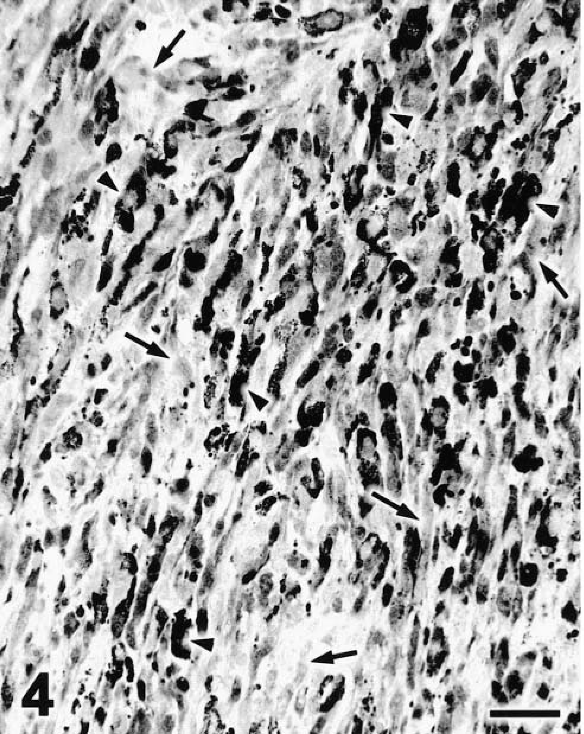
Dermis; cat. Melanoma. Spindle cell type. Dark cells (arrowheads) are negative for Melan A and contain only melanin. Lightly stained cells (arrows) are positive for Melan A and contain DAB product but no detectable melanin. EnVision+-peroxidase stain with Azure B counterstain. Bar = 27 µm.
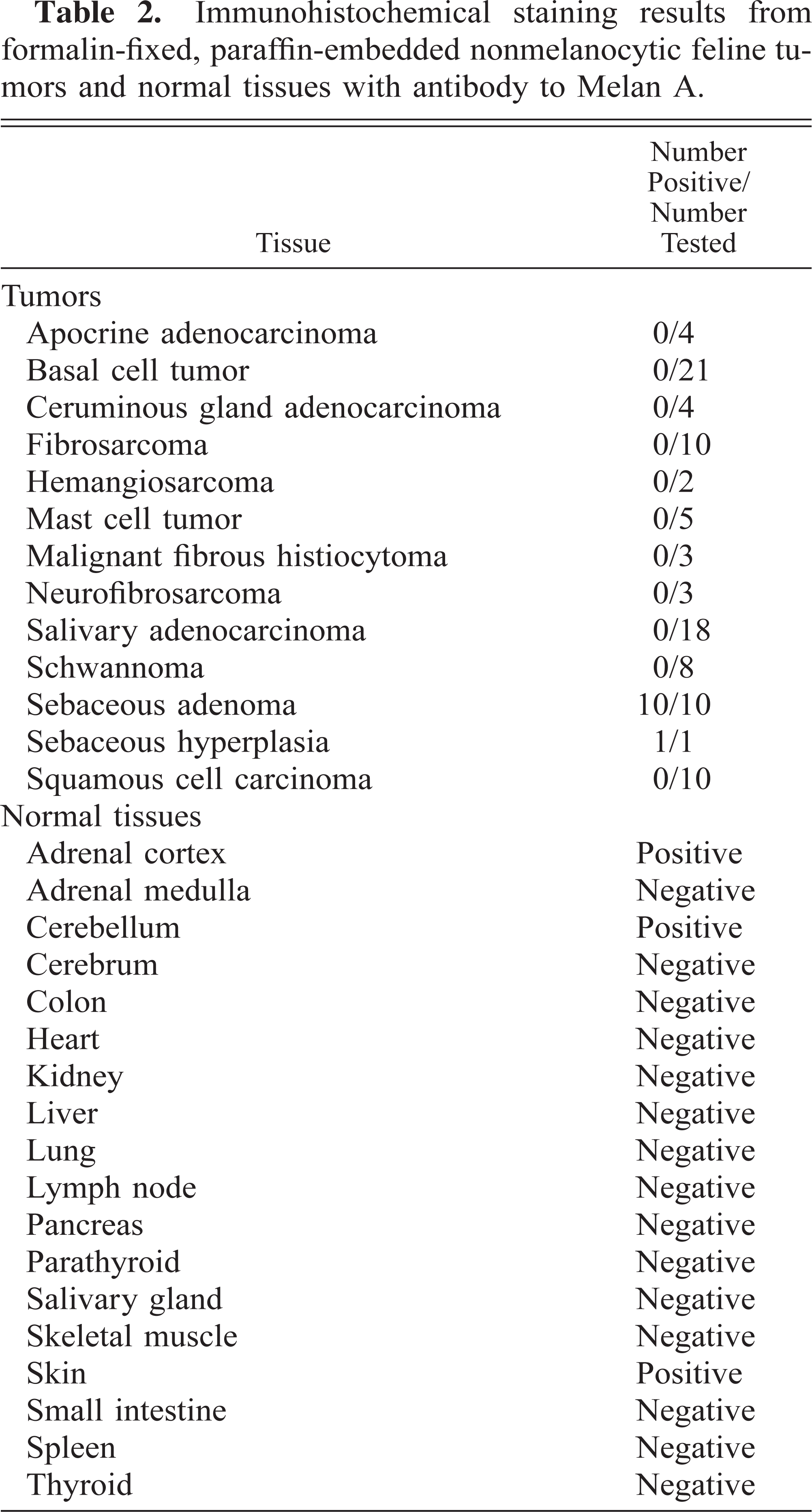
Although Melan A or a cross-reacting antigen has been detected in some normal tissues and nonmelanocytic tumors in humans and dogs, 4,9,15 the likelihood of confusing those with melanomas is low. Only three normal feline tissues were positive for Melan A (Table 2). The cytoplasm of cells in the adrenal gland cortex was strongly positive. Within the cerebellum, cell processes interpreted as those of basket cells surrounding Purkinje cells were multifocally and strongly positive. Also, there was mild granular staining of the cytoplasm of Purkinje cells and of unidentified cell processes in the molecular layer. Other parts of the brain were negative for Melan A. In the skin, melanocytes were positive, as were sebaceous glands. The only nonmelanocytic lesions positive for Melan A were sebaceous hyperplasia and sebaceous tumors (Table 2). Within the sebaceous gland, the most differentiated cells were positive and germinative cells were consistently negative. Only occasional dendritic/spindle cells, interpreted as melanocytes, were positive for Melan A in basal cell tumors, whereas tumor cells were negative.
Immunohistochemical staining results from formalin-fixed, paraffin-embedded nonmelanocytic feline tumors and normal tissues with antibody to Melan A.
Forty-two melanomas (87.5%) were positive for S100 protein. Three of six (50%) melanomas negative for S100 protein were amelanotic. The staining was usually both nuclear and cytoplasmic (Fig. 5). Nuclear staining was more intense than cytoplasmic staining. The nonspecific staining was stronger than with Melan A. Two tumors had fewer than 10%, 5 tumors had 11–30%, 9 tumors had 31–70%, and 24 tumors had >70% S100-positive cells. The intensity of staining in more than 80% (32 of 42) of the melanomas was moderate to high. The neoplastic cells in four of the S100-negative cases were mixed, with one spindle and one epithelioid. Only one tumor negative for S100 was positive for Melan A. The number of positive cells was slightly higher for S100 than for Melan A except in nine cases.
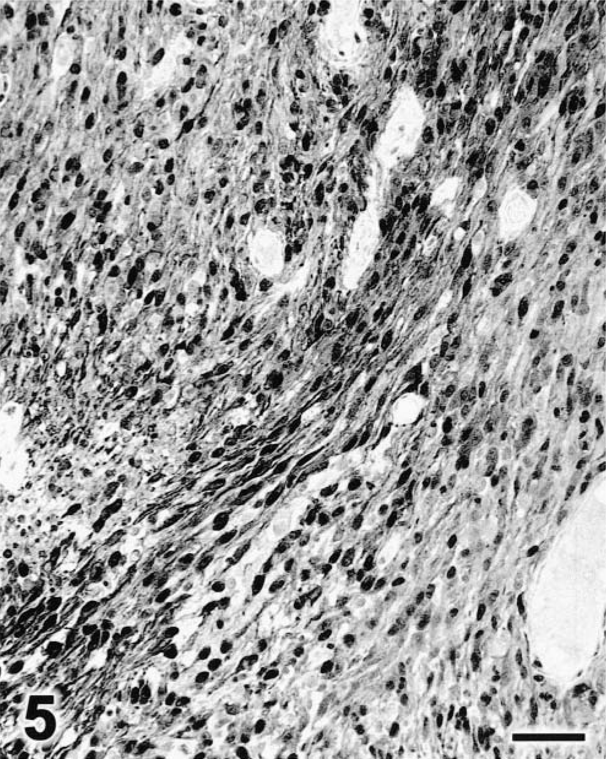
Dermis; cat. Melanoma. Spindle cell type. The nucleus (strongly stained) and cytoplasm (lightly stained) of numerous cells are positive for S100 protein. Streptavidin–peroxidase stain with Mayer's hematoxylin counterstain. Bar = 54 µm.
S100 protein is more sensitive than Melan A in detecting feline melanomas, especially amelanotic tumors, but its specificity is low, because it is also demonstrated in a variety of nonmelanocytic cells, including glial cells, neurons, Schwann cells, Langerhan's cells, macrophages, myoepithelium, chondrocytes, and their tumors. 17,19 The only nonmelanocytic tumors detected by Melan A antibody were benign tumors derived from sebocytes. No sebaceous carcinomas were available among the tumors of this study for testing the reactivity of Melan A. However, the staining of sebaceous adenomas was in the most differentiated sebocytes and not in the germinative cells, which are identical to the basal cells of the epidermis. 7 Staining of human or canine sebaceous tumors with Melan A has not been observed. 4,9,15 Basal cell tumors can be confused with melanomas in the cat when they are heavily pigmented. 6 However, basal cell tumors were consistently negative for Melan A in this study. The only positive staining in such tumors was in melanocytes. The presence of melanocytes has been described in basal cell tumors. 6 The proportion of feline melanoma cases positive for Melan A was lower than in canine oral melanomas. 16 In addition, the pattern of staining was always diffuse; canine oral melanomas may have a punctate/focal or mixed (diffuse and punctate) reaction. 16 The mixed cell type of melanoma was more likely to be negative for Melan A and S100 than other cell types. The use of Azure B as a counterstain is a simple and quick method to distinguish melanin from DAB product. In summary, immunohistochemistry for Melan A can help confirm a diagnosis of feline melanoma but its sensitivity is moderately low, thus necessitating the use of other markers (e.g. S100 protein).
Footnotes
Acknowledgements
We thank M. Beissenherz for technical assistance with immunohistochemistry and H. Wilson for photographic preparations.