
Abstract
The immunoreactivity of PNL2, Melan A, and protein gene product (PGP) 9.5 was compared with that of S100 protein in 50 formalin-fixed, paraffin-embedded equine melanocytic neoplasms. PNL2, PGP 9.5, and S100 protein were detected in all 50 neoplasms; none expressed Melan A. PNL2 was not expressed in 62 nonmelanocytic tumors (equine sarcoids, schwannomas, carcinomas, sarcomas, endocrine tumors, sex-cord stromal tumors, germ cell tumors, and leukocytic tumors) or in normal tissues other than epidermis. In summary, antibody PNL2 is a sensitive marker of equine melanocytic neoplasms and is more specific than S100 protein or PGP 9.5. In contrast, the monoclonal antibody to Melan A did not react with any of the equine melanomas.
The skin is the organ most commonly affected by neoplasia in horses, accounting for approximately 50% of all neoplasms. 22 Melanoma is one of the 3 most common cutaneous tumors of horses, making up 4% to 19% of the total number of tumors. 22 Several clinical forms of equine melanocytic tumors have been described: melanocytic nevus, dermal melanoma, dermal melanomatosis, and anaplastic malignant melanoma. 26 Despite the benign phenotype of many melanocytic proliferations, most equine melanocytic neoplasms, other than melanocytic nevus, should be considered at least potentially malignant. 9 Variants of melanocytic nevi similar to those in humans have been reported in horses. 20 Although the diagnosis of melanocytic neoplasms in horses is typically straightforward due to heavy pigmentation, equine melanomas may be amelanotic or poorly pigmented. 25,26 The heterogeneity in pigmentation can make diagnosis difficult in punch biopsy specimens. Furthermore, the variety of histologic appearances of equine melanocytic neoplasms makes them difficult to distinguish from mimics such as schwannoma and equine sarcoid. 21
Immunohistochemistry is widely used to confirm a diagnosis of melanoma in human and veterinary medicine. Melanoma markers have variable sensitivity and specificity. 4 In humans and dogs, commonly used melanocytic markers include Melan A, PNL2, S100 protein, and tyrosinase. 1,4,16,24,27 In horses, S100 protein is the marker consistently expressed in melanocytic neoplasms. 22 Tyrosinase expression has been detected by immunohistochemistry or molecular methods in equine melanomas, 11,23 but benign equine melanocytic tumors apparently do not express tyrosinase. 23 Monoclonal antibody HMB-45, which recognizes the melanocytic differentiation antigen gp 100, is also expressed in equine malignant melanomas. 23 Expression of CD44 has been reported in both benign and malignant equine melanocytic neoplasms. 23
S100 protein is a very sensitive marker for human and animal melanocytic tumors, but its specificity is poor. Many nonmelanocytic tumors and normal tissues, including nerve sheath cells, myoepithelial cells, adipocytes, and leukocytes, express S100 protein, which makes it unsuitable as a sole test for the diagnosis of melanoma. 10,14
Protein gene product (PGP) 9.5, a protein of the ubiquitin-proteasome system, is expressed in various cells, including neurons and neuroendocrine cells and their tumors. 15 PGP is moderately sensitive for human melanomas. 19
Melan A is a cytoplasmic protein recognized by cytotoxic T cells. 10 It is a very specific melanocytic differentiation antigen (95%–100%) and highly sensitive (75%–92%) in human melanomas. 10 Melan A has been used in the diagnosis of melanomas from animals, particularly dogs. 5,16,17,24 Its sensitivity in the dog is high (expressed in more than 92% of melanomas) and lower for feline melanomas (87%); 14,17 however, Melan A is also expressed in nonmelanocytic steroid-producing cells and tumors of the adrenal gland and gonads. 13 Melan A expression in 1 equine melanocytic neoplasm and melanocytic cell lines has been reported. 3,25
PNL2 is a monoclonal antibody that recognizes a fixative-resistant melanocytic antigen in humans, dogs, and some laboratory animals, 2,7,8,16,18,24 the exact structure of which has yet to be identified. 10 In humans, PNL2 also labels angiomyolipomas and mature myeloid cells, including neutrophils, but not other nonmelanocytic tumors, including various carcinomas and sarcomas. 2 The sensitivity of PNL2 in human melanomas is comparable to that of Melan A, particularly in the epithelioid variants, but it is very low for desmoplastic melanomas. 2,18 To our knowledge, monoclonal antibody PNL2 has not been used in the characterization of equine melanocytic neoplasms.
The aims of this study were to (1) document the immunoreactivity of monoclonal antibody to melanocytic marker PNL2, Melan A, and PGP 9.5 in equine melanomas; (2) compare their immunoreactivity with that of S100 protein; and (3) determine PNL2 cross-reactivity in equine normal tissues and nonmelanocytic tumors.
Methods
The Purdue University Animal Disease Diagnostic Laboratory and Colorado State University Veterinary Diagnostic Laboratory databases were searched for poorly pigmented or unpigmented excisional biopsies of equine melanocytic neoplasms. Fifty melanocytic neoplasms from 48 horses were selected, and histologic sections of each case were reviewed separately by 2 pathologists (C.B.F., J.A.R.-V.) to confirm the diagnosis. When there was disagreement in the diagnosis, the case was reevaluated and a consensus diagnosis based on the hematoxylin and eosin (HE) section was achieved among 3 pathologists (M.A.M., C.B.F., J.A.R.-V.). The neoplasms in this study were from the skin except for 2 cases in which only samples of metastatic melanoma (in the liver and lung of 1 case; spleen from the other) were available. The duration of formalin fixation for most of the samples was unknown. Tumors were histologically classified by the prevalent phenotype (epithelioid, polygonal, spindloid, mixed, pleomorphic) and following a modified classification based on tumor distribution and pleomorphism 26 in melanocytic nevi (tumor cells located in the superficial dermis or at the dermo-epidermal junction), dermal melanoma/dermal melanomatosis (single or multiple to confluent neoplastic nodules in the deep dermis), or anaplastic malignant melanoma (neoplastic cells with marked pleomorphism, high mitotic index, and invasion).
Four markers were used (Table 1): mouse monoclonal antibody antimelanoma marker, clone PNL2 (Santa Cruz Biotechnology, Santa Cruz, CA); mouse monoclonal antibody anti–Melan A, clone A 103 (Dako, Carpinteria, CA); rabbit polyclonal antibody anti-S100 protein (Dako) ; and rabbit polyclonal antibody anti–PGP 9.5 (Dako) following published procedures validated in canine tissues. 12,15,16 A non–avidin-biotin immunoperoxidase-diaminobenzidine (DAB) detection method (EnVision+; Dako) was used for all markers. To evaluate possible cross-reactivity of monoclonal antibody PNL2, normal equine tissues and various nonmelanocytic equine tumors, including equine sarcoids, schwannomas, and other mesenchymal, epithelial, and leukocytic tumors, were also evaluated with this marker (Table 2).
Antibody Reagents, Antigen Retrieval, and Detection Systems Used in Immunohistochemistry
HIER, heat-induced epitope retrieval with decloaker, 20 to 24 psi, 125°C, for 30 seconds.
PNL2 Immunoreactivity in Normal Equine Tissues and in Nonmelanocytic Neoplasms
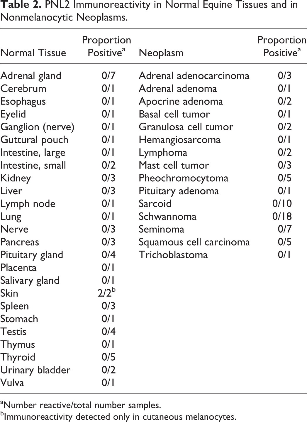
aNumber reactive/total number samples.
bImmunoreactivity detected only in cutaneous melanocytes.
Immunoreactivity was semi-quantitatively evaluated as 0 = detected in <5% cells, 1 = detected in 5% to 10% cells, 2 = detected in 11% to 50% cells, or 3 = detected in >50% cells, modified from a previous study. 16 This grading system was used to compare reactivity among different melanocytic markers in the same tumor. Evaluation of immunoreactivity for all 4 markers based on cell phenotype and location of tumor cells (superficial vs deep) was also done; for this purpose, present vs absent was recorded.
Results
The most common breeds represented in this study were Quarter Horse (16), Thoroughbred (7), and Arabian and Tennessee Walking Horse (5 each). The most common location for melanocytic neoplasms was the tail/perineum area (14), followed by the distal extremities (8) and the prepuce (4). Six horses had multiple skin tumors, and 2 had metastasis to internal organs (lung, spleen, or liver). Twenty-seven tumors were classified as melanocytic nevi, 16 as dermal melanomas/dermal melanomatosis, 5 as anaplastic melanomas, and 2 as nonclassified (visceral metastases). The most common phenotype was spindle (17 cases) followed by mixed (13), epithelioid (10), polygonal (9), and pleomorphic (1). The mitotic index (number of mitotic figures in ten 400× fields) was 0 in 22 tumors (including 14 of 17 spindle-cell tumors), 1 to 10 in 18 tumors, 11 to 20 in 6 tumors, 21 to 30 in 2 tumors (both of polygonal type), and >30 in 2 tumors (1 epithelioid, 1 mixed).
Immunoreactivity of Melanomas for PNL2, Melan A, and PGP 9.5
All 50 melanocytic tumors expressed PNL2, PGP 9.5, and S100 protein (Figs. 1 –4). However, Melan A expression was not detected in any tumor (Figs. 1b and 4a). The number of reactive cells varied by immunohistochemical marker, with the highest number of cells expressing PGP 9.5, closely followed by S100 protein and PNL2 (data not included). All tumors had cytoplasmic expression for PGP 9.5 and S100; nuclear expression for PGP 9.5 and S100 protein was observed in 45 of 50 and 43 of 50 tumors, respectively. Variation in PNL2 immunoreactivity by cell phenotype or cell location within the tumor was not observed in most cases; however, in 6 tumors, PNL2 expression was stronger in superficial cells, and in 1 case with mixed phenotype, PNL2 expression was stronger in polygonal than in spindle cells (Fig. 2b). Expression of S100 or PGP 9.5 was not dependent on cell phenotype or location. In the 2 cases with visceral metastatic melanoma, the immunoreactivity of the markers in lung, liver, or spleen was similar to that in the cutaneous tumors.
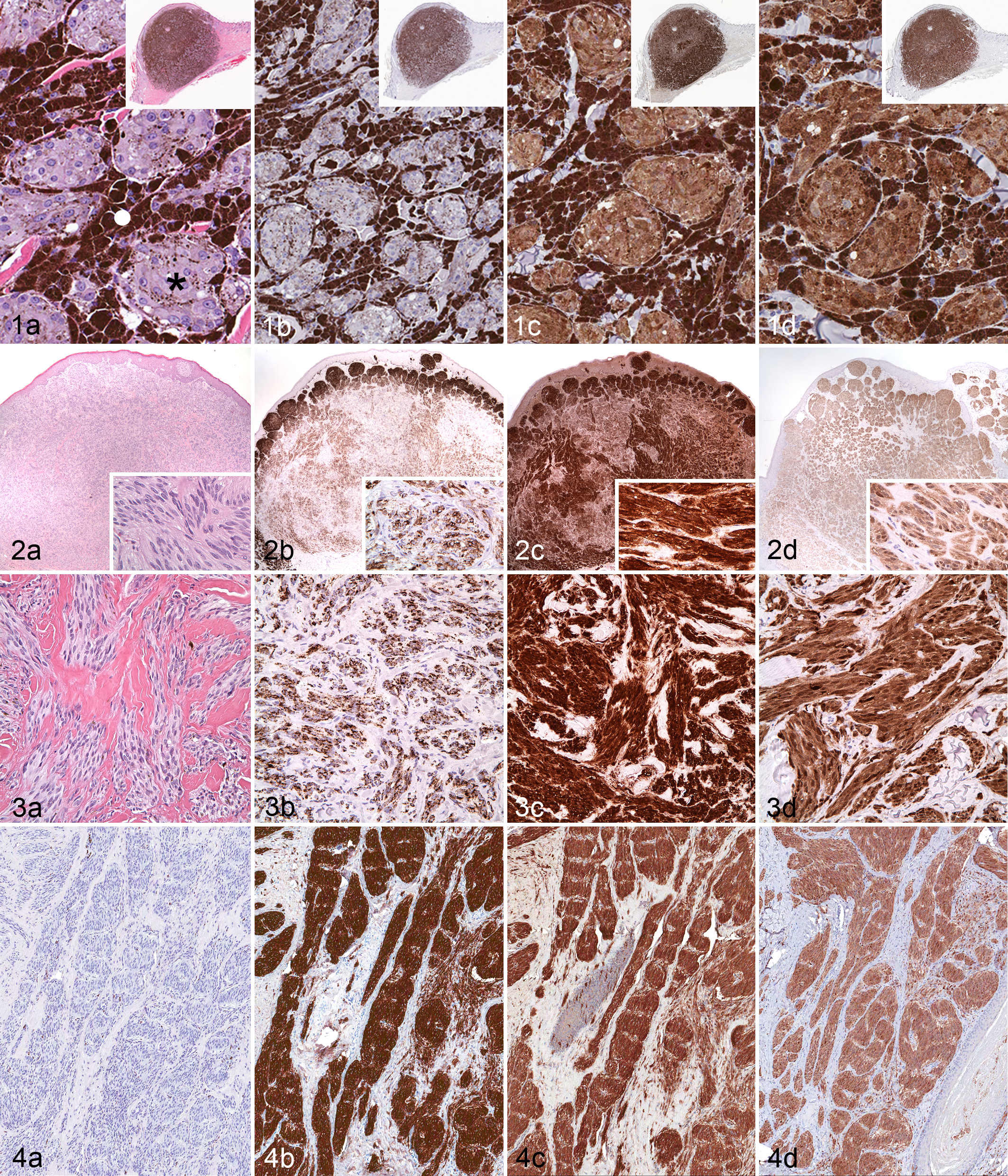
Skin; horse. Dermal melanomatosis. (a) Clusters of mildly pigmented polygonal cells (asterisk) surrounded by melanophages (solid circle). Hematoxylin and eosin (HE) stain. (b–d) Comparison of immunoreactivity with melanocytic markers. (b) Melan A. (c) PNL2. (d) S100 protein. There is no labeling for Melan A (brown color is melanin pigment present in melanocytes and melanophages). Both PNL2 and S100 protein are expressed in melanocytic tumor cells (diffuse cytoplasmic labeling). Insets: Low magnification of this dermal tumor, which is a well-circumscribed but unencapsulated nodule. Immunoperoxidase-diaminobenzidine (DAB), hematoxylin counterstain.
Immunoreactivity of Normal Tissues and Nonmelanocytic Neoplasms
With the exception of skin, none of the normal equine tissues or nonmelanocytic neoplasms expressed PNL2 (Table 2). In the skin samples, only melanocytes in the epidermis or epithelium of hair follicles expressed PNL2. The 62 neoplasms evaluated for cross-reactivity included benign and malignant epithelial, mesenchymal, and leukocytic tumors in a variety of body systems with special attention to equine sarcoid and schwannoma because of their histologic similarity to spindle cell melanoma.
Discussion
In this study, antibody PNL2 was highly sensitive and specific for equine melanocytic neoplasms. Although PNL2 antibody has been used successfully for human and rodent melanocytic proliferations, 2,8,18 to the authors’ knowledge, it has not been applied to equine melanocytic neoplasms. The number of melanomas that expressed PNL2 equaled the number reactive with S100 protein and PGP 9.5. PNL2 reactivity is low or undetectable in human spindle and desmoplastic melanomas. 2,18 In our study, although spindle cell melanomas were less reactive to PNL2 than were other phenotypes, the number of PNL2-expressing spindle cell melanomas was the same. Significant reduction of PNL2 reactivity in rodent spindle cell melanomas has not been observed. 7,8
None of the nonmelanocytic tumors or normal tissues (except for normal melanocytes) reacted with antibody PNL2. Based on these results, the specificity of PNL2 for equine melanocytes or melanocytic tumors appears to be 100%. No attempt was made to examine the cross-reactivity of S100 protein or PGP 9.5 with nonmelanocytic neoplasms, but it is well known that both markers are expressed in multiple cell types and their tumors in humans and animals. 10,14,15
In most of the melanocytic tumors, location of the neoplastic cells (superficial or deep) within the mass did not correlate with labeling intensity of the melanocytic markers, although in 6 cases, superficial neoplastic cells had more intense immunolabeling with PNL2 than those in deeper portions of the tumor. Similar results with PNL2 antibody have been reported in canine melanomas. 16 In human tumors, immunoreactivity for some melanocytic markers (eg, antibody HMB45) is progressively lost with increasing depth from the tumor surface. 10
Melan A is one of the most commonly used melanocytic differentiation markers for human and canine melanomas. 6,14,24 In this study, Melan A was not expressed in any of the equine melanocytic tumors or in normal melanocytes of equine skin, although expression of Melan A has been reported in an equine melanocytic tumor and in equine melanocytic cell cultures. 3,25 Immunoreactivity of Melan A was not evaluated in nonmelanocytic equine tumors in those studies. In at least one of the studies, 25 the same Melan A antibody clone was used, although details of the procedure were not included. The immunohistochemical procedure for this study has been validated in canine melanomas 14 and also has been successfully applied to other species, including cats and reptiles. 5,17 Although Melan A immunoreactivity was decreased in canine melanomas fixed in formalin >1 month, 16 it is unlikely that any samples in the current study were fixed longer than 2 to 3 weeks. We did not modify the published immunohistochemical technique for Melan A 14 to attempt to detect this marker in equine melanomas.
On the basis of the results of this study, we conclude that (1) immunohistochemistry on formalin-fixed, paraffin-embedded tissues with antibody to melanoma marker PNL2 is highly sensitive and specific for equine melanocytic neoplasms. (2) Antibody PNL2 sensitivity is similar to that of S100 protein and PGP 9.5. (3) The specificity of PNL2 for melanocytic neoplasms is superior to that of S100 protein or PGP 9.5. (4) Melan A was not detected in equine melanocytic neoplasms or in normal melanocytes. (5) Normal tissues (other than melanocytes) and nonmelanocytic tumors did not react with PNL2 antibody. (6) PNL2 antibody can be used as the sole marker of melanocytic origin in equine neoplasms.
Footnotes
Acknowledgements
We appreciate the technical assistance of the ADDL Histology Laboratory staff and Purdue University and Colorado State University pathologists in providing the diagnosis of melanocytic neoplasms.
Declaration of Conflicting Interests
The author(s) declared no potential conflicts of interest with respect to the research, authorship, and/or publication of this article.
Funding
The author(s) received no financial support for the research, authorship, and/or publication of this article.
Preliminary results from this study were presented as a poster (abstract N-20) at the 2012 annual meeting of the American College of Veterinary Pathologists; December 1–5, 2012; Seattle, WA.