
Abstract
PNL2 is a recently generated monoclonal antibody (mAb) that recognizes normal and neoplastic melanocytes. Although the antigen recognized by PNL2 remains unknown, recent studies of human and mouse melanomas have confirmed its usefulness as a diagnostic marker. In the current study, the immunoreactivity of PNL2 in canine melanomas was tested and compared with Melan A (A103). Validation of PNL2 was performed by Western blot analysis. PNL2 and Melan A immunoreactivity were tested on frozen samples of canine melanomas and on 69 formalin-fixed, paraffin-embedded melanocytic neoplasms. Normal canine tissues and nonmelanocytic neoplasms were included as negative controls. Western blot confirmed the presence of a protein recognized by the PNL2 antibody in canine melanomas. Immunohistochemically, PNL2 stained the melanocytic neoplastic cells with an intracytoplasmic, granular pattern. Among the melanocytic neoplasms tested, 62% stained positively with PNL2 and 59% with Melan A; 50.7% stained positively with both mAbs. The overall percentage of neoplasms that stained positively with at least 1 of these 2 antibodies was 68%. The extent of staining (i.e., the percentage of cells stained per specimen) was greater with PNL2 than with Melan A. With both mAbs, staining was most intense and diffuse in the epithelioid cell phenotype. Neither nonspecific staining nor staining in cells other than melanocytes was detected with either mAb. In contrast to human granulocytes, canine granulocytes were negative by both Western blot and immunohistochemical analyses. PNL2 mAb proved to be highly specific for the identification of formalin-fixed canine melanocytic neoplasms and should be a valuable diagnostic reagent.
Introduction
Histologic diagnosis of melanoma can be challenging, especially when pathologists are facing amelanotic or metastatic lesions. In consequence, immuno-histochemical staining is often necessary for a definitive diagnosis. For several years, antibodies against S100 proteins and HMB45 have been used extensively in human pathology. 12,21 In veterinary pathology, however, HMB45 monoclonal antibody (mAb) has been applied only rarely, and its use on canine tissues requires an elaborate antigen unmasking technique. 14,17,19 More recently, a number of novel mAbs that recognize formalin-resistant melanocytic differentiation antigens have become available in human and veterinarypathology, including Melan A/MART 1 (clone A103/M2-7C10), anti-tyrosinase (T311), and microphthalmia transcription factor (D5). 4,6,7,9
Melan A/MART 1 is one of the most extensively used mAbs in both human and veterinary pathology diagnostics. It is a melanocyte-differentiating protein that is recognized by tumor-infiltrating human cytotoxic T lymphocytes. Melan A is expressed by normal human melanocytes, benign nevi, melanomas, and, less frequently, desmoplastic human melanomas. In 2 independent studies, 1,2 benign melanocytic lesions had a higher overall percentage of positively stained cells compared with malignant lesions. In a single study on canine tissues, 7 the intensity of Melan A staining was also positively correlated with biologic behavior; more differentiated neoplasms staining more diffusely and intensely than amelanotic, undifferentiated ones.
A novel mAb recognizing melanocytes, designated PNL2, was recently generated. 16 Originally created to detect a subtype of human somatostatin, a few recent studies 8,10,11,16 have shown that PNL2 reacts with normal and neoplastic human and murine melanocytes in both benign and malignant lesions. Although the protein target of PNL2 remains to be identified, the results from these different studies suggest PNL2 is a very useful marker for the immunohistochemical diagnosis of melanocytic lesions.
The aims of the present study were to verify the usefulness of PNL2 in the immunohistochemical diagnosis of formalin-fixed, paraffin-embedded canine melanocytic neoplasms and to compare PNL2 and Melan A immunostaining. In the first step, the cross-reactivity of the antibody was investigated by Western blot (WB) in both human and canine melanomas. In the second step, the immunoreactivity of PNL2 in canine melanoma was assessed by immunohistochemistry.
Materials and methods
Western blot validation of PNL2 antibody against dog tissues
Western blot analysis was conducted using tissue homogenates of canine melanoma and neutrophils (PMNs). Human neutrophils, obtained from healthy donors and previously reported to specifically react with PNL2, 16 as well as samples of human melanoma, were used as positive controls.
Melanoma samples were collected by routine surgery and were immediately processed for protein extraction. An aliquot of 50–100 mg of fresh tissue was mechanically homogenized in 10-fold volumes (w/v) of cold lysis buffer (50 mM Tris-HCl, pH 7.6, 150 mM NaCl, 4% NP40, 2% Triton X-100, 1% zwitterion 3.14, 5 mM ethylenediamine tetra-acetic acid [EDTA]). A cocktail of protease inhibitors a was then added to each aliquot, which were left on ice for 30 min. To improve mincing, the tissues were mechanically homogenized again, and then centrifuged for 10 min at 14,000 × g. The supernatant was collected and protein concentration was determined by ultraviolet spectrophotometry (λ = A280). Dog and human PMNs (1 × 10⁷) were purified following previously described protocols. 20,22
Human circulating PMNs were purified from peripheral blood of healthy donors as previously reported 20 with some modifications. The PMNs were isolated by sedimentation in 2% dextran a in Hanks' balanced buffer 1% for 1 hr at room temperature and centrifuged a at 400 × g for 30 min. Remaining erythrocytes were removed from the pellet of PMNs by hypotonic lysis using red blood cell lysis buffer a at 37°C for 5 min. The PMNs were then washed twice with ice-cold phosphate buffered saline (PBS), counted with an automated hematology analyzer, and suspended in ice-cold PBS at a concentration of 1 × 10⁶ cells/ml. The purity of the neutrophils suspension was >95%, as determined by morphological evaluation. Cell viability was >98%, as determined by the Trypan blue exclusion test. The cells were subsequently centrifuged at 400 × g at 4°C and resuspended at a concentration of 1 × 10⁸ cells/ml. An aliquot of 100 μl of this suspension was used for WB analysis. Circulating canine PMNs were purified from peripheral blood of healthy animals as previously reported, 22 with some modifications. Heparinized whole blood was collected from the cephalic vein, and erythrocytes were depleted by dextran sedimentation (6%, w/v). The leukocyte-rich supernatant was then collected and further centrifuged on a Ficoll density gradient (1.077) at 400 × g for 30 min at 4°C. The PMN fraction was harvested, and contaminating erythrocytes were removed by lysis at 37°C for 5 min. The PMNs were then washed twice with PBS and processed as described for human PMNs.
Immunoreactivity was tested on different concentrations of tissue and PMN lysates ranging from 5 to 20 μg of total protein content that were electrophoresed in a 12% sodium dodecyl sulfate-polyacrylamide gel electrophoresis and transferred to a nitrocellulose membrane for Western blot analysis. Immunodetection was done for 1 hr at room temperature using PNL2 mAb at a concentration ranging from 1:100 to 1:2,000 of the stock solution. A horseradish peroxidase (HRP)-conjugated antimouse secondary anti-body a was used (1:2,000 dilution) to localize sites of PNL2 binding and the positive bands were detected using chemiluminescent HRP substrate. b
Immunohistochemistry
Immunohistochemical staining was performed initially on frozen melanoma specimens and subsequently on formalin-fixed, paraffin-embedded samples. Frozen samples were used to assess the intracellular localization of PNL2 in nondenaturing conditions (i.e., without antigen retrieval application).
Four primary canine melanomas (2 oral melanomas and 2 nail-bed melanomas), previously diagnosed by cytology, were surgically obtained and immediately frozen in liquid nitrogen (− 196°C). Serial frozen sections (7 μm thick) were prepared, fixed in cold acetone (−20°C) for 2 min, and then immunostained with anti-Melan A (clone A 103, mouse IgG1) c and anti-PNL2 (clone PNL2, mouse immunoglobulin G1 kappa) d mAbs using a standard avidin-biotin-peroxidase complex (ABC) method. 5 Endogenous peroxidase activity was blocked with 0.3% hydrogen peroxide in 0.01 M Tris buffer saline (TBS; pH 7.4) for 30 min. Slides were then rinsed in TBS and incubated at room temperature (RT) for 30 min with TBS containing 10% normal horse serum to block nonspecific protein binding. Finally, slides were incubated overnight in a humidified chamber at 4°C with the primary antibodies. Various serial dilutions were tested for both primary antibodies, and a dilution of 1:25 was selected as providing optimal immunostaining results.
After three 5-min rinses in TBS, the tissue sections were incubated with antimouse biotinylated secondary antibody e for 30 min at RT. After washing 3 times in TBS, the tissue sections were incubated with ABC for 30 min at RT, and then rinsed 3 times in TBS. The chromogen, 3-amino-9-ethylcarbazole, e was then applied for 15 min. After rinsing in tap water, slides were counterstained with Mayer hematoxylin f for 1 min and mounted with glycerin jelly. g
Sixty-nine formalin-fixed, paraffin-embedded canine melanocytic neoplasms submitted between 2002 and 2004 were selected from the archives of the section of Veterinary Pathology of the University of Milan (Milan, Italy). Clinical information, including signalment data and tumor location, was available for all samples. Hematoxylin and eosin-stained slides were reviewed, and the cell type (epithelioid, spindle, mixed) was recorded. The predominant cell type was assigned for neoplasms in which one cell type constituted less than 5% of the tumor mass.
Normal canine tissues evaluated for PNL2 and Melan A immunostaining (tissue microarrays).
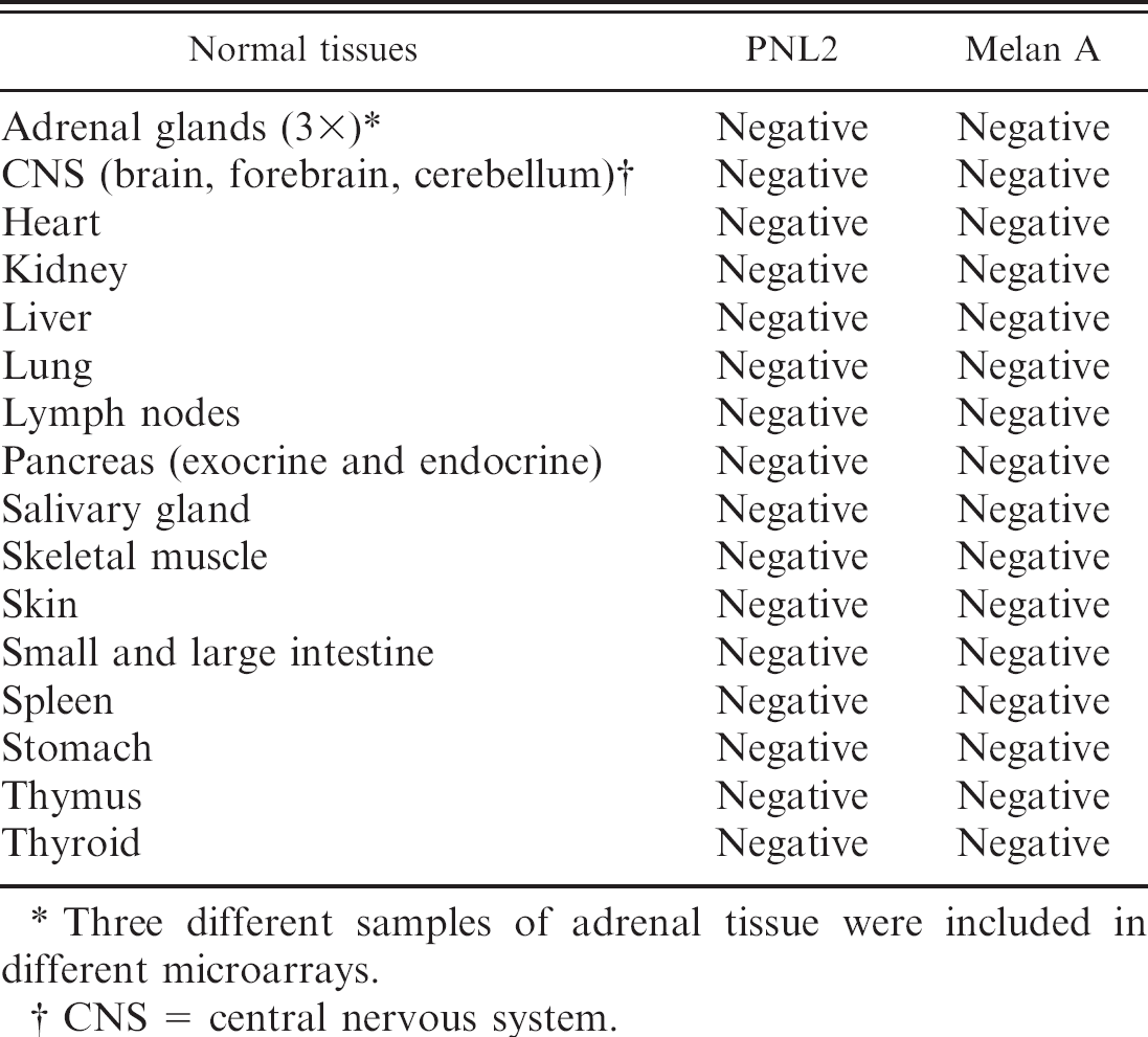
Three different samples of adrenal tissue were included in different microarrays.
CNS = central nervous system.
Serial sections were obtained from paraffin blocks and immunostained with anti-Melan A and anti-PNL2 mAbs (dilution 1:25), using the same ABC protocol used for frozen sections. After dewaxing in xylene and rehydration through a descending series of ethanol solutions, endogenous peroxidase activity was blocked with 0.3% hydrogen peroxide in methanol for 45 min. Antigen retrieval was performed using various methods of heat retrieval and buffer pH concentrations that were tested on positive control tissues (i.e., neoplasms for which a nitrogen-frozen portion was previously obtained and which had positive immunostaining). Optimal staining on control tissues was obtained by heating slides in a pressure steamer for 10 min in an EDTA buffer solution (pH 8.0); this protocol was subsequently applied to all tissue specimens. After antigen retrieval, the tissue sections were allowed to cool for 40 min and were then rinsed in TBS.
Because PNL2 is a protein of unknown function whose expression has never been previously investigated in the canine specimens, a wide variety of normal canine tissues and nonmelanocytic neoplasms was included in the immunoassay as negative controls. Normal canine tissues were included to exclude cross reactivity of PNL2 with proteins normally expressed in these tissues. Nonmelanocytic neoplasms were included to test the specificity of PLN2 for melanoma.
Microarrays of normal canine tissue were created by assembling multiple samples (5 mm × 5 mm × 3 mm each) of formalin-fixed tissue in the same paraffin block. These normal tissues are listed in Table 1. Because Melan A has been reported to be positive in the adrenal cortex, multiple samples of adrenal tissue were included in different microarrays.
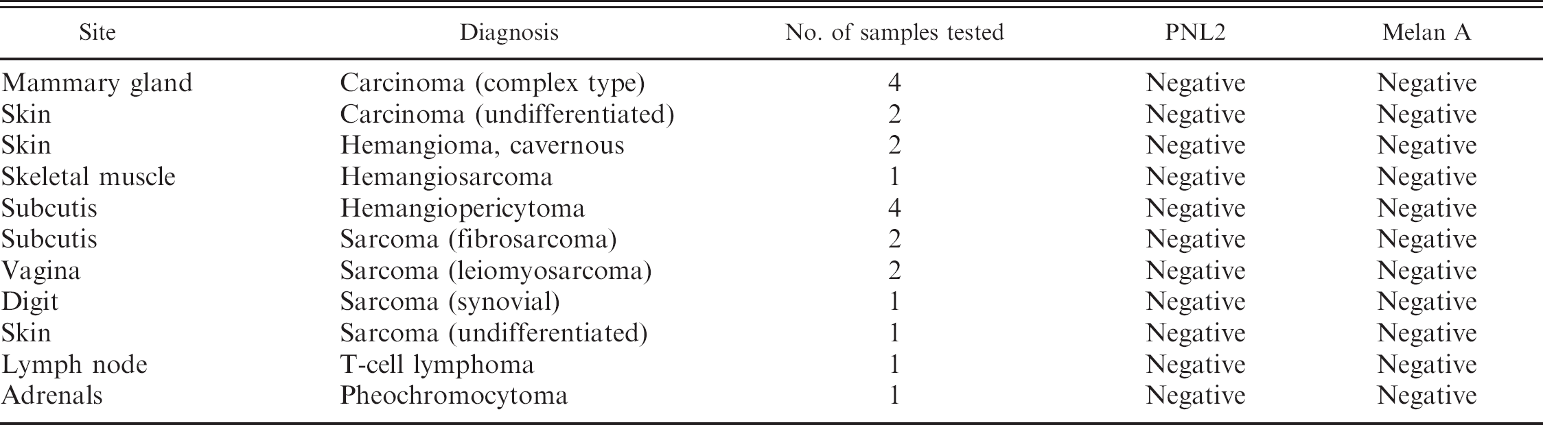
For immunoassay of nonmelanocytic neoplasms, conventional tissue sections were used. The neoplasms assayed are listed in Table 2. The presence of immunostaining, its localization within neoplastic cells, its distribution, and its specificity for neoplastic cells were evaluated. Moreover, the intensity of the antibody staining was subjectively graded as weak, moderate, and intense. The extent of immunostaining, defined as the percentage of neoplastic cells stained, was also semiquantitatively assessed for each antibody and graded as negative (no cells stained) or variable degrees of positivity (<25% of the cells stained, 26–50% of the cells stained, 51–75% of the cells stained, or >75% of the cells stained).
Results
Western blot
The reactivity of PNL2 was validated against dog melanoma and human granulocytes, the latter serving as positive controls. Results are presented in Figure 1A. Western blot analysis revealed that PNL2 reacted with at least 3 major bands with apparent molecular weights (MW) of >120, 31, and <10 kDa, and a minor band at 62 kDa when tested against whole dog melanoma protein extract. Human granulocytes produced 2 major bands, with a MW of 100 and 31 kDa, and a minor band at 62 kDa. The comparison of PNL2 immunoreactivity against dog and human melanoma is shown in Figure 1B. In both tissues samples, a low MW band (<10 kDa) could be detected. Figure 1B also demonstrates that immuno-reactivity was not detected in the homogenate obtained from canine granulocytes.
Nonmelanocytic neoplasms evaluated by PNL2 and Melan A immunostaining (conventional tissue sections were used).
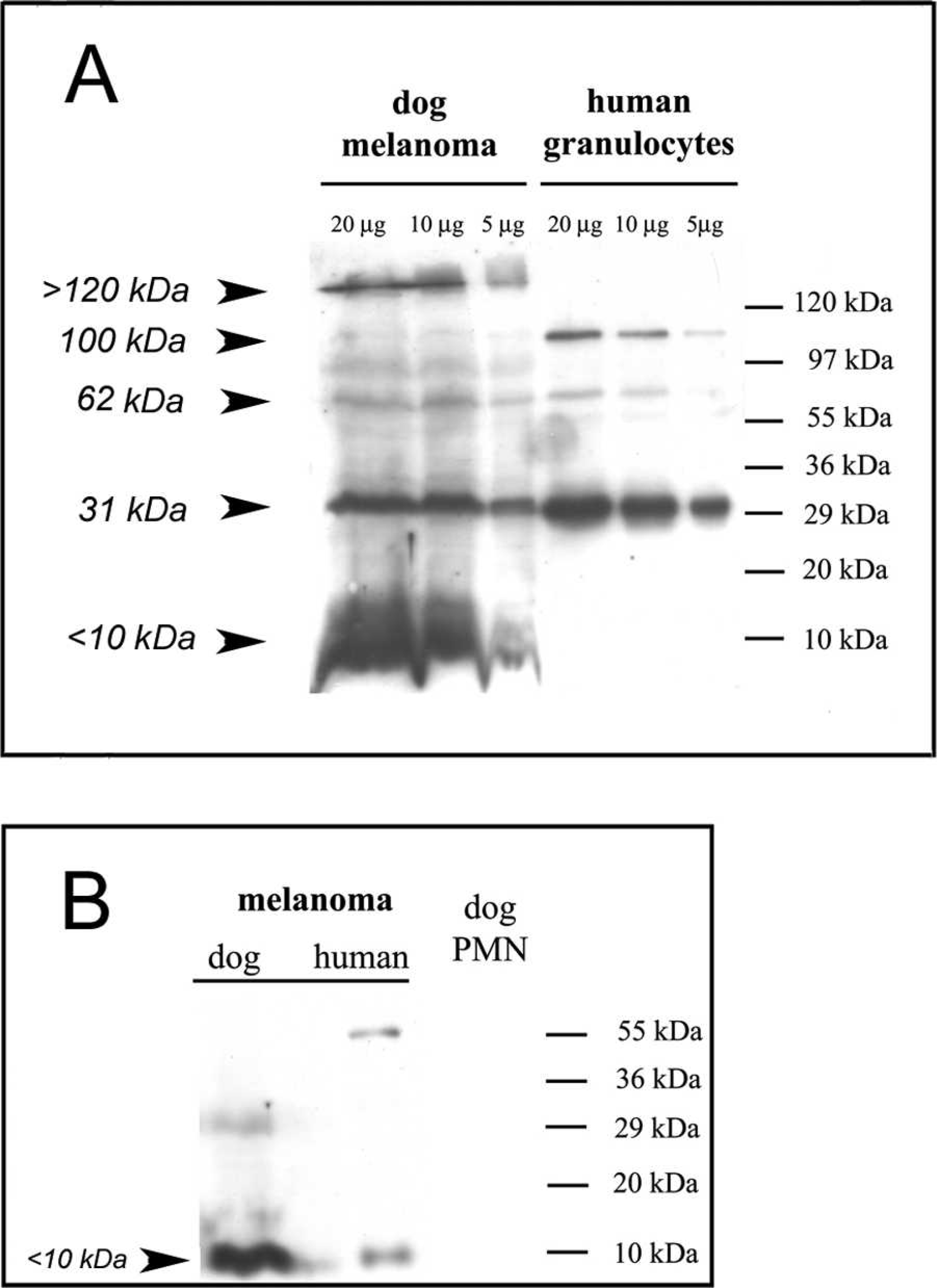
Western blot analysis of canine and human melanoma and of blood granulocytes; immunoblot probed with an anti-PNL2 monoclonal antibody.
Immunohistochemistry on frozen tissues
Canine frozen melanoma specimens had positive staining with both PNL2 and Melan A. The immunostaining was always cytoplasmic with both mAbs. Specifically, immunostaining for PNL2 was intense, finely granular, and cytoplasmic in neoplastic cells. No background staining or staining of stromal tissue, blood vessels, or intravascular granulocytes was observed.
Paraffin-embedded tissues
Melanocytic neoplasms from 69 dogs were examined. Sixty-eight specimens were primary neoplasms (39 cutaneous, 17 oral cavity, 9 mucocutaneous, 3 epibulbar), and 1 neoplasm was metastatic to a lymph node. The cutaneous neoplasms were distributed as follows: 9 eyelids, 7 limbs, 8 trunk, 8 head/neck, 6 nail-bed, and 1 mammary skin. Twenty-eight neoplasms were classified as benign (melanocytomas), and 41 were classified as malignant (melanomas).
Results of PNL2 and Melan A immunostaining of benign and malignant neoplasms.
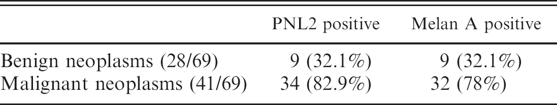
PNL2 immunostaining was present in 43 out of 69 melanocytic neoplasms (62%), and Melan A immunostaining was present in 41 out of 69 (59%) neoplasms. The overall percentage of melanocytic neoplasms that stained positively for at least 1 of the 2 antibodies tested was 68% (47/69), while 51% of neoplasms stained positively for both mAbs. Both PNL 2 and Melan A stained fewer benign neoplasms (9/28, 32%) than malignant melanomas (32/41, 78% PNL2 positive; 34/41, 83% Melan A positive; Table 3). No differences were observed between PNL2 and Melan A immunostaining when the percentage of positively stained samples was compared with the primary site of the neoplasm.
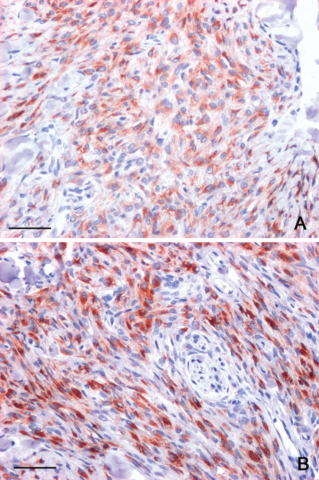
The staining pattern for both PNL2 and Melan A was exclusively cytoplasmic and limited to cells of melanocytic lineage. Nonspecific staining was not observed in necrotic areas or in cells other than melanocytes. When present, PNL2 and Melan A immunostaining was always intense (Fig. 2A, B).
The extent of immunostaining, evaluated as the percentage of cells stained, is presented in Figure 3. In general, PNL2 stained >75% of neoplastic cells in a greater number of neoplasms (19/43) than Melan A (9/41). When neoplasms were grouped by cell type, PNL2 immunostaining was observed in 14 out of 29 spindles and 28 out of 40 epithelioid cell melanomas; whereas Melan A stained 15 out of 29 spindles and 25 out of 40 epithelioid cell melanomas. As a general rule, the neoplasms that stained positively for both antibodies showed a similar distribution of immunostaining (i.e., the same cells, evaluated in serial sections, stained positively). A remarkable exception was melanocytes at the dermoepidermal junction, which had more frequent and intense immunostaining with PNL2 than with Melan A.
All normal canine tissues and nonmelanocytic neoplasms lacked staining with both antibodies (Tables 1, 2). No PNL2 immunostaining was observed in canine granulocytes within blood vessels of normal or neoplastic tissues.
PNL2 consistently stained normal and hyperplastic melanocytes in skin sections, whereas Melan A failed to stain normal cutaneous melanocytes. Melan A did stain infrequent, large, intraepidermal, star-shaped melanocytes that were occasionally present in proximity to cutaneous melanocytic neoplasms.
Formalin-fixed, paraffin-embedded tissue sections of eyelid dermal canine melanocytoma immunostained with monoclonal anti-Melan A (
Discussion
The differentiation of melanomas from a variety of poorly undifferentiated neoplasms is often challenging for pathologists. Thus, there is a need for melanocytic markers that are resistant to formalin fixation and readily detectable using routine immunodiagnostic procedures. In recent years, an increasing number of antibodies have become commercially available for routine diagnosis of melanomas in both veterinary and human pathology. In veterinary pathology, the most extensively used antibodies are S100 and Melan A because of their high sensitivity (S100) and specificity (Melan A). Although S100 has a sensitivity approaching 90% in several studies, it has low specificity. 4,7,15,18 In contrast, Melan A is considered highly specific but has been reported to stain polygonal cell melanomas more consistently than spindle cell melanomas in both canine and human neoplasms. 6,13,15
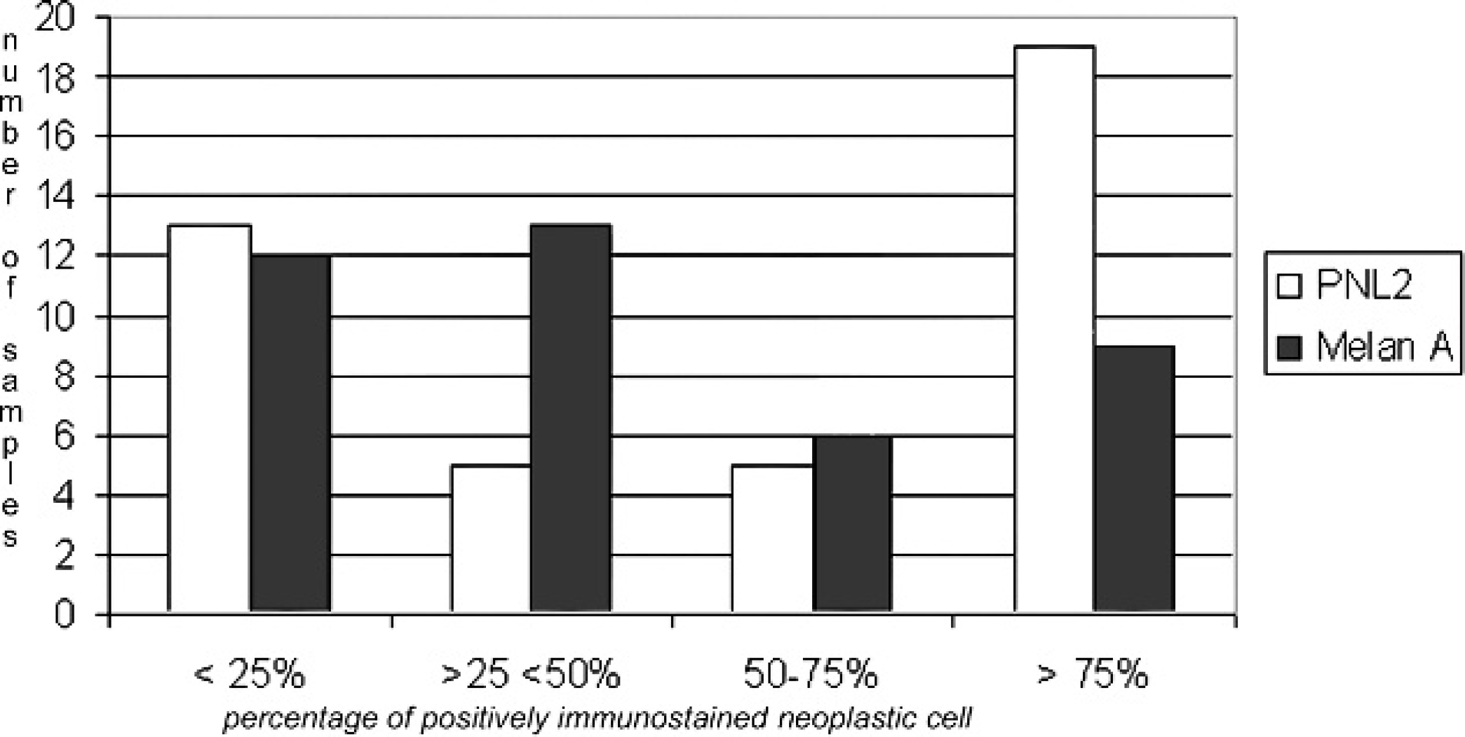
Graphic representation of the extent of immuno-staining (percentage of cells stained with each antibody tested). PNL2 stained more than 75% of neoplastic cells in a greater number of tumors (19/43) than did Melan A (9/41).
PNL2 is a mAb that has recently been introduced as a reagent for immunohistochemical identification of human melanocytes in formalin-fixed, paraffin-embedded tissues. 16 Although the discovery of PNL2 as a marker of melanoma cells was fortuitous and the exact role of the PNL2 protein is still unknown, its value in the diagnosis of human and albino rat melanomas has been confirmed by multiple studies. 3,8,10,11
To the authors' knowledge, this is the first study reporting the use of PNL2 to detect canine melanocytic antigens. Immunostaining of canine and human melanoma extracts with PNL2 revealed similar reactivity, suggesting that PNL2 reacts with the same protein in both species. Interestingly, human granulocytes reacted with PNL2 (a result that is consistent with previous publications 16 ) in the present study, but dog granulocytes did not cross-react with this mAb. This result was confirmed by the absence of immunostaining of canine granulocytes in both frozen and formalin-fixed tissues, suggesting a different distribution of PNL2 protein in dogs.
Evaluation of a large number of formalin-fixed canine specimens demonstrated high specificity of PNL2 for canine melanocytes, staining 62% of the specimens tested. In the present cohort of cases, the percentage of melanocytic neoplasms stained by PNL2 (62%) was only slightly higher than those positive for Melan A (59%). Interestingly, a higher percentage of neoplasms (68%) were identified as melanocytic neoplasms when both mAbs were applied than when either antibody was used alone.
When compared with Melan A, PNL2 detected a higher percentage of neoplasms where more than 75% of the neoplastic cells stained (44% with PNL2 vs. 22% with Melan A). This may confer an important advantage when small biopsy samples are examined. Unfortunately, both PNL2 and Melan A stained epithelioid cell melanomas more frequently than spindle cell melanomas. Thus, the ability to differentiate spindle cell amelanotic melanomas from other spindle cell sarcomas is still unresolved. Notwithstanding, the high specificity and sensitivity of PNL2 make this novel mAb an important addition to the panel of antimelanoma antibodies for the routine diagnosis of canine melanocytic neoplasms in formalin-fixed, paraffin-embedded tissue sections.
Acknowledgements
The authors are grateful to Dr. Gabrina Tragni for her invaluable help, and Dr. Andrea Iachetti, who provided the frozen specimens used in the present study.
Footnotes
a.
ACCUSPIN™ System-HISTOPAQUE®-1077, Sigma-Aldrich, St. Louis, MO.
b.
Millipore SAS, Molsheim, France.
c.
Novocastra™, Leica Microsystems GmbH, Wetzlar, Germany.
d.
Dako Denmark A/S, Glostrup, Denmark.
e.
Vector Laboratories Inc., Burlingame, CA.
f.
Diapath SpA, Martinengo, Bergamo, Italy.
g.
Kaiser's glycerol gelatin, Merck KGaA, Darmstadt, Germany.