
Abstract
A urachal abscess was diagnosed in a 2-month-old, crossbred heifer that was presented for a distended abdomen and clinical signs of choking. Cultures of the mucopurulent exudate, obtained from within the mass on necropsy, yielded Haemophilus somnus. This is the first known documented report of H. somnus isolated from a urachal abscess.
Haemophilus somnus, a gram-negative, nonmotile pleomorphic bacterium, is frequently isolated from cattle with pneumonia, septicemia, and thromboembolic meningoencephalomyelitis. 3,7 H. somnus has also been isolated with frequency from normal and diseased female bovine reproductive tracts, 12,14 male bovine reproductive tracts, 10 bovine semen, 11 and aborted bovine fetuses. 20 There have been reports of isolates from mastitis, 9 conjunctivitis, 13 and otitis 15 in cattle. This report describes isolation of H. somnus from an urachal abscess in a young calf.
A 2-month-old, crossbred heifer was presented to the ambulatory unit of Boren Veterinary Medical Teaching Hospital, Oklahoma State University, College of Veterinary Medicine. On the previous day, the owner had noticed that the animal was salivating, was not eating, and was lying down and getting up frequently. Physical examination revealed a markedly distended abdomen and hypersalivation. Initial treatment consisted of the placement of a nasogastric tube. The tube could be passed only about 10 cm distal to the oropharynx into the esophagus because it was blocked by an unidentified obstruction. A small amount of water was passed through the tube, allowing the tube to be massaged through the esophagus. When the tube was passed into the rumen, the animal lay down and started agonal breathing. At this point, the tube was removed. A trocar was placed through the skin and into the rumen, and excessive amounts of gas were released. Because of the animal's poor response to treatment and the owner's financial constraints, the calf was euthanatized, and a necropsy was performed. On necropsy, a 3 × 6–cm piece of chicken wire was found occluding the esophageal lumen and preventing ingesta and gas from passing. There was a large 35 × 30 × 25–cm fluctuant, caudal abdominal mass attached dorsally to the urinary bladder by a 3-cm remnant of the urachus and adherent to the ventral abdominal wall near the umbilicus (Fig. 1). The mass did not communicate with the urinary bladder. Reddish-gray mucopurulent exudate mixed with large fibrin clumps filled the mass. Aerobic and anaerobic cultures of the exudate yielded H. somnus in pure culture. No lesions were observed in the lungs, brain, reproductive tract, or urinary tract. The umbilical stalk was already desiccated and detached, and there were no openings found in the abdominal wall. A diagnosis of urachal abscess was made on the basis of the location of the mass. A relationship between the esophageal obstruction and the urachal abscess was not apparent, and these appeared to the author to be separate entities.
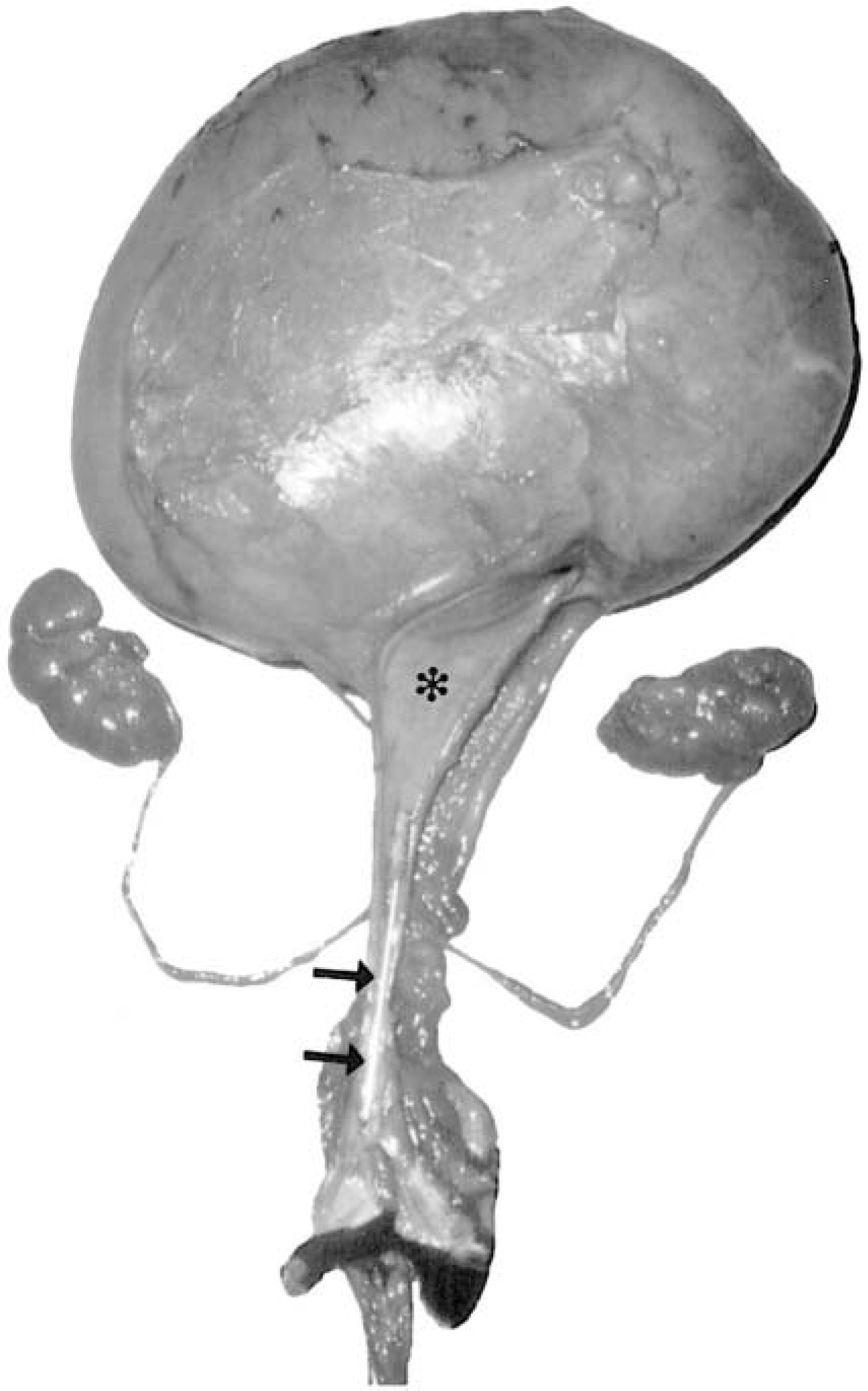
Urachal abscess and associated urinary tract. Note the size of the abscess in comparison to the normal kidneys. A 15-cm probe is present in the urethral lumen for anatomical orientation (arrows), and partially extends into the urinary bladder (asterisk).
Sections of the urachal abscess wall were fixed in 10% formalin, routinely processed, and stained with hematoxylin and eosin. Microscopic examination of the sections revealed an outer wall of fibrous connective tissue surrounding a layer of granulation tissue infiltrated by neutrophils, macrophages, lymphocytes, and plasma cells. The luminal surface was composed of necrotic cellular debris, fibrin, and degenerate neutrophils. No bacteria were seen in sections stained by Gram's and Warthin-Starry's methods.
The urachus, which is remnant of the allantois, extends from the apex of the urinary bladder to the umbilicus. Normally, after birth of the calf, the urachus and associated umbilical arteries collapse and degenerate in the free edge of the bladder's median ligament. 16 The external umbilical stalk thins, desiccates, and is gone by 3–4 weeks of age. Any delay in the closure of these structures can lead to infection.
Umbilical infections are common in newborn calves. 2,17 It has been reported that infection of the urachus is the most common infection related to the umbilicus in cattle. 8 Common problems encountered with the urachus in animals and humans include patent urachus, urachal abscess (i.e., infected urachal cyst), urachal sinus, and vesicourachal diverticulum. 1,4,17 Clinical entities associated with urachal abscesses include umbilical hernia, infected umbilical stalk, 5,18 cystitis, 6 and septicemia. 19 None of these was present in this case.
Urachal abscesses caused by Actinomyces pyogenes, Escherichia coli, Proteus sp., Enterococcus sp., Streptococcus sp., or Staphylococcus sp. have been documented in calves. 2,8 Pathogenic organisms, including H. somnus, can enter the urachus through the umbilical stalk because of a contaminated environment or birth canal, or by way of the urinary tract, in utero through ascending infection from the vagina or cervix, or hematogenously. 2,17 There are two possible sources of H. somnus in this case. Either the cow had an infected birth canal that allowed the calf's umbilicus to become infected, or the calf's umbilicus contacted a contaminated environment. If this calf became infected in utero or hematogenously, a disseminated infection would be more likely than an isolated infection.
In conclusion, because H. somnus has the ability to infect umbilical remnants, this infection should be included in the differential diagnosis in cases of umbilical infections in calves.
Footnotes
Acknowledgements
I thank Dr. Gregory Campbell for his assistance with computer imaging.