
Abstract
Epulides account for 0-7.8% of tumors in surveys of feline oral neoplasms. A review of the literature revealed no reports of multiple epulides in cats. Multiple, concurrent epulides were diagnosed microscopically in 13 cats. Fibromatous and ossifying epulides were diagnosed in 11 of 13 cats and fibromatous epulides were diagnosed in 2 of 13 cats. Microscopically, these epulides were nonencapsulated, well-vascularized, infiltrative, highly cellular neoplasms that expanded the gingiva and were composed of haphazardly arranged, spindle-shaped to stellate cells amid a dense, collagenous stroma. Osseous foci were a feature in the fibromatous and ossifying epulides. The mitotic rate was low and there was marked hyperplasia of the overlying gingiva with a prominent downgrowth of epithelial cords. These tumors recurred in 8 of 13 cats following surgical excision. While uncommon, multiple epulides in cats have a high incidence of recurrence but do not appear to have metastatic potential.
Keywords
Epulides, tumors of periodontal ligament origin, are common in dogs but occur infrequently in cats. 1 2 5–13 They account for 0–7.8% of tumors in surveys of feline oral neoplasms. 5 6 9 11 12 Histologically, they are characterized by a dense, well-vascularized stroma populated by stellate cells with abundant fibrillar collagen and resemble the periodontal ligament. Multiple epulides have been described in dogs, but multiple, concurrent epulides have not been reported in cats. Excisional biopsies from 13 cats with multiple gingival masses that were diagnosed microscopically as epulides of periodontal origin were reviewed. Microscopic and clinical features of multiple epulides from 13 cats are described.
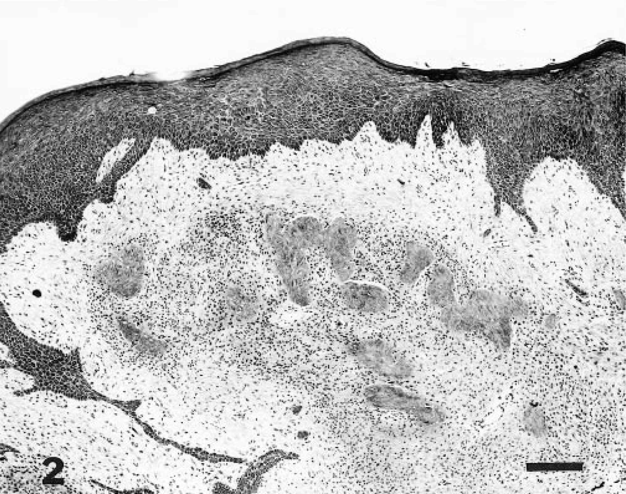
From 1995 to 1997, seven biopsies from cats with multiple epulides were examined by one pathologist (FYS) as a consultant for Marshfield Veterinary Diagnostic Laboratory, Marshfield, Wisconsin. Four were examined by one of the authors (RRD) from the Veterinary School at the University of Wisconsin and two were retrieved from the archives at the Armed Forces Institute of Pathology. Criteria for inclusion in this report included a clinical description of multiple gingival lesions, material (paraffin-embedded tissue and/or glass slides) available for review of the histopathology, and microscopic features consistent with epulides of periodontal ligament origin. The prevalence of this lesion cannot be determined since the number of cat lesions or cat tumors examined over the collection period is not known. The referring veterinarians or owners were contacted for follow-up information. Case summaries are presented in Table 1. Eight cats were Domestic Shorthair cats, one was a Persian, and in four cases, the breed was not specified. Five of the cats were castrated males, three were intact males, three were spayed females, and the sex was not specified in two cats. The cats ranged in age from 1 to 15 years. The FeLV status was reported to be negative in four cats and unknown in the remaining nine. The number of lesions was recorded in four cases (Nos. 1, 4, 7, 9) and ranged in number from 3 to 12. The size and location of the gingival masses were not specified.
Case summaries of 13 cats with multiple epulides.∗
M/C = castrated male; F/S = spayed female; M = male; F = female; N/S = not specified; DSH = Domestic Shorthair; F = fibromatous epulis; F/O = fibromatous and ossifying epulis.
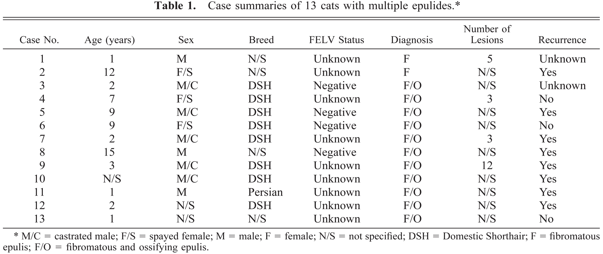
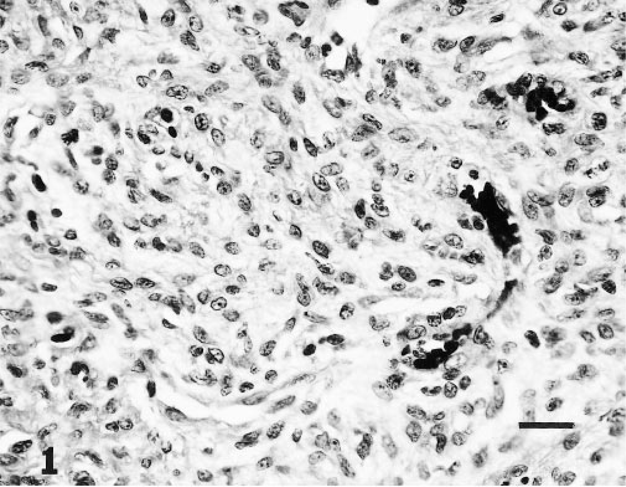
Microscopically, these epulides were nonencapsulated, poorly demarcated, infiltrative, well-vascularized, highly cellular neoplasms. They expanded the gingiva and were composed of haphazardly arranged, spindle-shaped, occasionally stellate cells surrounded by a moderate amount of fibrillar collagen. Neoplastic cells had indistinct borders and a small amount of eosinophilic, often microvacuolated, cytoplasm. Nuclei were irregularly oval, occasionally angular, with finely stippled chromatin and one small, variably distinct nucleolus. Anisokaryosis was not evident and mitotic figures were rare (Fig. 1). In 11 cases (Nos. 3–13), these cells multifocally surrounded foci of dense, brightly eosinophilic material (interpreted as dental hard substance) that was occasionally mineralized (Fig. 2). All tumors exhibited marked epithelial hyperplasia of the overlying gingiva, often with prominent downgrowth of anastomosing epithelial cords. There was multifocal erosion and ulceration with an associated neutrophilic inflammatory infiltrate. Low to moderate numbers of perivascular lymphocytes and plasma cells admixed with fewer macrophages were scattered throughout the neoplasms.
Fibromatous and ossifying epulis; cat No. 3. A moderate amount of fibrillar collagen surrounds neoplastic cells that are spindle-shaped and have a small amount of eosinophilic, often vacuolated cytoplasm. The nuclei are irregularly oval with finely stippled chromatin and one small nucleolus. HE. Bar = 17 µm.
Fibromatous and ossifying epulis; cat No. 5. Proliferative periodontal ligament stroma with foci of dental hard substance. HE. Bar = 170 µm.
Clinical follow-up information was available from 11 of the 13 cats. Eight of these cats had one or more recurrences at the surgical sites 3 months to 8 years after the initial surgery. One cat (No. 2) was euthanized 3 months after surgical excision when the tumors recurred. Case No. 4 had had no recurrence 13 months after excision. Subsequent to the excision of a single epulis, multiple, concurrent epulides were removed three times over a 39-month period in one cat (No. 5). Epulides did not recur in case No. 6; however, 8 months after the epulides were excised, the cat presented with an abdominal mass and was euthanized. A necropsy was not performed. Tumors recurred in one cat (No. 7) within 10 months of the initial surgery. Multiple, concurrent epulides were excised four times over the course of 8.5 years in case No. 8. The cat received prolonged glucocorticoid therapy for systemic mast cell tumors and was euthanized 6 months after the last surgery. Surgical excision was performed three times during a 27-month period in case No. 9. This cat was vaccinated but not tested for FeLV. The clinicians felt that the lesions were more proliferative with each recurrence in case Nos. 8 and 9. Remission of the epulides was attempted with glucocorticoid therapy following biopsy in five cats (Nos. 8, 9, 11, 12, 13). Significant regression of the lesions was noted with glucocorticoid therapy in one cat (No. 13); no response was observed in the four other cats (Nos. 8, 9, 11, 12). No information about tumor recurrence was available for the remaining two cats.
Although fibromatous epulis has been reported in cats, this is the first report of multiple, concurrent epulides in this species. The hallmark of fibromatous and ossifying epulis is a well-vascularized collagenous stroma populated by stellate cells. 1 2 7 8 10 12 The ossifying epulis contains osteoid or cementin-like or dentin-like material often referred to as dental hard substance. 2 7 8 10 12 We consider the fibromatous and ossifying epulis to be a histologic variant of the fibromatous epulis. The histologic features of canine epulis and multiple feline epulides are similar; however, in general, the feline tumors detailed in this report were more cellular. The apparent human counterpart to fibromatous epulis in dogs is the peripheral odontogenic fibroma. Unlike its canine counterpart, this neoplasm is rare. 8
There are conflicting views on the biologic behavior and treatment of canine fibromatous epulis in the literature. Head stated that canine fibromatous and ossifying epulides recur following surgery but did not indicate the frequency, 10 while Barker et al. stated that excision is curative. 2 Bostock and White reported three recurrences in 17 cases of canine fibromatous and ossifying epulis following excision of the tumor without removal of the underlying bone. 4 Gardner recommended complete excision of the canine fibromatous epulides but did not feel that the en bloc resection of bone and extraction of associated teeth favored by Bjorling et al. was necessary. 3 8 Gardner further stated that these lesions have no tendency to recur and that those that recurred were most likely secondary to incomplete excision. In our experience (RRD, FYS), canine fibromatous epulides are usually successfully treated by local excision and recurrence occurs infrequently, whereas recurrence was observed in 8 of 11 (72%) of the cats in this series for which follow-up information was available.
In summary, although the number of cases is too small to definitively assess the biological behavior of multiple feline epulides, certain statements about the biological behavior and treatment can be made. These tumors were found in cats that ranged from 1 to 15 years of age. Six of the 12 cats whose age was reported were 3 years old or younger, suggesting a higher prevalence in young cats. There was no sex predilection. The FeLV status was negative in the four cats tested, suggesting that there is no association between this lesion and feline leukemia virus infection. Glucocorticoid therapy was not beneficial in treating four of five cats. Unlike canine fibromatous epulides, multiple feline epulides appear to recur frequently after surgical excision; consequently, wide surgical excision is indicated.
Footnotes
Acknowledgements
We would like to thank Ms. Robin-Anne V. Ferris, MFS, and Mr. Prinya Yoophasook for photographic support. We are grateful to the following veterinary clinicians and pathologists for providing tissues, slides, and clinical data in this study: Drs. M. P. Fender, S. M. Dial, P. H. Rowland, C. G. McLeod, A. L. Kincaid, C. A. Schiller, D. Nyren, C. Apker, W. Damitz, and D. E. Clausen. Portions of this work were presented as a poster at the 47th Annual Conference of the American College of Veterinary Pathologists, December 1996, Seattle, Washington. (The opinions or assertions contained herein are the private views of the authors and are not to be construed as official or as reflecting the views of the Department of the Army or the Department of Defense.)