
Abstract
Fibrolipoma is defined as a typical lipoma transected by variable amounts of paucicellular and collagenous fibrous components. Oral and lingual fibrolipomas are well-recognized histological entities in human medicine that are slightly more prevalent in females, occur most commonly after the fourth decade, and arise from the buccal mucosa. The documentation of this neoplasm in the oral cavity is lacking in veterinary medicine. Through a multi-institutional retrospective compilation of cases submitted to diagnostic pathology services, here we describe the clinical and pathologic features of oral fibrolipomas in dogs. A total of 112 cases of oral fibrolipomas in dogs were retrieved. The mean age was 10.1 years (range 2–16 years, ±2.63 years standard deviation), with an average tumor size of 1.7 cm (range 0.2–8 cm, ±1.1 cm standard deviation). The most common location was the tongue (57.1%, 64/112), followed by the buccal mucosa (15.2%, 16/112), sublingual area (8.0%, 9/112), gingiva and lip (4.5%, 5/112 each), and palate (1 case). The anatomical location of oral fibrolipomas only differed significantly among the dog breeds (P < .001) but not among sex, age, anamnesis, or reason for submission. The tumor was most commonly reported in males (69.7%, 78/112), and in 62.5% (70/112) of the cases, the tumor was an incidental finding. Fibrolipoma should be considered a differential diagnosis when considering benign lingual and other oral soft tissue masses in dogs.
Lipoma is a benign adipocytic neoplasm that is commonly recognized in subcutaneous tissues of dogs and less frequently in other species. 17 Benign neoplastic adipocytes are indistinguishable from nonneoplastic cells.8,17 Histological variants of lipoma include fibrolipoma, angiolipoma, chondrolipoma, osteolipoma, chondro-osteoblastic lipoma, lipoleiomyomas, spindle cell lipoma, infiltrative lipoma, and angiolipoma. 17 Fibrolipoma is defined as a typical lipoma transected by variable amounts of paucicellular and collagenous fibrous components.17,4 Similar to conventional lipoma, fibrolipoma has a good prognosis after surgical removal.8,17 Oral and lingual fibrolipomas are well-recognized entities in human medicine,13,14 but detailed descriptions are lacking in veterinary medicine. To the authors’ knowledge, oral fibrolipomas have not previously been reported in veterinary medicine. Here, we describe the clinical and pathologic features of oral fibrolipomas in dogs through a multi-institutional retrospective compilation of cases submitted to diagnostic pathology services.
Materials and Methods
A retrospective analysis was performed of biopsy and necropsy cases submitted between 2010 and 2023 to diagnostic pathology services from the following diagnostic laboratories and academic institutions: Histopatovet, Auburn University, University of Georgia, University of Tennessee, Specialty Oral Pathology for Animals, Antech Diagnostics, Mars Petcare Science & Diagnostics, and Tufts University. The inclusion criteria for the case selection included a final diagnosis of oral fibrolipoma based on the following definition: A neoplasm composed of aggregates of well-differentiated adipocytes often transected by variable amounts of broad, paucicellular collagen bundles. 17 The following search terms were used: “oral,” “lingual,” “sublingual,” “buccal,” “gingival,” “palatal,” “labial,” “mucosa,” and “fibrolipoma.” We defined oral fibrolipomas using a previous definition for oral neoplasms arising within the oral and buccal cavity, pharynx, gingiva, dental structures, tongue, salivary glands, and lips. 11 Relevant signalment and clinical information, including breed, age, sex, anatomic location, dimensions (diameter), and the anamnesis, were retrieved from each case, when available. Anamnesis is defined as the information obtained from the owners to establish the patient’s medical history and included relevant aspects like the reason for submission and the timeline of lesion progression. Descriptive statistics were used to describe continuous data (age, dimension of the tumor), and frequencies and percentages were calculated on categorical data (sex, anatomic location, anamnesis) using the IBM SPSS Statistics software package (International Business Machines Corp., Armonk, New York). A McNear’s chi-square test was performed to evaluate the statistical significance of the correlation between the categorical data. An alpha value of P ≤ .05 was considered statistically significant.
Results
A total of 112 cases in dogs were retrieved (Supplemental Table S1). All tumors were diagnosed as oral, lingual, or sublingual fibrolipomas; however, 4 cases were reported as “infiltrative fibrolipomas” (cases 91, 96, 103, and 112) and 1 case as an “intramuscular fibrolipoma” (case 99). The fibrolipomas presented as proliferations of benign adipocytes transected by fibrous tissue (Fig. 1).
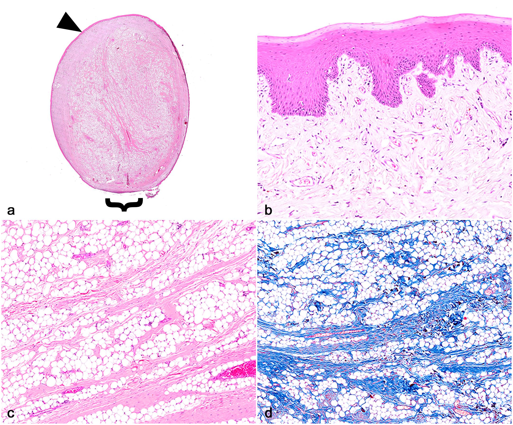
Sublingual fibrolipoma in a 10-year-old, spayed female, Bull Terrier dog. Case 1. (a) Subgross image of the mass. The submucosa is expanded by a well-demarcated, partially encapsulated neoplasm that is covered by hyperplastic mucosal epithelium (arrowhead) and has a narrow base (bracket). Hematoxylin and eosin (HE). (b) The mucosal epithelium is irregularly hyperplastic with rete pegs. HE. (c) The neoplasm consists of broad sheets of mature adipocytes transected by bands of cell-poor, mature collagen arranged as haphazard or arborizing bundles. HE. (d) The fibrous bands are highlighted with Masson’s trichrome stain.
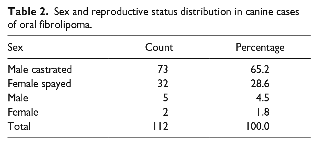
In 18.8% (21/112) of the dogs, the breed was not available; however, in the accessions with the breed information available, mixed breeds were the most common (21.4%, 24/112), followed by Labrador Retriever (10.7%, 12/112), Husky (5.4%, 6/112), Boxer (5.4%, 6/112), and Bichon Frise (3.6%, 4/112; Table 1). The remaining breeds were represented by 3 or less individuals. Oral fibrolipomas were reported in dogs with an average age of 10.2 years, ranging from 2- to 16-years-old (±2.6 years standard deviation). In terms of sex, 69.7% (78/112) of the cases were males (73 castrated males and 5 intact males), while only 30.3% (34/112) were females (32 spayed females and 2 intact females; Table 2). The most common oral location was the tongue, with 57.1% (64/112) of the cases, followed by the buccal mucosa (15.2%, 17/112), sublingual area (8.0%, 9/112), gingiva and lip (4.5%, 5/112 each), and palate (1 case; Fig. 2). In 9.8% (11/112) of the submitted cases, the location in the oral cavity was not otherwise specified (Table 3).
Distribution of frequency and percentage of dog breeds with the diagnosis of oral fibrolipoma.
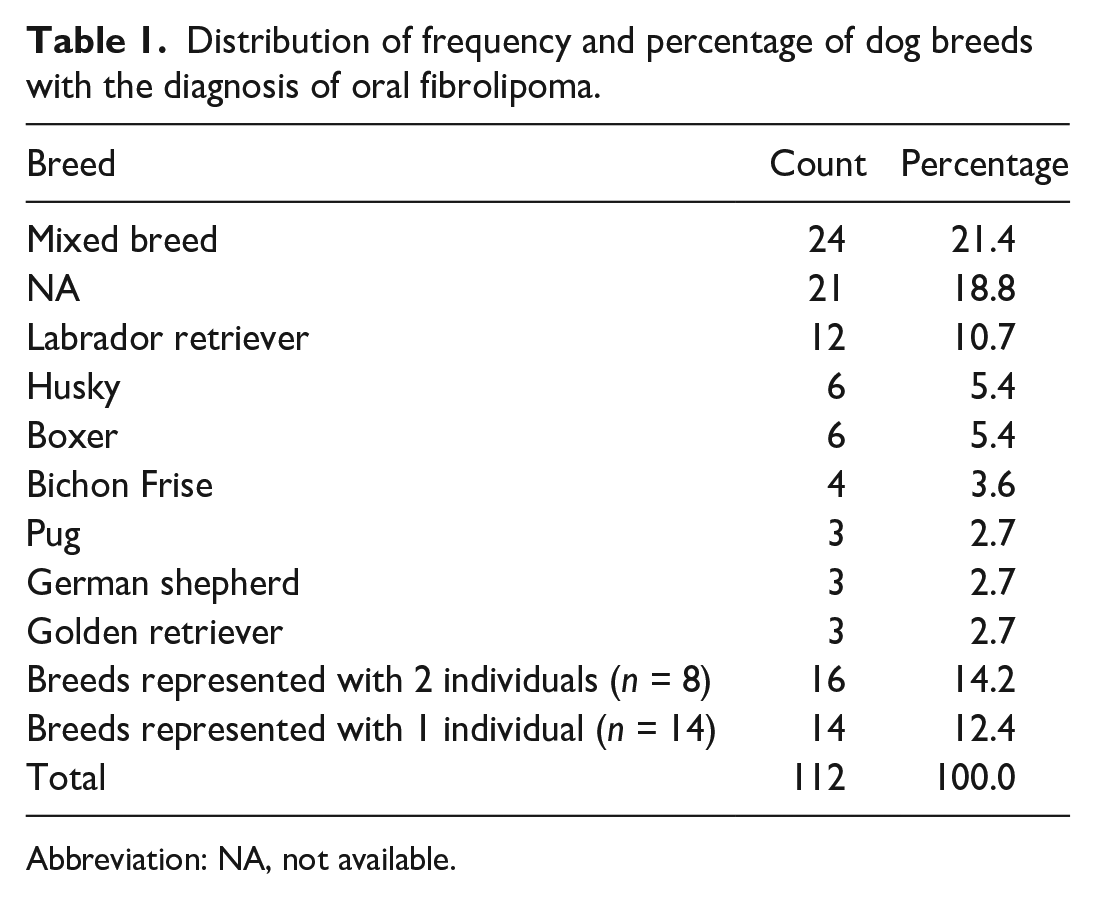
Abbreviation: NA, not available.
Sex and reproductive status distribution in canine cases of oral fibrolipoma.
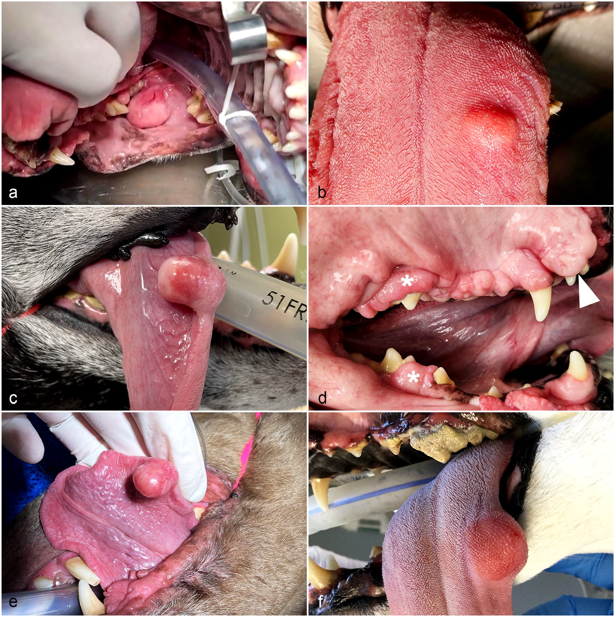
Macroscopic features of canine oral fibrolipomas. (a) A 2-cm diameter pedunculated sublingual mass in a 10-year-old, spayed female, Bull terrier dog. Case 1. (b) A 1.6-cm diameter, sessile, dorsal lingual mass in a 16-year-old, castrated male, pug dog. Case 20. (c) A 2.6-cm diameter, right ventral lingual mass in a 9-year-old, castrated male, Border collie dog. Case 21. (d) A 3-cm diameter, multilobulated, pale pink mass within the buccal mucosa (arrowhead), and labial and dorsal gingiva in an 8-year-old, castrated male, boxer dog. This dog also has gingival hyperplasia (asterisks). Case 24. (e) A 0.7-cm diameter, raised, pink, pedunculated lingual mass within the ventral surface in a 9-year-old, castrated male, Chesapeake Bay retriever dog. Case 25. (f) A 1.8-cm diameter, sessile, submucosal mass within the left caudal aspect of the tongue in a 13-year-old, spayed female, Siberian husky dog. There is marked dental calculus and periodontal disease. Case 26.
Distribution of frequency and percentage of oral fibrolipoma anatomical locations in dogs.
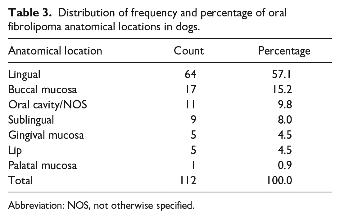
Abbreviation: NOS, not otherwise specified.
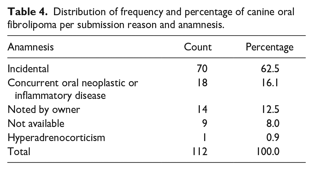
In 62.5% (70/112) of the cases, the presence of the tumor was reported as an incidental finding during physical examination, dental cleaning, or determined by the veterinarian (Supplemental Table S1, Table 4). In 18 cases (16.1%), the fibrolipoma occurred with a concurrent inflammatory disease (stomatitis, gingivitis, or periodontal disease) or other neoplastic or nonneoplastic proliferative condition, including oral malignant melanoma, squamous cell carcinoma, osteosarcoma, fibroepithelial polyp, and fibromatous hyperplasia of the gingival ligament (Supplemental Table S1, Table 4). In 12.5% (14/112) of the cases, oral fibrolipoma was noted by the owners with a reported progression ranging from several weeks to a year (Supplemental Table S1, Table 4). The anamnesis or submission reason was unavailable in 8.0% of the cases (9/112). The tumor dimensions ranged from 0.2 to 8 cm in diameter, with a mean diameter of 1.7 cm (±1.1 cm standard deviation; Supplemental Table S1). The anatomical location of the tumor only differs significantly among the breeds (chi-square test,P < .001), but not in dogs from different sexes (chi-square test, P = .823), ages (chi-square test, P = .919), and anamnesis or reason for submission (chi-square test, P = .353).
Distribution of frequency and percentage of canine oral fibrolipoma per submission reason and anamnesis.
Discussion
Benign and malignant adipocytic tumors located in the oral cavity or the tongue have been described sporadically in dogs. Lipomas represented 1.5% (18/1196) of the total lingual lesions in dogs in a retrospective study. 3 Another publication documented a single case of a lingual infiltrative lipoma, in a retrospective study of lingual tumors in 42 dogs. 20 There is a single case report describing a chondrolipoma, a rare adipocytic tumor in the tongue of the dogs. 5 Liposarcoma is sporadically documented within the oral cavity, with a predilection for the tongue.3,18 We obtained no cases of oral fibrolipomas diagnosed in dogs and cats in a comprehensive search of Google, PubMed, CAB Direct, Web of Science, and Scopus, using search terms “oral fibrolipoma,” “lingual fibrolipoma,” “dog,” and “canine,” suggesting that this condition has not been reported in dogs.
The lack of oral and lingual fibrolipomas reported in veterinary medicine supports its rarity at these anatomical locations. In human medicine, selected case reports describe this benign adipocytic tumor as a distinctive “rare histological entity” within the oral cavity and tongue.13,14 In humans, oral fibrolipomas are more commonly reported after the fourth decade of life and are identified within different anatomical locations including the buccal mucosa and vestibule, alveolus, gingiva, tonsil, lips, tongue, palate, sublingual space, and retromolar location.13,14 The buccal mucosa and tongue are considered the first and second most common locations for oral fibrolipomas in humans.7,9 In our cases, the mean age of the dogs with this lesion was 10.2 years, supporting that older dogs are more commonly affected. However, the most common location recorded in canine patients was the tongue followed by the buccal mucosa.
In human patients, oral fibrolipomas are slightly more prevalent in females.21,13,6 Tumors are asymptomatic and slow growing, but some can impede mastication and speech.13,14 Medical attention is requested by the patients due to functional impairment or aesthetics. 13 In our cases, the majority of the dogs were male and the tumor was an incidental finding in 62.5% of the cases. Only one case reported that the dog had difficulty ingesting hard pellets (case 37). These findings confirm that canine oral fibrolipomas are incidental and subclinical, like their human counterpart.
Human fibrolipomas are oval, sessile or pedunculated, soft to firm masses with a yellow cut surface.7,9,14,13 Most of the reported tumors are less than 3 cm in diameter but can grow more. 21 In dogs, the average size of tumors was 1.7 cm, with an exceptional case in which the sublingual tumor reached a diameter of 8 cm in a Rottweiler (case 52). The histological features reported in humans are straightforward, with the majority of the neoplasm composed of well-differentiated lobules of mature adipocytes dissected by bands of fibrous connective tissue.13,14 All the cases included in our study fulfilled this histological criteria. In humans, oral lipomas are classified into 3 subtypes: diffuse affecting deeper tissues, superficial, and encapsulated.14,16 Such classifications have not been described for fibrolipomas in veterinary medicine, unlike infiltrative and intramuscular lipomas which are recognized histological entities. 17 We identified 3 cases of infiltrative fibrolipoma and 1 case of intramuscular fibrolipoma in the tongue. We speculate that their biological behavior mimics their adipocytic counterpart; however, this remains unknown.
The pathogenesis of lipomas and fibrolipomas is obscure. Inciting factors postulated in humans include an inherited predisposition, degeneration of the adipose tissue, hormonal imbalances, infarction, previous infections, or chronic irritation that leads to rearrangement of the adipose tissue and fibrosis. 13 Oral fibrolipomas are proposed to be caused by metabolic imbalances, anomalous location of fetal adipose cells, hormone therapy, or trauma.14,21 The anomalous location of adipocytic fetal cells and sequelae or chronic panniculitis with extensive fibrosis are possible factors in the pathogenesis of oral fibrolipomas. In most of the cases included in this study, the fibrolipomas presented as an incidental finding without an underlying disease. The previous existence of a lipoma with extensive fibrosis secondary to the trauma incited during mastication, injuries, or chronic inflammation within the oral cavity might contribute to the development of this lesion. However, determining if fibrolipomas are true neoplasms or abnormal storage of adipocytic cells due to the aberrant fetal migration has not been established. This case series does not suggest a pathogenesis of oral fibrolipomas in dogs, since the history of each case is limited.
The location of the tumors differed only among the dog breeds; however, one must be careful in the interpretation of these findings. For more than 40% of the animals in this study, the breed was not available or was undetermined (mixed breed). The tongue was the most common location among the mixed breed dogs (58%; 14/24) and Labrador Retrievers (58%; 7/12). Many breeds were represented with a small number of individuals, for example, 8 breeds were represented with only 2 individuals (Collie, Beagle, Jack Russell Terrier, Border Collie, Rottweiler, Havanese, Boston Terrier, and Bull Terrier), and fibrolipomas were located in the tongue in 56% (9/16) of these cases. An additional 14 breeds were represented with only 1 individual, and fibrolipomas were located in the tongue in 50% (7/14) of them. Thus, our statistical significance is derived from the small number of individuals per breed category. We can conclude that the tongue is the most common anatomical location, and oral fibrolipomas are present in a wide range of dog breeds.
Diagnosis in humans relies on histopathology, imaging techniques (computed tomography, ultrasound, or magnetic resonance imaging), and cytological evaluation of fine needle aspirates.7,1,10,22,19 The histological features of fibrolipomas in dogs are straightforward; however, benign neoplastic and nonneoplastic lesions within the tongue of dogs should be considered macroscopic differential diagnoses.2,3,11,15,20,12
The treatment of choice is surgical removal; since fibrolipomas in human patients are amenable to surgical excision and rarely recur, the prognosis is considered favorable in these cases.13,14 One of the biggest limitations of our case series is the lack of follow-up information from each case. If similar to their human counterpart and to other types of lipomas in the skin, oral fibrolipomas are likely to be cured by surgical excision. 17
Supplemental Material
sj-pdf-1-vet-10.1177_03009858241273238 – Supplemental material for Oral fibrolipoma in dogs: Retrospective case series study and comparative review
Supplemental material, sj-pdf-1-vet-10.1177_03009858241273238 for Oral fibrolipoma in dogs: Retrospective case series study and comparative review by Daniel Felipe Barrantes Murillo, Alexis Berrocal, Cynthia Bell, Daniel R. Rissi, Linden E. Craig, Erin A. Graham, Emily J. Brinker and Tatiane Terumi Negrão Watanabe in Veterinary Pathology
Footnotes
Authors’ contributions
DFBM and TTNW designed the study and AB contributed to the supervision of the study. DFBM, AB, CB, DRR, LEC, EAG, EJB, and TTNW collected the data and performed the histopathological evaluations. DFBM performed the data analysis. DFBM, AB, and TTNW wrote the manuscript with the collaboration of all the authors. All the authors reviewed and critically edited the manuscript. All authors read and approved the final manuscript.
Declaration of Conflicting Interests
The author(s) declared the following potential conflicts of interest with respect to the research, authorship, and/or publication of this article: Cynthia Bell is the owner of Specialty Oral Pathology for Animals, Alexis Berrocal is the owner of Histopatovet, and Tatiane Terumi Negrão Watanabe is employed at Antech Diagnostics, Mars Petcare Science & Diagnostics. The remaining authors declared no potential conflicts of interest concerning the research, authorship, and/or publication of this article.
Funding
The author(s) received no financial support for the research, authorship, and/or publication of this article.
ORCID iDs
Supplemental Material for this article is available online.
References
Supplementary Material
Please find the following supplemental material available below.
For Open Access articles published under a Creative Commons License, all supplemental material carries the same license as the article it is associated with.
For non-Open Access articles published, all supplemental material carries a non-exclusive license, and permission requests for re-use of supplemental material or any part of supplemental material shall be sent directly to the copyright owner as specified in the copyright notice associated with the article.