
Abstract
The brain of a 6-year-old Holstein cow, which showed progressive neurologic symptoms during several months, was examined by histopathologic methods. Many round or oval-shaped cytoplasmic inclusions were observed, mainly in neurons of the temporal lobe and the hippocampus. Those inclusions were faintly eosinophilic with hematoxylin and eosin and positive with Bielschowsky's silver stain. Immunohistochemically, the inclusions were recognized by antiubiquitin and antiphosphorylated tau antibodies. Ultrastructurally, the inclusions were globular and well demarcated from the rest of the cytoplasm, lacked limiting membranes, and were mainly composed of straight fibrils about 15 nm in width. The structure of the inclusions was similar to that of Pick bodies in Pick's disease of humans. The pathogenesis of this bovine condition is not known.
A 6-year-old Holstein–Friesian cow was presented with a progressive neurologic disease. Initially, the cow had only occasionally shown fine tremors or spasms from head to shoulder. In recent months, she began to thrash all limbs violently and then fall down suddenly in apparent agony. Such behavior appeared every 3 or 4 days, with episodes lasting a few minutes each time. The cow returned to normal immediately after, and both rumination and milking occurred without problems. During the seizures, she did not lose consciousness. The course of her symptoms deteriorated day by day such that after 4 months, their duration reached 15–20 minutes and occurred frequently each day. Except for definite ataxia, her behavior was normal. However, she had numerous skin abrasions. No hematologic or clinical chemistry abnormalities were found. On the basis of the progressive nature of the disease, the owner decided to eliminate her from the herd.
Immediately after meat inspection, the brain was removed and fixed with 15% neutral buffered formalin. From the frontal to the occipital lobe, 1-cm-thick coronal slices were taken from each hemisphere, and every sliced face was embedded in paraffin by conventional methods. Seven-micrometer-thick serial sections were stained with hematoxylin and eosin (HE), Klüver–Barrera (for myelin and Nissl's bodies), Holzer, Congo red, periodic acid–Schiff (PAS), and Bielschowsky's silver stains. Immunohistochemical studies, by an indirect immunoperoxidase method utilizing avidin-biotin complex on paraffin sections, were carried out with rabbit antiubiqutin polyclonal antibody (DAKO, Clostrop, Denmark) and mouse antiphosphorylated tau monoclonal antibody (Sigma Immuno Chemicals, St. Louis, MO). For electron microscopic examination, small pieces of tissue sampled from the formalin-fixed hippocampus were postfixed in 1% osmium tetroxide in 0.1 M phosphate buffer (pH 7.3) and embedded in epoxy resin. Ultrathin sections were stained with uranyl nitrate and lead citrate and examined with a Hitachi H-600 transmission electron microscope at an accelerating voltage of 75 kV. The brains of two age-matched Holstein cows were used as controls.
Grossly, in the afflicted cow the volume of the parietal and temporal lobes in each hemisphere had decreased moderately, although no overt atrophy of the gyri was apparent. The affected brain weighed 450 g; each control brain weighed about 480 g. Gross examination of the coronal slices revealed that the white matter had decreased slightly in volume compared with controls, that there was ventricular dilatation, and that the gray matter showed slight atrophy in the parietal and temporal lobes. No significant changes were observed in other organs or tissues.
Histologically, the remarkable findings in the affected cow were the presence of numerous neuronal cytoplasmic inclusions. These bodies were faintly eosinophilic and homogeneous with HE, varied in shape from round to oval or crescentic, and tended to displace the nucleus to one side. The inclusions were observed most frequently in the parahippocampal gyri, the fascia dentata (Fig. 1), and the external granular layers of the temporal lobes (Fig. 2). They also occurred rarely in the substantia nigra and the frontal lobe. No neuronal inclusions were formed in the occipital lobe, caudate nucleus, or thalamus. The inclusions were positive with Bielschowsky's silver stain (Fig. 3) but were negative with PAS and Congo red stains. In the temporal and frontal lobes, there was a laminar loss of neurons in several regions of the external granular and pyramidal layers. Some of the surviving neurons were atrophied, whereas other swollen ones were scattered in the gray matter. Astrogliosis was observed diffusely throughout the white matter. In the subcortical nuclei, a slight decrease in neurons was observed in comparison with controls. The degenerative neurons in the cortex or subcortical nuclei lacked inclusions but often had swollen axons and finely vacuolated cytoplasm and contained many silver-positive granules. Except for the occipital lobes, little demyelination was observed. In the hippocampus, there was an increase in fibrous astrocytes, and neurons with swollen somata or exhibiting central chromatolysis were seen occasionally. The neuronal inclusions were recognized by both antiphosphorylated tau and antiubiquitin antibodies.
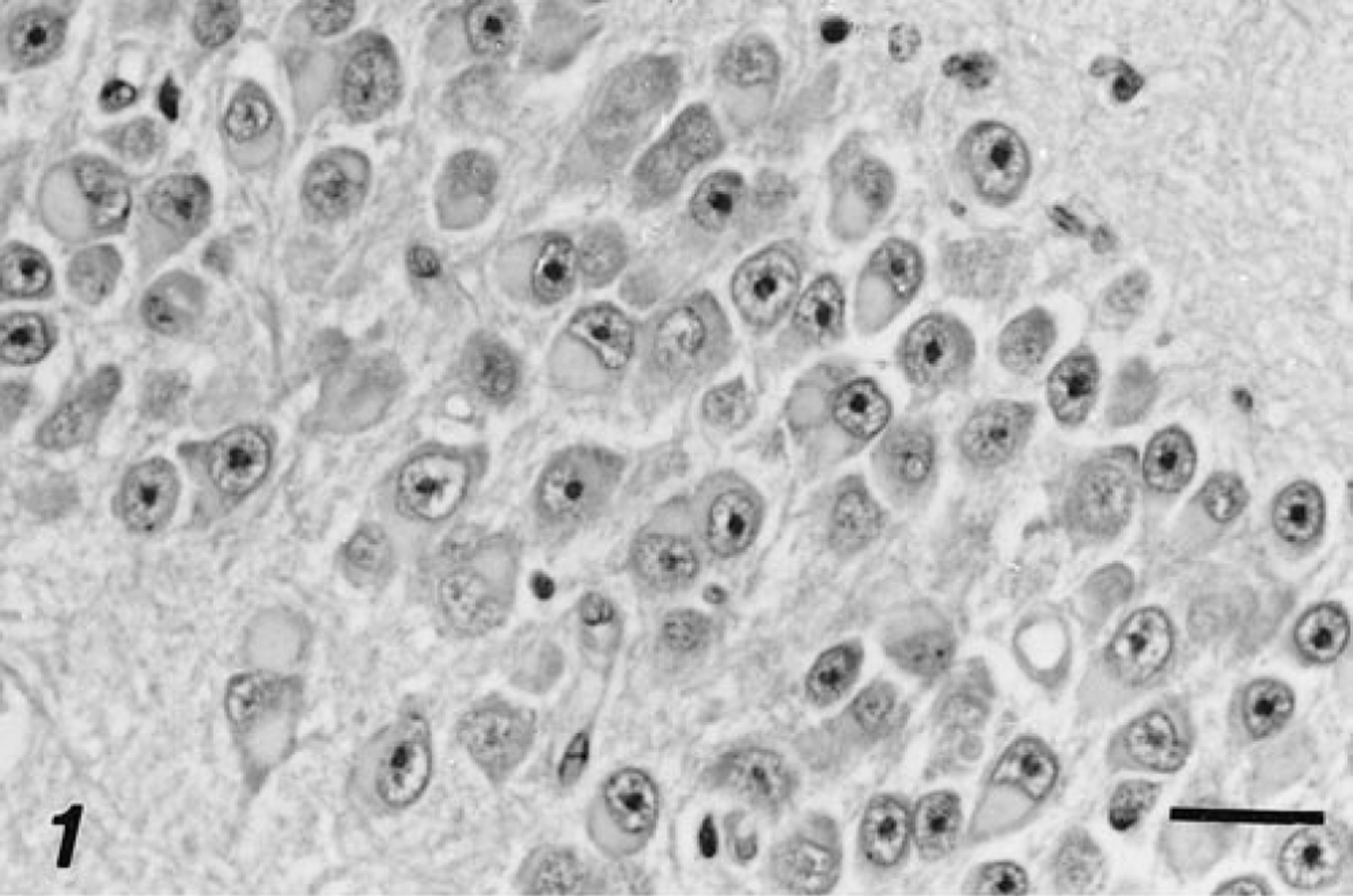
Fascia dentata; cow. The neuronal inclusions are faintly eosinophilic, homogeneous, and varied in shape but typically globular and tend to displace the nucleus to the periphery in the granular cells. HE. Bar = 25 μm.
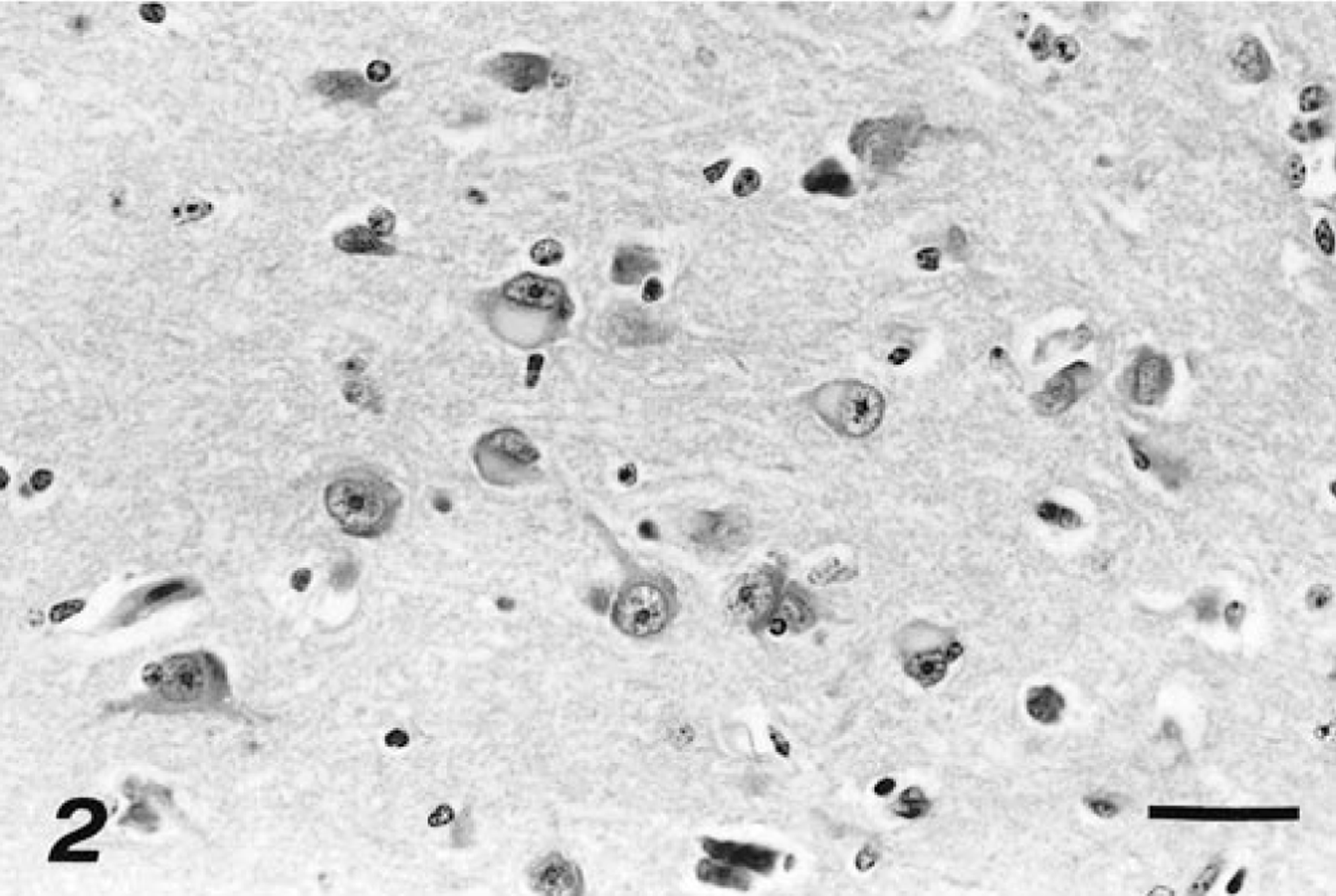
Pyramidal layer of the hippocampus; cow. Some neurons contain round or oval inclusions in their perikarya. HE. Bar = 38 μm.
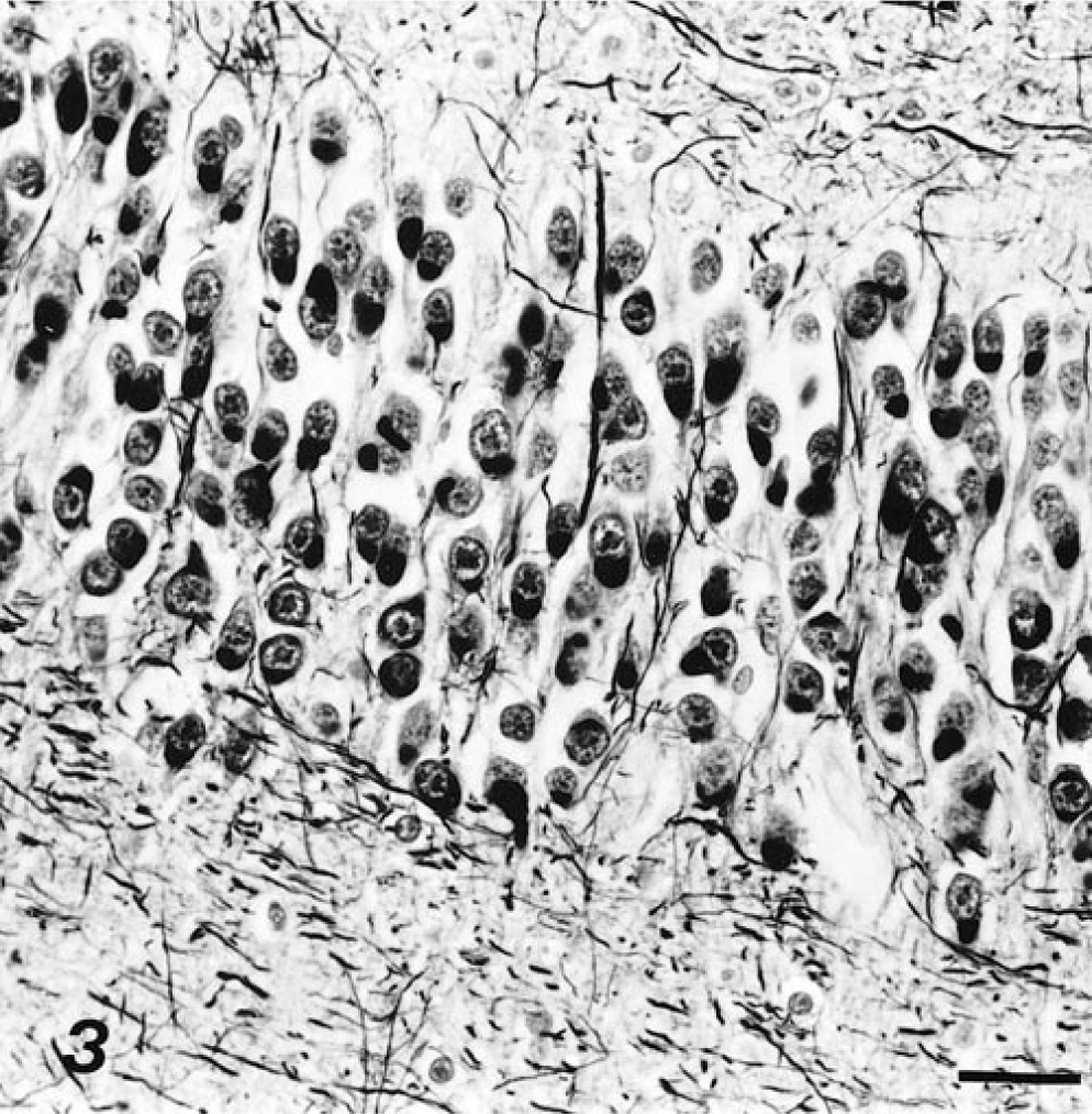
Fascia dentata; cow. Numerous argyrophilic globules are visible in the granular cells. Bielschowsky's silver methods. Bar = 26 μm.
By electron microscopy, the argyrophilic inclusions were well-demarcated globular deposits in the cytoplasm. All were non-membrane bound and contained straight fibrils mixed with cytoplasmic organelles, mainly ribosomes and vesicles (Fig. 4). The fibrils were about 15 nm in diameter, varied in length, and lacked side branches and periodic twists (Fig. 5). In particular, spiral or periodic structures comparable with those found in Alzheimer's neurofibrillary tangles were not observed. The proportion of fibrils among cytoplasmic organelles was varied.
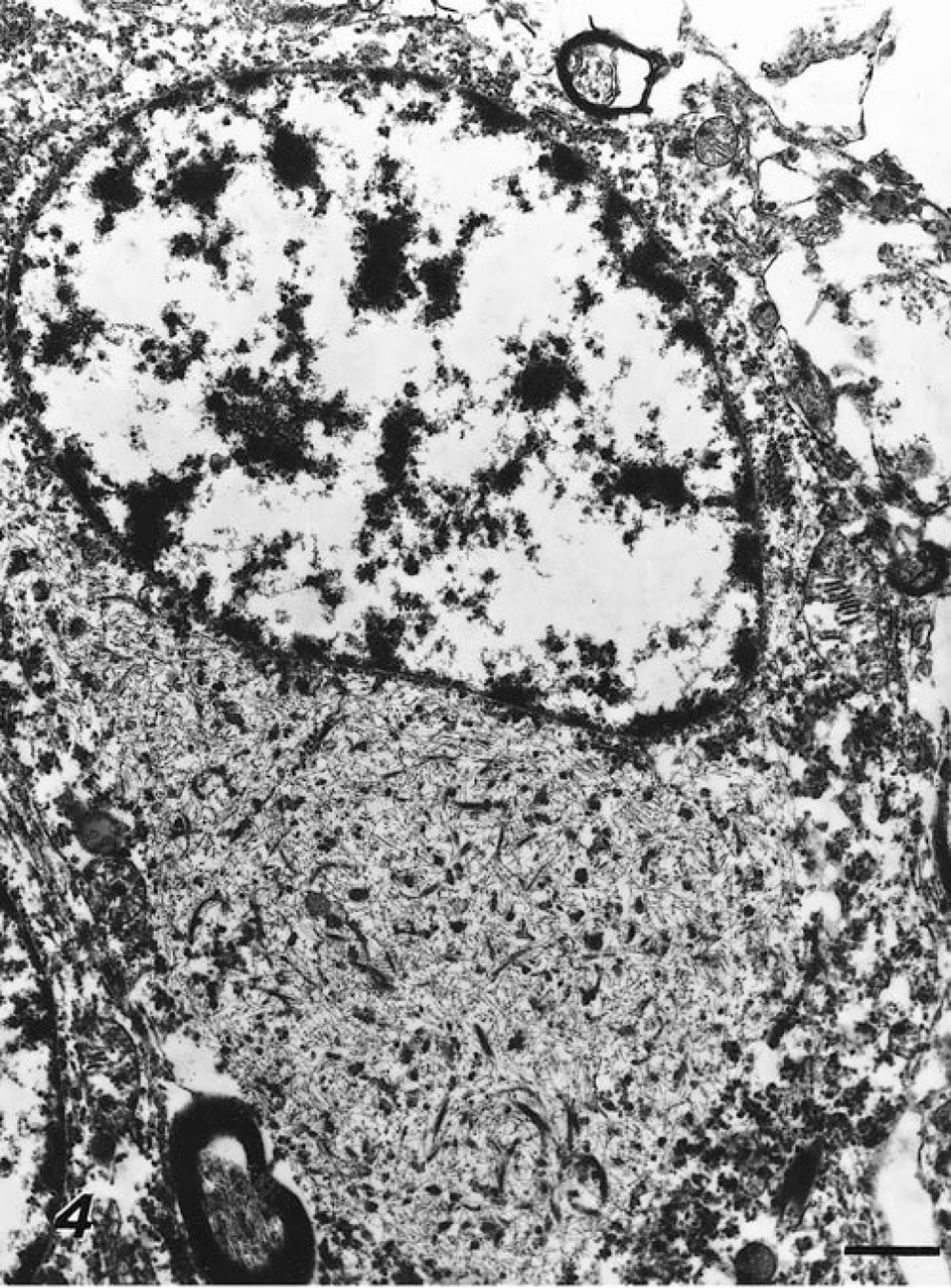
Fascia dentata; cow. The neuronal inclusion is well demarcated from the rest of the cytoplasm, lacks a membrane, and mainly consists of straight fibrils. The nucleus is displaced to the periphery and indented. Uranyl nitrate and lead citrate. Bar = 1.2 μm.
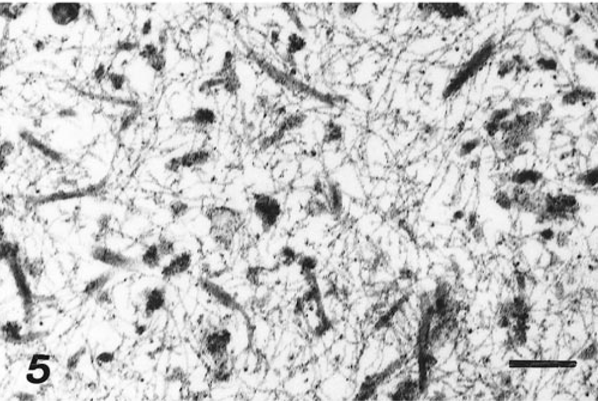
High magnification of the central portion of the structure illustrated in Fig. 4. Bar = 350 nm.
A review of the literature revealed no instance of spontaneous encephalopathy with argyrophilic inclusions reported in animals. In Pick's disease, a form of presenile dementia in humans, neuronal argyrophilic inclusions (Pick bodies) are found in the brain of about half of all patients. The histopathologic, immunohistochemical, and ultrastructural characteristics of the neuronal inclusions in this cow's brain resembled those of Pick bodies. 1 3 5–8 Besides Pick's disease, primary cerebral cortical degeneration with fibrillary inclusions in neuronal cytoplasm also occurs in Alzheimer's disease and diffuse Lewy body disease in humans. 3 Alzheimer's neurofibrillary tangles are basophilic or eosinophilic in HE stain and positive for Congo red and mainly consist of paired helical filaments in electron microscopic preparations. Lewy bodies are eosinophilic inclusions surrounded by clear halos in HE stain, do not react with tau protein, and ultrastructuraly consist of 9-nm fibrils that are arranged radially at the rim. Thus, the nature of the inclusions in this bovine case differs from those of Alzheimer's neurofibrillary tangles and Lewy bodies. Furthermore, encephalopathy with dementia in humans, including Pick's disease, is determined on the basis of complicated clinical symptoms and morphologic diagnostic schemes. 2–4, 6 9 Although the cow in the present case showed progressive neuronal symptoms, it was impossible to determine that she suffered dementia in the clinical sense. Thus, it seems improper to apply the names of those human diseases directly to a case of spontaneous encephalopathy in an animal, let alone infer a comparable pathogenesis.
Generally, in domestic animals, their deaths are not necessarily correlated with their natural life spans because most livestock are slaughtered. For this reason, chronic progressive diseases of the nervous system will not be readily found among livestock. Our report of a natural case of encephalopathy with argyrophilic inclusions in a cow indicates that such disorders occur and may be detected in animals, thereby suggesting their sporadic occurrence in other animals made available for laboratory studies.