
Abstract
Mannheimia granulomatis was isolated for 10 months from the milk of a cow with elevated somatic cell counts. The infection was self-limiting. Phenotypic and molecular characteristics of the isolate were determined.
Keywords
In May 2007, a combined milk sample from all 4 quarters from a cow in the second month of pregnancy in her second lactation was submitted to the laboratory due to a high somatic cell count (SCC), enumerated on a commercial counter, a of 1,008,000. Gel formation at the California mastitis test (http://www.infovets.com/demo/demo/dairy/d100.htm) was observed only in the sample from the left forequarter, indicating that this quarter was the source of the high SCC in the combined sample.
The significance of SCC as an indicator of udder infection and its deleterious impact on dairy products has been reviewed. 8 Briefly, SCC higher than 200,000/ml may indicate an intramammary infection. A SCC higher than 600,000 is often caused by major mammary pathogens, such as Staphylococcus aureus and Streptococcus spp. A high SCC causes significant economic damage to the dairy industry by decreased product quantity and quality, due primarily to proteolytic activities. Moreover, the shelf life of milk and dairy products is shortened. The bulk tank SCC is an indication of the prevalence of subclinical mastitis in the herd, because milk from cows suffering from clinical mastitis is discarded.
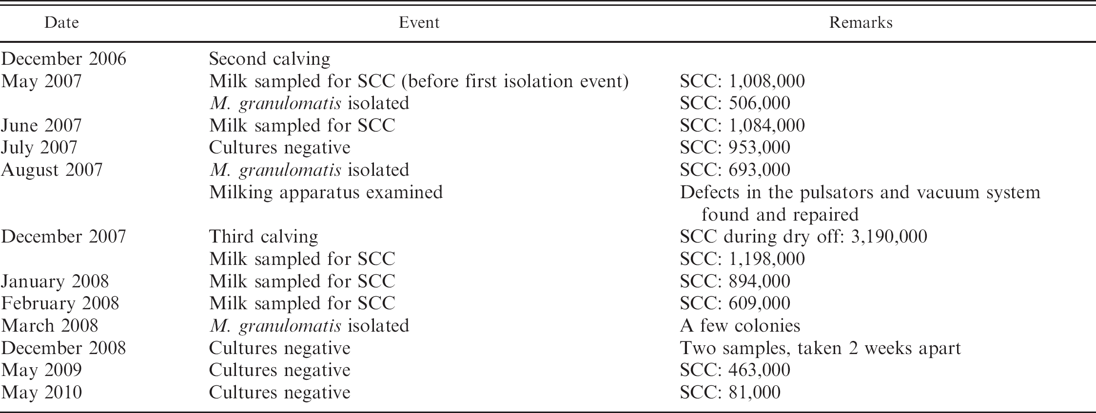
The cow in the current study was located on a family-managed dairy farm consisting of 48 Israeli Holstein dairy cows, 18 of which were primiparous cows. The management on the farm and the hygienic conditions were very good. Milking hygiene included predipping, wiping, and postmilking teat dip. The farm was under continuous monitoring by the National Service for Udder Health and Milk Quality. A microorganism, later identified as Mannheimia granulomatis, was isolated in pure culture from the left forequarter of the cow in question. According to the policy adopted in Israel to abstain from antibacterial therapy in cases of subclinical mastitis, except for infections with specific bacteria in which a positive outcome is expected, the cow was not treated. It kept shedding the microorganism, exclusively from the same quarter, at least until March 2008. Elevated SCC was found throughout this period, even in a sample from which M. granulomatis was not isolated. Mannheimia granulomatis was not isolated from other cows on the farm in spite of the affected cow's not being isolated. In fact, between April and June 2007, 40 milk samples from other cows, all from subclinical mastitis cases, were submitted to the laboratory; 26 yielded no growth, and 1 was contaminated. The following bacteria were isolated from the other samples: coagulase-negative Staphylococcus spp. (9), Streptococcus dysgalactiae (1), Enterococcus faecalis (1), Escherichia coli (1), and Corynebacterium bovis (1). A detailed timeline of the event and follow up are presented in Table 1.
Bacteriologic examination of the milk samples and presumptive identification of bacteria were performed as previously described. 10 The isolate stained Gram negative, was oxidase and catalase positive, did not grow on MacConkey agar, was hemolytic on 5% sheep blood agar, and was inert and nonmotile in semisolid glucose–cystine–trypticase agar. The following phenotypic characteristics were found.
Gram-negative rods. b Positive results: potassium nitrate reduction, esculin, 4-nitrophenyl-βD-galactopyranoside. Negative results: indole production, D-glucose (fermentation), L-arginine, urea, gelatin, assimilation of D-glucose, L-arabinose, D-mannose, D-mannitol, N-acetylglucosamine, D-maltose, potassium gluconate, capric acid, adipic acid, malic acid, trisodium citrate, phenylacetic acid. Identification: Mannheimia haemolytica (85.9%, T = 0.56) at low discrimination with Brevundimonas vesicularis (11.7%, T = 0.53).
Corynebacteria. c Positive results: potassium nitrate reduction, 2-naphtyl-phosphate, 2-naphthyl-βD-galactopyranoside, esculin, fermentation of D-glucose, D-ribose, D-xylose, D-mannitol, D-maltose, and D-saccharose. Negative results: pyrazine carboxamide, pyroglutamic acid-β-naphthylamide, naphthol ASBI-glucuronic acid, 2-naphthyl-αD-glucopyranoside, 1-naphthyl-N-acetyl-βD-glucosamine, urea, gelatin, fermentation of D-lactose and glycogen.
Anaerobes. d Positive results: 4-nitrophenyl-βD-galactopyranoside, 4-nitrophenyl-βD-glucopyranoside, potassium nitrate reduction, 2-naphtyl-phosphate, L-arginine-β-naphthylamide, L-alanyl-L-alanine-β-naphthylamide, L-glycine-β-naphthylamide. Negative results: urea, L-arginine, 4-nitrophenyl-αD-galactopyranoside, 4-nitrophenyl-βD-galactopyranoside-6-phosphate-2CHA, 4-nitrophenyl-αD-glucopyranoside, 4-nitrophenyl-αL-arabinofuropyranoside, 4-nitrophenyl-βD-glucuronide, 4-nitrophenyl-N-acetyl-βD-glucosamine, glutamic acid decarboxylase, 4-nitrophenyl-αL-fucopyranoside, indole production, L-proline-β-naphthylamide, L-leucyl-L-glycine-β-naphthylamide, L-phenylalanine-β-naphthylamide, L-leucine-β-naphthylamide, pyroglutamic acid-β-naphthylamide, L-tyrosine-β-naphthylamide, L-histidine-β-naphthylamide, L-glutamyl-L-glutamic acid β-naphthylamide, L-serine-β-naphthylamide, fermentation of D-mannose and D-raffinose.
Bacteriologic and somatic cell count (SCC; cells/ml milk) follow-up of the cow affected by subclinical mastitis caused by Mannheimia granulomatis.
Enterobacteriaceae. e Positive results: esculin, para-nitro-phenyl-phosphate, para-nitro-phenyl-α-β-glucoside, para-nitro-phenyl-β-galactoside, para-nitro-phenyl bis-phosphate, para-nitro-phenyl-arabinoside, fermentation of mannitol, saccharose, and sorbitol. Negative results: L-arginine, urea, para-nitro-phenyl-xyloside, para-nitro-phenyl-phosphorylcholine, para-nitro-phenyl-glucuronide, pa-ra-nitro-phenyl-N-acetyl glucosamide, proline nitroanilide, γ-L-glutamyl p-nitroanilide, p-nitro-DL-phenylalanine, glycine, citrate, malonic acid, triphenyl tetrazolium chloride, lysine, arabinose, mannose, melibiose, rhamnose, adonitol, galactose, inositol. Identification: Mannheimia haemolytica, 0.9899.
When the same analyte was tested with different kits, results were consistent, except for D-glucose fermentation. This reaction was negative with the Gram-negative rod kit b and positive with the corynebacteria kit, c which was confirmed by performing the tests in triplicate.
Results lead to a tentative identification of the isolate as M. granulomatis. To confirm this identification, 2 isolates from 2007 were submitted to molecular identification by 16S ribosomal DNA (rDNA) sequencing. Briefly, total bacterial DNA was extracted from colonies using a commercial kit, f according to the manufacturer's instructions. Polymerase chain reaction amplification of the first 800 base pairs of the 16S rDNA was performed with the following universal primers 6 : forward sequences (4F: TTGGAGAGTTTGATCCTGGCTC), reverse sequences (801R: GGCGTGGACTTCCAGGGTATCT). The polymerase chain reaction was started with an activation step at 90°C for 15 min, followed by 35 cycles of denaturation at 94°C for 30 sec, annealing at 50°C to 60°C for 90 sec (60°C for the first 5 cycles, 55°C for next 5 cycles, and 50°C for the last 25 cycles), and extension at 72°C for 4 min, ending with a final extension step at 72°C for 10 min.
The amplicon product was verified with agarose gel electrophoresis, purified according to the manufacturer's instructions, g and sequenced using primer 4F on an automated DNA sequencer. h Obtained sequences were aligned and compared with archived NCBI sequences and with the Bioinformatics Bacterial Identification database (http://pbil.univ-lyon1.fr/bibi/). In addition, a partial sequence of the rpoB gene of the original isolate was obtained as previously described. 5
The sequence obtained from the molecular analysis of the 16S ribosomal RNA (rRNA) gene was deposited in GenBank (accession no. GQ844959). A match of 99% was found between this sequence and the 16S rRNA sequence from M. granulomatis strain W4672/1 16S and M. granulomatis strain ATCC 49244 16S. The sequence obtained from partial amplification of rpoB was deposited in GenBank (accession no. GQ856691). A match of 97% was found between this sequence and the rpoB sequence of M. granulomatis strain ATCC 49244.
In vitro antibacterial susceptibility testing was performed by the disk diffusion method, according to Clinical and Laboratory Standards Institute standards. 7 Inhibition zones were determined with an imaging instrument. i Interpretive criteria applied were those defined for M. haemolytica. The isolate was susceptible to all the antibacterial drugs tested except tetracyclines (Table 2).
The Pasteurellaceae family has undergone substantial changes in the past decade due to the application of molecular taxonomical tools. The genus Mannheimia comprises M. haemolytica, M. glucosida, M. ruminalis, M. varigena, and M. granulomatis. 2 The latter species has been associated with a potentially fatal infection defined as bovine focal proliferative fibrogranulomatous panniculitis, or “lechiguana” in south Brazil. 9 Isolates from this syndrome were shown to produce leucotoxin. 11 In addition, the microorganism was isolated in Australia from cattle with suppurative bronchopneumonia, jaw abscess, and actinobacillosis-like lesion of the tongue 3 ; in Denmark from a hare with pneumonia 1 ; and in Sweden from various infections, especially in the respiratory tract and the mouth, in Western roe deer (Capreolus capreolus). 4
Disk susceptibility testing results for the Mannheimia granulomatis strain isolated from the case of subclinical bovine mastitis. *
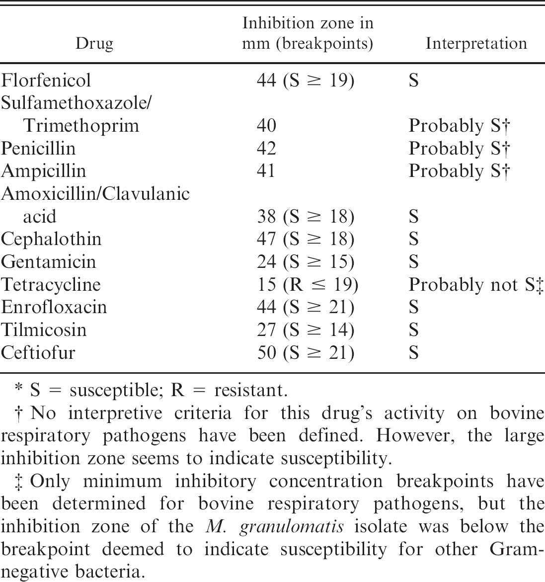
S = susceptible; R = resistant.
No interpretive criteria for this drug's activity on bovine respiratory pathogens have been defined. However, the large inhibition zone seems to indicate susceptibility.
Only minimum inhibitory concentration breakpoints have been determined for bovine respiratory pathogens, but the inhibition zone of the M. granulomatis isolate was below the breakpoint deemed to indicate susceptibility for other Gramnegative bacteria.
To the authors' knowledge, this is the first report of M. granulomatis causing subclinical mastitis in a cow. Although direct causality between the microorganism's presence and the high SCC counts was not established, the fact that they were both found in the same single quarter suggests the possibility that they are related. The fluctuations in SCC observed during the follow-up period are an accepted phenomenon. 8 The source of the microorganism's introduction to the farm and possibly to Israel is unknown. The intensive routine monitoring of Israeli dairy cattle by the National Service for Udder Health and Milk Quality makes its previous presence in the country very unlikely. Because this is thus far the only case in Israel, its importance relative to other causes of environmental or infectious mastitis cannot be established.
The fact that this microorganism was previously not isolated from similar cases may be due to its similarity to other Mannheimia spp. and Actinobacillus spp. Thus, the possibility of misidentification cannot be excluded, especially because some commercial kits that do not include M. granulomatis in their databases would identify the isolates as M. haemolytica. In fact, strains identified in Australia as M. granulomatis were previously thought to belong to other Mannheimia spp. or Actinobacillus spp. 3 Consequently, M. granulomatis should be considered as the possible etiology of typical actinobacillary lesions of the jaws and tongue and of infections caused by M. haemolytica, such as pneumonia and udder infections.
Footnotes
a.
Fossomatic™, FOSS, Hillerød, Denmark.
b.
API® NE, bioMérieux SA, Mercy l'Etoile, France.
c.
API® CORYNE, bioMérieux SA, Mercy l'Etoile, France.
d.
ID 32 A, bioMérieux SA, Mercy l'Etoile, France.
e.
BD BBL™ Crystal™, BD Diagnostic Systems, Sparks, MD.
f.
DNeasy Blood and Tissue Kit, Qiagen GmBH, Hilden, Germany.
g.
MEGAquick-spin™, iNtRON, Gyeonggi-do, Korea.
h.
ABI PRISM 3700, Applied Biosystems Inc., Foster City, CA.
i.
Biomic V![]() -104063871002200627" href="#bibr3-104063871002200627" id="a-41">
3
, Giles Scientific Inc., Santa Barbara, CA.
-104063871002200627" href="#bibr3-104063871002200627" id="a-41">
3
, Giles Scientific Inc., Santa Barbara, CA.