
Abstract
Mammary gland lesions characterized primarily as mammary teat atresia were observed in a Limousin beef cattle herd in eastern Texas. Atresia of multiple teats per mammary gland was reported in firstcalf heifers at the time of calving. Pathogens were not identified in eight mammary glands collected at slaughter. Histology of affected glands demonstrated superficial and deep perivascular inflammation and fibrosing dermatitis of teat and mammary gland skin that resulted in formation of the atretic lesions of glandular tissue. Institution of a horn fly (Haematobia irritans irritans (L.)) control program using insecticide-impregnated ear tags was associated with elimination of the problem from the herd.
Mastitis in prepartum heifers is not a common clinical entity. 2 , 5 , 12 , 15 , 17 Mastitis associated with suckling by penmates or abnormal hormonal conditions in heifers has been reported. However, most studies of heifer mastitis involve peripartum mastitis of dairy heifers contaminated by environmental pathogens such as Arcanobacterium pyogenes, Corynebacterium bovis, Staphylococcus aureus, Streptococcus spp., Escherichia coli, and Mycoplasma spp. Because of the infrequency of its occurrence, heifer mastitis in beef cattle has not been studied.
Veterinarians were called to investigate a mastitis outbreak in a purebred Limousin cattle herd. A year before calling for veterinary assistance, the rancher had implemented improvements in the nutrition, nematode parasite control, and mineral supplementation programs, including a loose-salt mineral supplement to correct a copper deficiency. These measures did not correct the mastitis problem. Mastitis was reported to affect first-calf heifers at calving, but was not observed in older cows maintained on the same pastures and subject to the same management practices. The problem was reported to have occurred over three breeding seasons. Except for four heifers purchased as replacement heifers, affected animals had been born and raised on the farm. At the time of present consultation, the frustrated owner had sold or slaughtered a group of 31, 21- to 25-month-old heifers (14 of which had “mastitis”) that represented his entire heifer calf crop for a breeding season. Mammary glands were collected from eight heifers at slaughter, 2–4 weeks after transferring their healthy calves to nurse cows. The glands were dissected and cultured for aerobic bacterial and mycoplasmal pathogens. Glandular and teat tissue from each quarter as well as mammary gland skin were fixed in buffered formalin, paraffin-embedded, and prepared for examination by light microscopy.
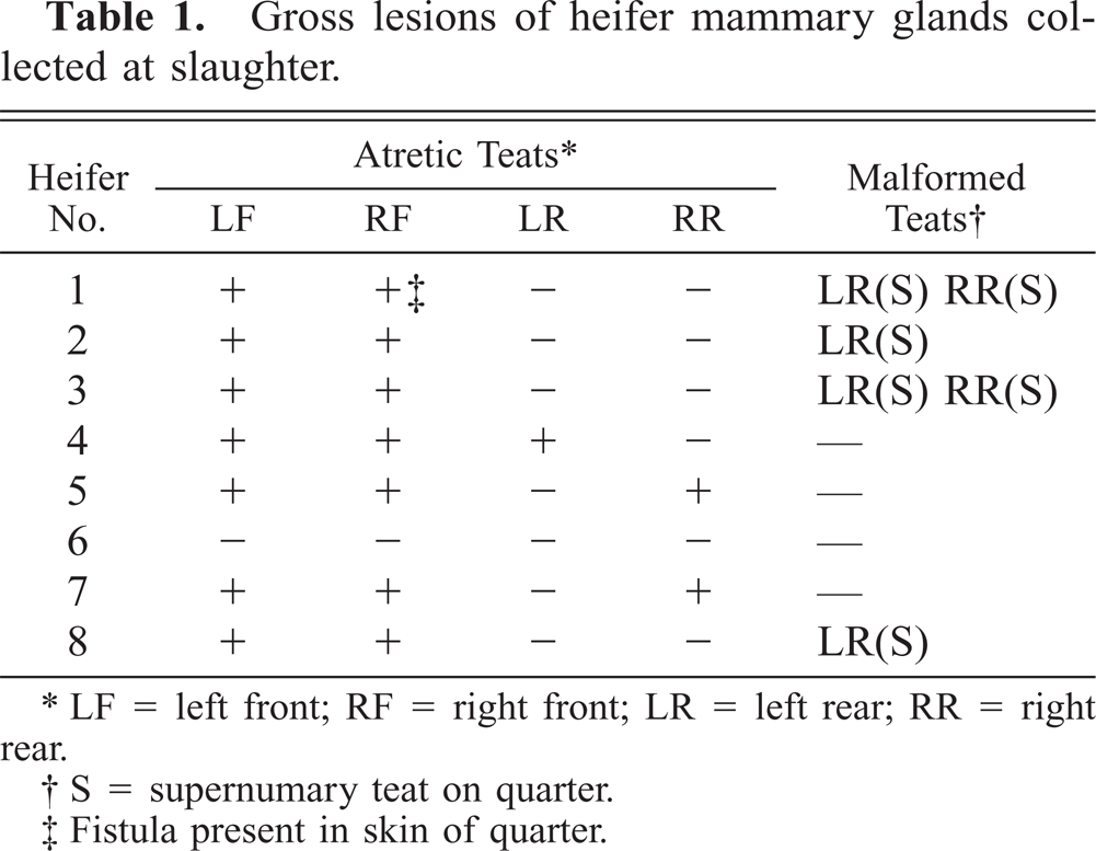
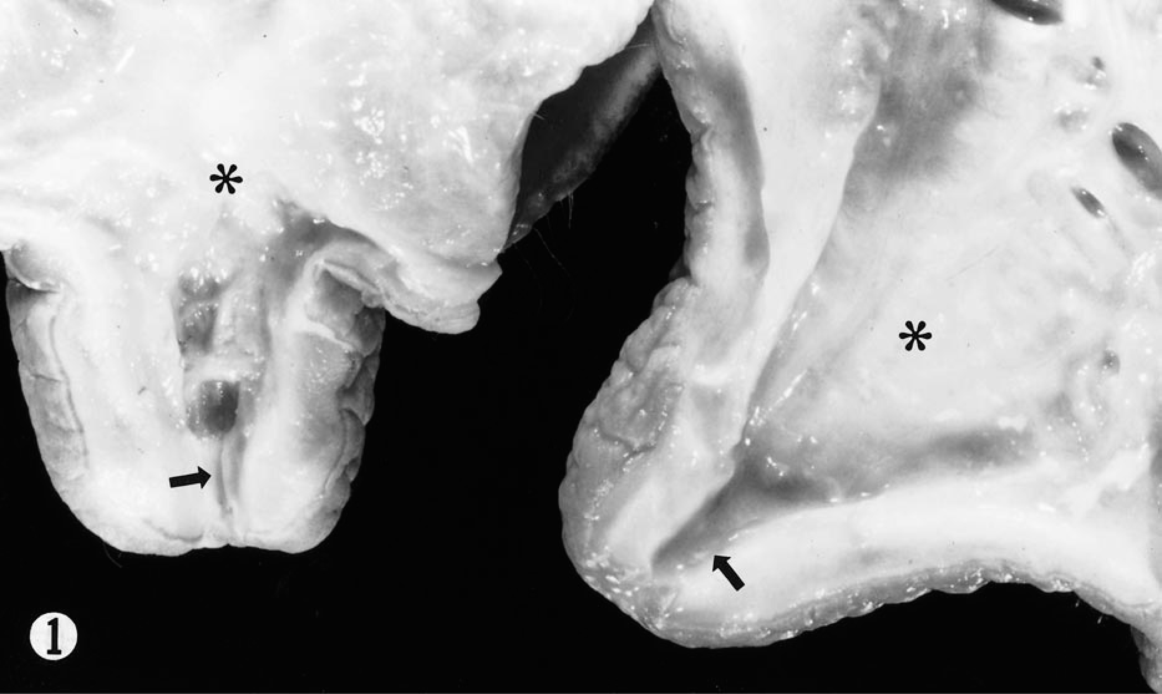
Of the eight glands collected at slaughter, one gland had grossly patent teats, but the mammary tissue was fibrotic. Each of the seven remaining glands had either two or three abnormal teats in which a cord of tissue was palpable in the area of the teat canal (Table 1), and from which no secretion could be expressed. In all of these seven glands, both front teats were abnormal, and in three glands, one rear teat was abnormal. In affected teats, the firm tissue that was palpated as a cord seemed to be mature connective tissue (Fig. 1), and no patency of the papillary duct was demonstrable by dissection. The connective tissue was continuous with a thickened, white, fibrous ventral dermis and subcutis of the mammary gland skin (Fig. 2). The mammary gland drained by atretic teats was fibrotic but not edematous or hyperemic, and no exudate was present. Several supernumerary teats were observed in the rear quarters, and none of these were continuous with the lactiferous sinus. One front quarter with an atretic teat contained a draining fistula into the adjacent mammary gland from which Pasteurella hemolytica and a beta-hemolytic Streptococcus sp. were isolated. No mycoplasmal or bacterial pathogens were isolated from cultured mammary tissue from all other quarters with atretic teats. No other defects were noted on any of the other seven mammary glands.
Gross lesions of heifer mammary glands collected at slaughter.
∗ LF = left front; RF = right front; LR = left rear; RR = right rear.
† S = supernumary teat on quarter.
‡ Fistula present in skin of quarter.
Mammary gland; Limousin heifer. An atretic teat from a front quarter (left) compared to a teat from the more-normal, patent, rear quarter (right) from the same gland. In the teat from the front quarter, the lactiferous sinus (asterisks) and the papillary duct (arrows) are occluded by fibrosis.
Mammary gland; Limousin heifer. Two affected front quarters from a 2-year-old heifer 4 weeks after calving are sectioned through the teat and lactiferous sinus. The atretic teats are adjacent to a thick band of subcutaneous fibrosis that extends to fill the lactiferous sinus. Connective tissue is increased in the mammary gland.
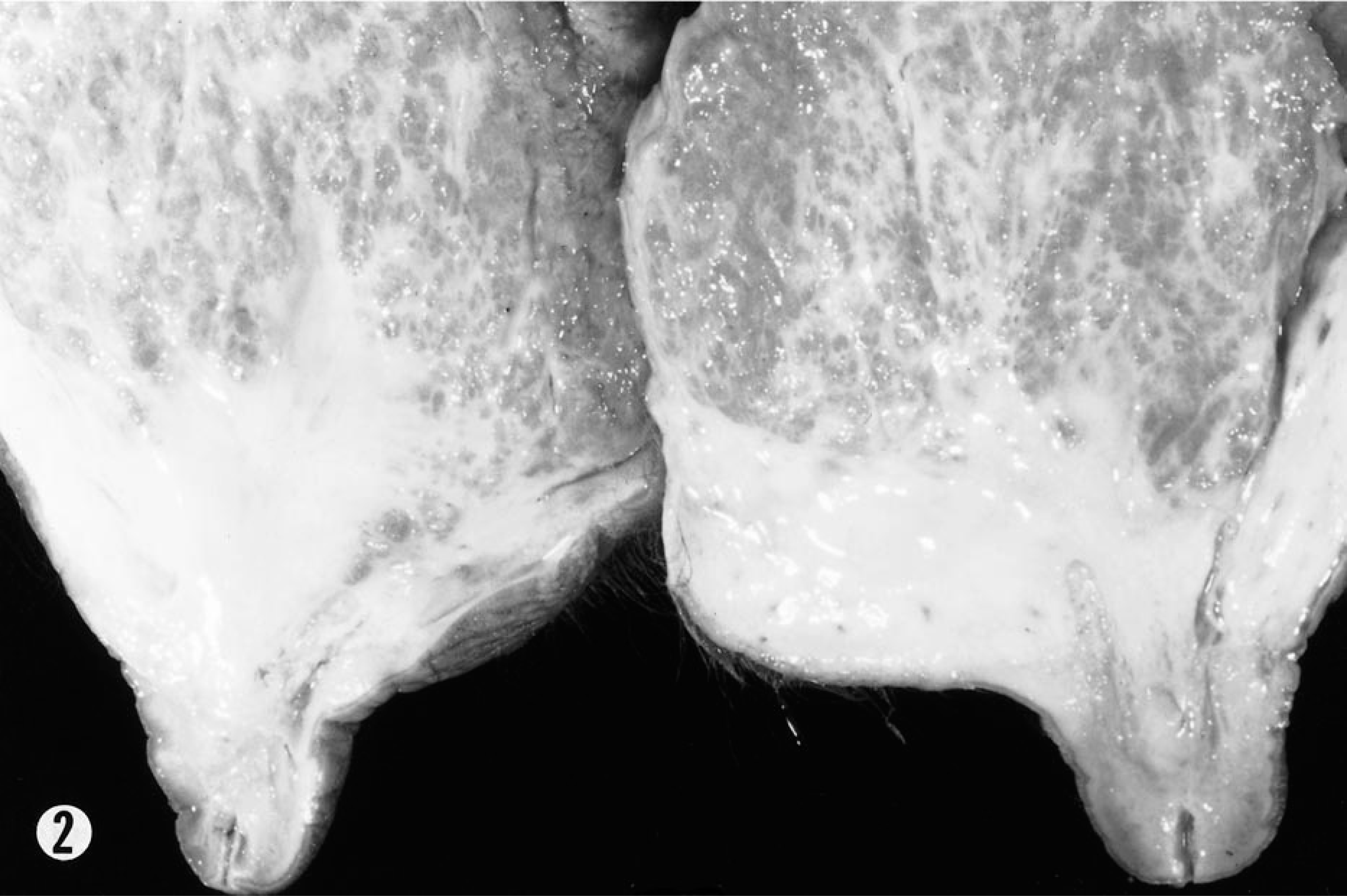
Histologic changes in the mammary tissue consisted of fibrosis with no exudate or neutrophilic exocytosis, and no lymphoid follicle formation occurred around ducts or in the interstitium. The papillary duct of affected teats was replaced by mature, fibrous connective tissue. The teat skin of all glands had a superficial and deep, perivascular, and interstitial dermatitis with perivascular mast cells and a few eosinophils, but this lesion was more severe in the skin of atretic teats. The mammary gland skin was thickened by collagenous bands separated by a loose, fibrovascular stroma infiltrated by many mast cells with occasional, admixed eosinophils (Figs. 3, 4). A diagnosis of allergic or arthropod-induced dermatitis with thelitis was made.
Mammary gland; Limousin heifer. The dermis is expanded by bands of collagen separated by loose connective tissue, dilated lymphatics, and blood vessels. An inflammatory infiltrate accompanies loose, perivascular connective tissue. HE. Bar = 80 µm.
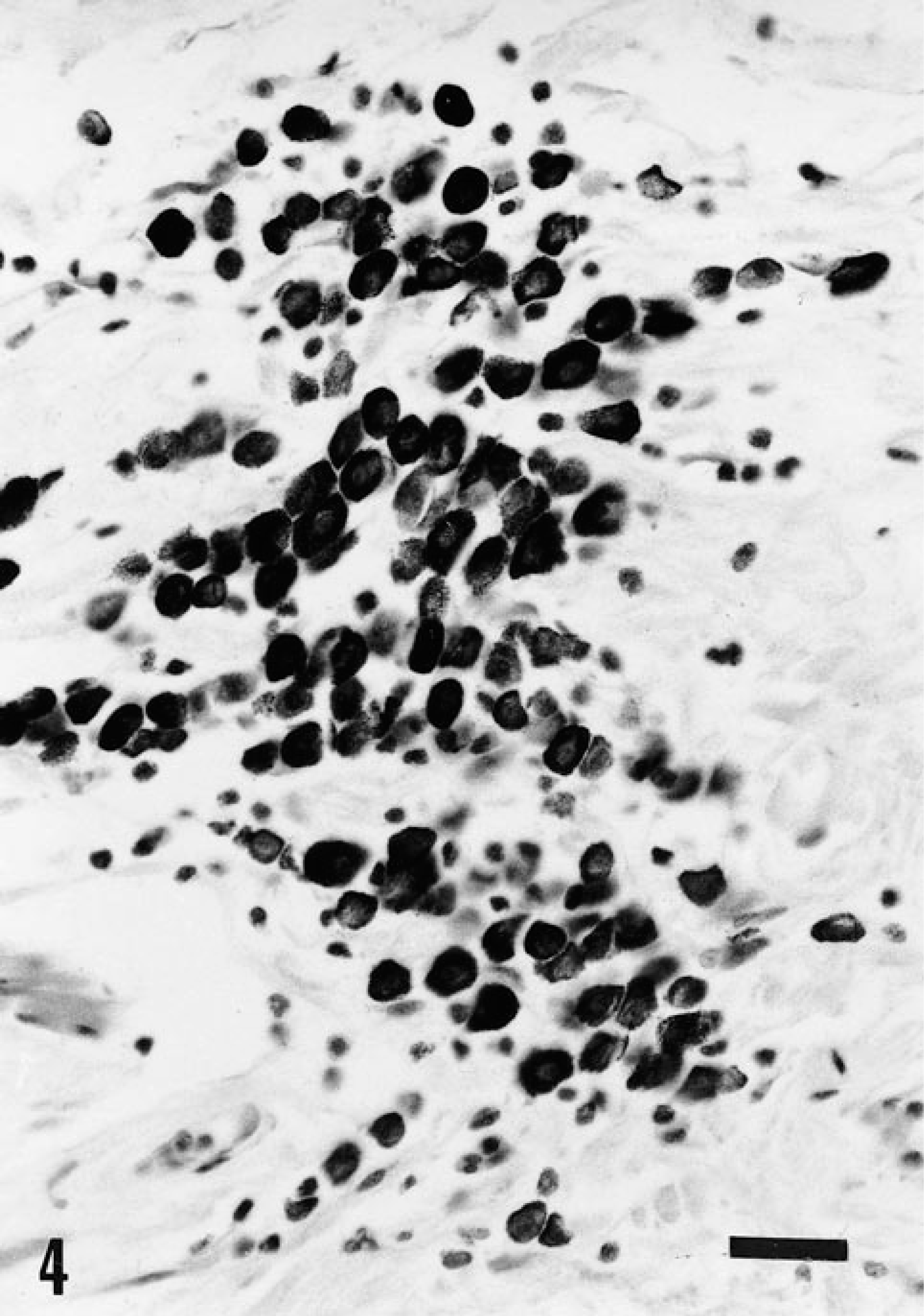
Mammary gland; Limousin heifer. The loose perivascular connective tissue contains nests of mast cells between bands of connective tissue in the thickened skin of quarters with teat atresia. HE. Bar = 30 µm.
On an initial visit, biting flies, primarily Haematobia irritans irritans (L.), the horn fly, were noted to be numerous on the backs and undersides of the cattle. The horn fly is a blood-feeding pest that feeds on soft tissue areas around the tailhead, belly, and udder. 9 Most animals observed had a scaly dermatitis visible in the nonhaired skin where flies would light. Horn fly numbers were observed to be greater than the number of flies associated with economic losses in cattle, established to be approximately 250 horn flies per animal. In order to test the hypothesis that fly or insect hypersensitivity was a key feature in the pathogenesis of the teat atresia syndrome, it was proposed that insecticide-impregnated ear tags (permithrin 10%, piperonyl butoxide 13%) be used for fly control in the herd. Because the problem affected the growing heifers and no adult cattle were noted to have affected teats, the herd incidence of dermatitis in heifers was examined before application of the ear tags. Dermatitis was noted on the teats, particularly the front teats and in the nonhaired perianal area. Ten heifers in midgestation were biopsied. Biopsies of mammary skin demonstrated an eosinophilic, perivascular dermatitis with a mild infiltration of mast cells. After initiating insecticide ear tag use, horn flies were no longer seen on clinical rechecks by the authors to swarm on the cattle, and teat and perianal dermatitis abated. In the subsequent six calving seasons after instituting fly control, no further cases of teat atresia have been observed clinically.
Bacterial mastitis associated with horn flies as vectors was among the initial differentials. 1 , 2 , 5 , 6 , 14 In heifers, horn flies have been associated with Staphylococcus spp.–induced mastitis in primigravid, dairy heifers as well as with “summer mastitis,” an acute suppurative infection of the mammary gland associated primarily with Arcanobacterium pyogenes. Both conditions present with indurated quarters having a fetid purulent secretion. Summer mastitis most commonly affects the anterior quarters and is often seen in pastures surrounded by woodlands (an environment favoring horn fly development). Although the present cluster of cases occurred in a wooded area, pyogenic inflammation and abscesses were not consistent lesions observed in the mammary glands collected at slaughter, and the herd's problem was not believed to be mastitis due to pyogenic organisms. One of the examined glands had a draining fistula, but that change was not characteristic of the remaining glands. Therefore, the fistulated gland was concluded to be the result of a separate process, perhaps a metastatic abscess in an occluded quarter.
The herd's breeding records were examined to investigate the contribution of an undefined genetic factor to the pathogenesis of the lesion. No evidence was found of linebreeding. However, because dermatitis seemed to be involved in the pathogenesis of the teat atresia, the fact that two calves (a bull and a heifer) in this herd were diagnosed with photosensitive dermatitis due to congenital protoporphyria during the previous year was considered significant. Protoporphyria in Limousin cattle is characterized by an exanthomatous dermatitis of skin in areas that are sparsely haired (e.g., muzzle, ear pinna, tailhead, and teats) and the ventral tongue exposed while suckling. 16 , 19 The dermatitis is most severe in newborn and suckling calves and is not progressive as animals age. The two protoporphyric calves of the previous season were examined, and macroscopically, a mild dermatitis persisted only on the ear pinna. The mammary glands were normal, and the protoporphyric heifer did not have abnormal teats. Blood protoporphyrin level testing of the dams of heifers with mastitis that were sold in the three seasons demonstrated that only three of the dams were carriers and none of the sires were carriers. Therefore, an involvement of protoporphyria in the herd's mammary gland problem was discounted.
“Blind” quarters, quarters that do not connect to a lactiferous sinus and end in a cul de sac, have been reported with low incidence in first-lactation dairy heifers. 3 , 4 Some of these represent quarters that have been suckled prepartum (especially rear teats accessible to pen mates), quarters that had produced little milk and actually had stopped lactation prematurely, and quarters that had scarred due to mastitis. However, in sporadic cases the blind-ending teats were concluded to have resulted from failure of ductular and glandular tissue development. 3 Histologically in such cases, glandular tissue is absent, and grossly, the blind quarters are smaller than adjacent normal glands. In reported cases, genetic studies of blind quarters resulting from agenesis or aplasia did not confirm a hereditary basis for the lesion. We found no data in the literature on blind quarters in beef cattle heifers. The affected glands of the present study did not seem to be cases of an agenesis or secondary to trauma or mastitis.
The teat atresia was concluded to have resulted from a chronic, fly-induced dermatitis. The nature of the histologic lesions, 10 , 20 and the resolution of the problem through the use of insecticide-impregnated ear tags suggested that insects were critical in the pathogenesis of the lesions. In these range cattle on improved pasture, no other causes of the condition could be identified. Excessive numbers of mast cells in these chronic lesions are an interesting feature. Mast cells have been associated with chronic, arthropod-induced skin lesions of mice and foxes. 8 , 11 The role of the mast cell is somewhat controversial in that these cells are commonly associated with fibrosis, but whether they are a cause of fibrosis or are a result of a stimulus for fibrosis is unclear. 7 , 18 Eosinophils were also seen in the lesions, particularly in the skin biopsies from heifers before ear tags were used, and one should remember that eosinophil degranulation may also play a critical role in fibrosis following inflammation. 13 Only horn and stable flies were identified as arthropod nuisances in the pasture. The fact that cranial teats were severely affected also supports the diagnosis of arthropod-induced dermatitis. The cranial teats are closer to the ground in growing heifers, and it is believed that they were more accessible to the activity of the flies than the caudal teats. The proximity of heifers' teats to the ground may aid in explaining the failure of the condition to develop in mature cows. Although the glands sampled represented lesions of only one of three seasons, they were from cattle that the owner selected as having lesions characteristic of the herd's problem. The glands were obtained from cattle 2 to 4 weeks after having their calves weaned; however, if the lesions had been induced by pathogens of mastitis, some inflammation would have been expected in the interstitium or secretions. The lesions were chronic, and they presumably developed in the skin of developing mammary glands of the yearling heifers. The condition was not observed to develop in mature cows. No genetic links to the disease were found except that it occurred in a herd of purebred Limousin cattle. Given that common management practices are utilized on cattle ranches in the area, one would expect more farms to report the problem; however, no other ranches were known to be affected. The authors only heard reports of sporadic cases of teat atresia but were not invited to investigate these reports. The relationship between the insects and this herd problem of teat atresia is still unclear.