
Abstract
Lameness related to growth plate lesions is an important problem in the beef industry. This article describes the macroscopic and microscopic lesions in the distal metatarsal physis of bulls from an association of farmers in northeastern Italy. The metatarsal bones of 62 bulls (12 with severe lameness and 50 without lameness), average age 16.44 ± 1.72 months, were examined at the abattoir. The animals came from the same geographic area and shared intensive husbandry practices and a diet based on maize starch. A total of 124 metatarsal bones were sectioned, and the distal metaphyseal growth plate was grossly examined. Twenty-three cases, including 12 lame and 9 nonlame animals with visible lesions on macroscopic examination, and 2 controls (a total of 46 physes) were examined microscopically. Eight of 12 bulls with severe lameness had a chronic purulent physitis in at least 1 limb. Segmental thickening of the hypertrophic zone, consistent with osteochondrosis (OC), was present contralaterally (n = 3 cases) and bilaterally (n = 3 cases) in 6 of these animals. In the group of nonlame bulls, 19 of 50 (38%) had similar segmental thickening of the physis consistent with OC. In the remaining bulls, minor findings included partial closure of the physis and a variable degree of metaphyseal hyperemia. A high incidence of OC was found in both lame and nonlame fattening bulls. It is likely that lame animals were clinically more severe due to secondary hematogenous implantation of bacteria, resulting in a purulent physitis and severe lameness that required emergency slaughter in some cases.
Keywords
Lameness is considered an important welfare and economic problem in the beef cattle industry. It can occur due to a number of causes, and many factors may interact to interfere with normal locomotion. 19 Lameness caused by lesions localized to the distal metacarpus, metatarsus, radius, and tibia is common in cattle. 3,19 Young beef bulls selected for rapid weight gain are particularly susceptible to developing lesions in the growing skeleton, particularly physeal lesions such as osteochondrosis (OC), which may lead to lameness and angular limb deformities. 2,3 Alternatively, an acute onset of severe lameness, with pain and swelling around the growth plate, may suggest a septic physitis or osteomyelitis. 1
The process of bone growth at the physis is highly regulated: chondrocytes proliferate, undergo hypertrophy, and then die or transform into osteoblasts. The cartilage extracellular matrix is invaded by blood vessels, osteoclasts, bone marrow cells, and osteoblasts that deposit bone on the remnants of calcified cartilage matrix. 2,3,11 A number of factors may interfere with this tightly regulated process, including damage to the blood supply, infection, and deficiencies of phosphorus, calcium, copper, or vitamin D, potentially resulting in lameness, angular limb deformities, and bone fragility. 3
There are no recent reports in the literature describing growth plate lesions in fattening bulls. The aim of this article is to describe the macroscopic and microscopic lesions found in the distal metatarsal physis of lame and nonlame fattening bulls from an association of farms in the Veneto region, northeastern Italy.
Materials and Methods
The bulls examined came from an association of farmers, in the same geographic area, who share animal husbandry practices. The bulls were a mix of beef breeds, including 40 Aubrac, 14 Limousine, 5 Charolaise, 2 Croise (Charolaise × Salers, and Charolaise × Aubrac), and 1 fattening Friesian bull. All the bulls were held on a hard slatted floor. The diet was provided once a day as a total mixed ration for ad libitum intake, based on 10% feed refusal, and drinking water was also available ad libitum. Daily dry matter intake was 10 ± 0.4 kg per animal. All bulls were fed a similar finishing diet composed of 14.2% ± 0.4% crude protein, 35.5% ± 1.5% starch, and 29.9% ± 2.9% neutral detergent fiber (as a percentage of the dry matter), mainly composed of ensiled maize starch. The diet was supplemented with Bull 100 (Soggia Mangimi, Padua, Italy), which contained 197 mg/kg E4 (cupric sulfate, pentahydrate), a copper compound approved as feed additive for animals, dispensed at 1 kg per head daily. Problems related to nutritional imbalances had never been reported on the farms.
Animals were transported and slaughtered according to the European Union regulations on animal welfare and hygiene. The official veterinary inspector of the Italian Ministry of Health oversees slaughter practices.
Of the 62 bulls examined at the abattoir, 12 animals underwent emergency slaughter because of severe lameness (group A), while 50 did not present with any documented signs of lameness (group B). Lame animals at clinical examination had leg weakness, mild swelling of soft tissues around the distal metatarsal region, variable regional hyperthermia, pain on palpation, and difficulty maintaining an upright position. In all cases, lameness was localized to the distal metatarsal growth plate and had an acute onset that progressively worsened with time. The age of nonlame animals (group B) was 16.7 ± 1.5 months, and the weight was 710.2 ± 41.5 kg (mean ± standard deviation). The age of group A animals was 15.3 ± 2.2 months, and the weight was 524.6 ± 87.5 kg. Of the 12 bulls in group A, 8 presented with grade 3 lameness, defined as marked gait asymmetry and severe symmetric abnormality, 7 while 4 had grade 4 lameness defined as recumbency. 7
The metatarsal bones, in particular the distal growth plate, of 62 bulls were examined at the abattoir. Metatarsal bones of both rear limbs were removed from each carcass, collected from the abattoir, and moved to the dissecting room of the Department of Veterinary Medical Sciences at the University of Bologna for examination.
The bones were cleaned of soft tissues, sectioned sagittally, and trimmed into at least two 6- to 10-mm-thick slabs that included the distal metatarsal growth plate. The bones were grossly examined by a pathologist, and pictures of every slab were taken to document any lesions. Microscopic examination was performed for 23 cases, including 12 lame and 9 nonlame animals with visible lesions on macroscopic examination and 2 controls, for a total of 46 physes. The 2 controls belonged to group B, were normal on visual inspection, and were selected randomly. The bone slabs were fixed in 10% neutral buffered formalin for at least 48 hours. For each case, the slabs were decalcified using 2 techniques: 1 slab with a commercial decalcifying solution with rapid action (a hydrochloric acid and formic acid salt solution; Histo-Decal rapid, Histo Line Laboratories, Milan, Italy) and one with 10% EDTA in deionized water (pH 7.4). Once decalcified, the slabs were trimmed into cassettes, dehydrated in graded alcohol, and embedded in paraffin wax. Then, 4-µm-thick sections were cut with a microtome and stained with hematoxylin and eosin (HE).
Results
Gross Pathology
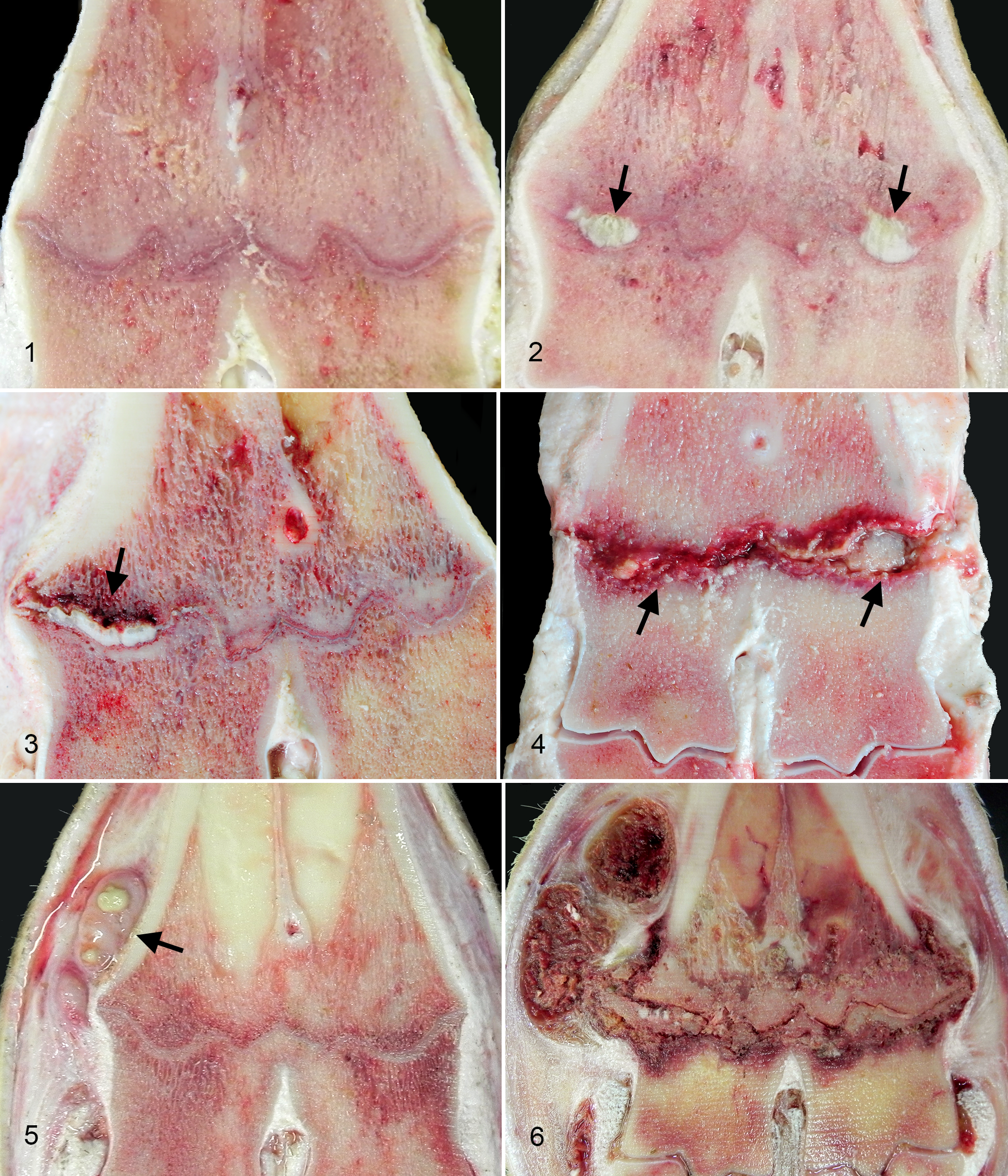
A total of 62 animals were examined at slaughter. In 21 animals (35%), both the right and left physes were grossly normal (Fig. 1). In 16 animals bilaterally and in 9 animals unilaterally, there was segmental to diffuse, mild to severe thickening of the physis, characterized by the presence of white to opalescent, firm tongue of thickened physeal cartilage that protruded into the metaphysis (Fig. 2), occasionally associated with adjacent hemorrhage (Fig. 3). Mild to moderate hyperemia of the growth plate and adjacent trabecular bone was found in 6 animals bilaterally and 7 animals unilaterally. In 7 cases unilaterally and in 1 case bilaterally, the physis was multifocally to diffusely replaced and expanded by a creamy, yellow to brown, purulent and hemorrhagic exudate that also extended irregularly into the adjacent bone of the epiphysis and metaphysis, consistent with necropurulent physitis (Fig. 4). In 1 limb of 3 cases, there were abscesses along the tendons adjacent to the growth plate (Fig. 5). Partial replacement of the physis with bone was found in 4 animals (2 bilateral and 2 unilateral) in association with moderate reddening of the adjacent cartilage. There were no skin wounds in the area adjacent to the distal metatarsal physis. The macroscopic findings are summarized in Table 1.
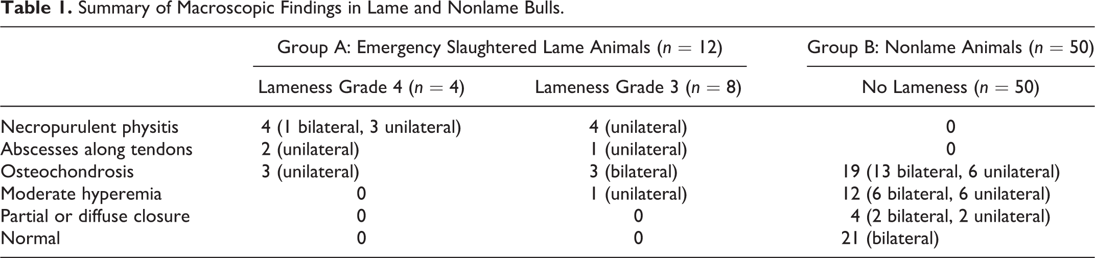
Summary of Macroscopic Findings in Lame and Nonlame Bulls.
In the group characterized clinically by severe lameness (group A, 12 bulls), the distal metatarsal physis was severely disrupted in at least 1 limb. In the 4 animals with grade 4 lameness, 1 had a severe bilateral suppurative and necrotizing physitis and osteomyelitis, while the other 3 animals had a similar physitis that only affected the right limb. In the contralateral left limb, there was segmental thickening of the physis with a tongue of cartilage protruding into the metaphysis. In 2 physes, the suppurative physitis was continuous with multifocal abscesses along the tendons (Fig. 6).
Similarly, 4 of the 8 bulls that presented with grade 3 lameness also had severe necropurulent unilateral physitis at the metatarsal growth plate. The contralateral physis and the physes of 3 other animals had segmental to diffuse, mild to severe thickening of the growth plate, similar to that described in 3 animals with grade 4 lameness. In the remaining bull with grade 3 lameness, both physes were only mildly diffusely hyperemic, but a focal abscess was present along tendons adjacent to the right metatarsus.
Fifty bulls did not show any signs of lameness at slaughter (group B). Of these, 29 (58%) animals had macroscopic physeal lesions and 21 animals were grossly normal. In 12 of 50 cases (24%, 6 bilateral and 6 unilateral), the main finding was moderate to severe hyperemia beneath the growth plate and between adjacent trabeculae of metaphyseal bone. Partial replacement of the physis with bone was found in 4 animals (2 bilateral and 2 unilateral) in association with moderate reddening of the adjacent cartilage.
In 19 of 50 cases (38%; 13 bilateral and 6 unilateral), there was segmental to diffuse, mild to severe thickening of the physis, with protrusion of a 3-mm to 10-mm tongue of physeal cartilage into the metaphysis, similar to that described above in the contralateral physes of lame animals with purulent physitis. In the 6 animals with unilateral physeal thickening, the contralateral physis either had moderate hyperemia or was normal.
Histopathology
A total of 23 cases (46 physes) were selected for histological examination. Bilateral metatarsal bone samples were selected for histological examination from all 12 lame animals, 9 nonlame animals with macroscopic lesions (12 physes with segmental thickening, 4 physes with moderate hyperemia, and 2 with partial closure), and 2 nonlame animals with no macroscopic lesions. In these last cases, the growth plate was microscopically normal. The microscopic findings are summarized in Table 2.
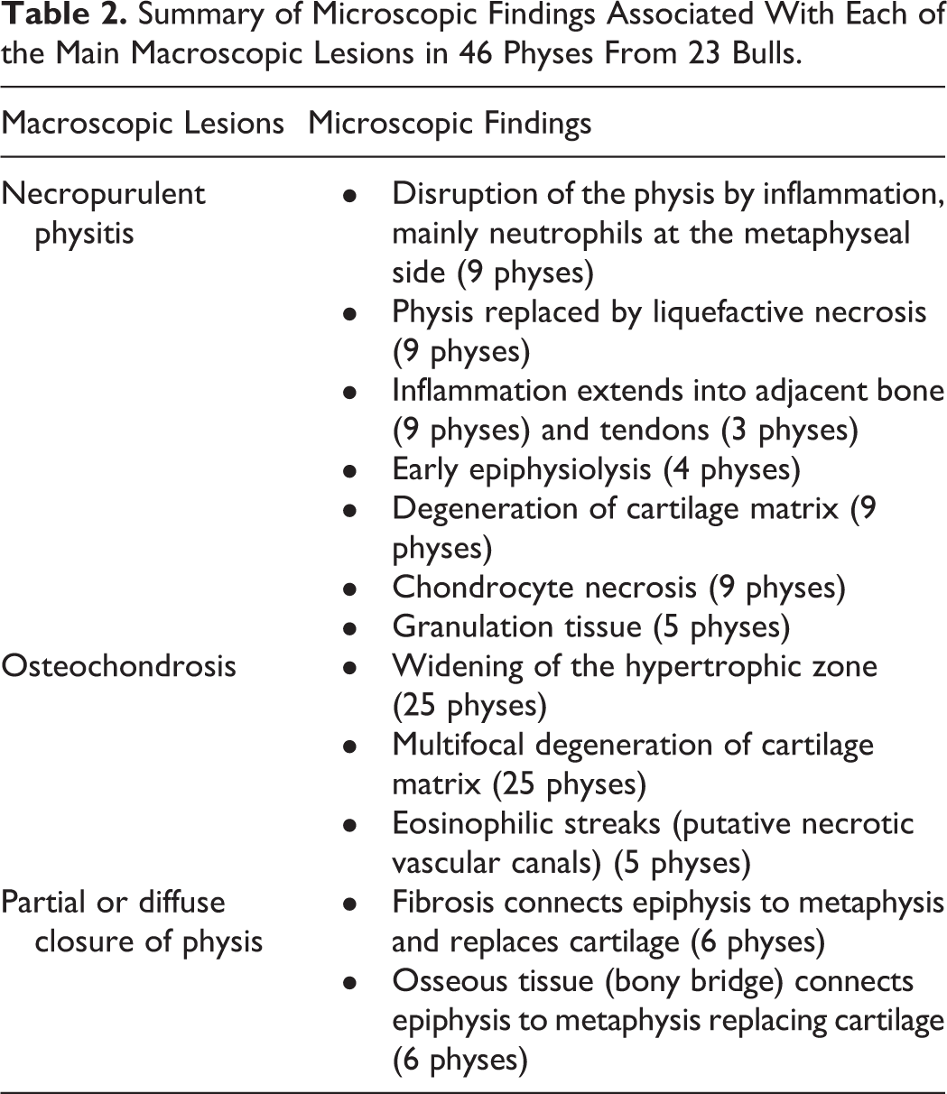
Summary of Microscopic Findings Associated With Each of the Main Macroscopic Lesions in 46 Physes From 23 Bulls.
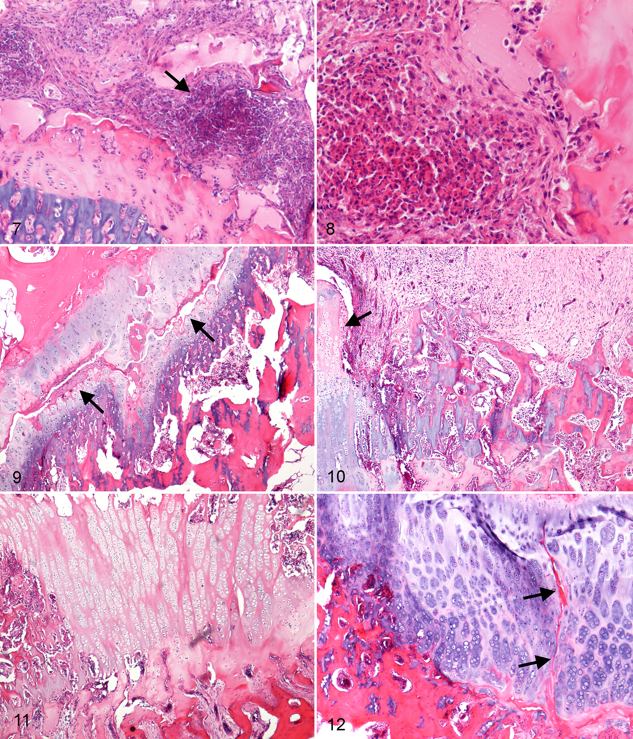
Microscopic examination of the metatarsal slabs from the lame animals with severe purulent and necrotizing physitis, 1 bilateral and 7 unilateral, showed that the 9 physes were disrupted and replaced by a multifocal inflammatory infiltrate, composed predominantly of numerous degenerate neutrophils with a low to moderate number of lymphocytes and plasma cells (Figs. 7, 8). Most of the inflammatory infiltrate was present on the metaphyseal side and multifocally extended between adjacent bone trabeculae. Extensive areas of the physis consisted of eosinophilic amorphous material, karyorrhectic debris, and hemorrhage. In 4 physes, between the proliferative and hypertrophic zones, there was a large continuous horizontal cleft filled with fibrin, necrotic debris, and eosinophilic fibrillar material (Fig. 9). The remaining cartilage of the physis, when present, was composed of thick areas of eosinophilic degenerate cartilaginous matrix surrounded by large islands of disorganized hypertrophic chondrocytes, frequently characterized by chondrocyte nuclear pyknosis, karyolysis, and loss (necrosis) and separated by the inflammatory infiltrate. In some cases, the inflammatory exudate replaced groups of hypertrophic chondrocytes and filled the spaces between the cartilage spicules of the primary spongiosa. The inflammatory area in 5 physes was delineated by a thick band of plump fibroblasts and numerous newly formed small vessels that were irregularly orientated to the collagen bundles, consistent with granulation tissue (Fig. 10). Overall, 8 of 12 (75%) of the lame animals had either unilateral or bilateral purulent physitis.
Mild to severe, segmental to diffuse thickening of the physis, corresponding to the thickened tongues and plugs of cartilage seen grossly, was found in 19 of the 50 nonsymptomatic animals (for a total of 32 physes in the nonlame group), 6 of the 8 animals with grade 3 lameness, and 3 of 4 animals with grade 4 lameness (for a total of 12 physes in the lame group). Concurrent with the lesions of OC, in 6 lame animals, there was purulent inflammation and growth plate necrosis in the contralateral limb.
The physeal thickening seen at postmortem examination was microscopically examined in 13 physes from lame animals and 12 physes from nonlame cases. In these cases, there was severe segmental to more diffuse thickening of the physis, with widening of the hypertrophic zone characterized by extremely long columns of hypertrophic chondrocytes (Fig. 11) that in some areas were arranged in groups and clusters, consistent with physeal OC. The retained cartilage tongues and islands were often encased in osteoid in the primary spongiosa. Cartilage matrix of samples decalcified with the EDTA solution was eosinophilic, indicative of matrix degeneration. The resting and proliferative zones of the physis appeared normal. Eosinophilic streaks within the retained cartilage were occasionally present, possibly representing collapsed necrotic vascular canals (Fig. 12). Rarely, areas of focal hemorrhage were found among hypertrophic chondrocytes.
Occasionally, concurrent with the lesions of OC, in some areas of severely damaged cartilage, the growth plate was replaced or interrupted by an area of bone formation that partially or totally connected the epiphysis to the metaphysis, forming a bony bridge. Complete or multifocal closure of the growth plate was present in 4 nonlame animals and was characterized by replacement of cartilage with fibrous and osseous tissue.
Four physes, in which diffuse hyperemia was observed grossly, were essentially normal microscopically other than mild multifocal disorganization of the chondrocyte columns and moderate congestion of metaphyseal blood vessels.
Two different decalcifying solutions were used in preparation of the bone sections, as both solutions have advantages and disadvantages with regard to speed and section quality (Figs. 7–12). Samples that underwent rapid decalcification with the Histo-Decal (acid-based) solution had better preservation of tissue architecture due to the absence of artifactual clefting when cutting histologic sections. However, the staining quality was poor due to the aggressive decalcification, and this meant that evaluation of the cartilage matrix and some cellular detail was difficult. On the other hand, the cartilage matrix and cellular detail were better evaluated in the samples slowly decalcified by the EDTA solution, which preserved the staining quality of the extracellular matrix, as well as cellular and nuclear details. However, the latter solution required a longer time of decalcification prior to paraffin embedding and sectioning with the microtome. Moreover, it was not always possible to obtain a high-quality slide without artifactual clefting.
Discussion
In this study, the metatarsal physes of 62 bulls were examined, and the most common lesion was metaphyseal OC, which was found in 25 animals. The other main lesion was a necropurulent physitis that was found in 8 animals. Thirty-five percent of animals had no lesions.
Twelve bulls were clinically characterized by the presence of grade 3 or 4 lameness, while 50 bulls presented without lameness at the slaughterhouse. In the lame animals, 2 main types of lesions were found in the distal metatarsal physis: a purulent and necrotizing physitis and osteomyelitis, as well as segmental thickening of the physis consistent with physeal osteochondrosis. In nonlame animals, similar to that found in lame animals, the most common lesion (38% cases) was also consistent with physeal osteochondrosis.
All of the bulls with grade 4 lameness and half of the bulls with grade 3 lameness had a purulent and necrotizing physitis and osteomyelitis. This lesion is common in the long-bone physes of calves and has been associated with rapid growth and trauma. 2,3 Bacterial osteomyelitis and physitis develop by direct implantation of bacteria into the affected bone, as may occur with trauma, or as a result of hematogenous spread with direct embolization. Bacterial embolization can occur either via cartilage canal blood vessels or in the blind-loop vessels at sites of endochondral ossification, with subsequent physeal cartilage invasion by bacteria and inflammatory cells. 2 In this study, similar to that reported by Verschooten et al, 22 we found that purulent lesions developed in the same areas as osteochondrosis lesions. It is difficult to determine whether the purulent physitis was due to either posttraumatic or hematogenous infection. In the first, a wound or fracture responsible for the bone infection is usually found. None of the bulls with purulent physitis had visible cutaneous wounds at the time of diagnosis. However, the presence of a primary cutaneous wound that had already healed cannot be excluded, and unfortunately bacterial culture of the bone lesions was not possible. Verschooten et al 22 reported the clinical, radiographic, and microbiological findings of bone infections in the appendicular skeleton of 445 cattle and found that physeal-type hematogenous osteomyelitis was 5 times more common than posttraumatic osteomyelitis. However, tissues were not examined histologically by Verschooten et al. 22 Nonetheless, together with the absence of a cutaneous wound and the greater likelihood that physeal osteomyelitis occurs due to hematogenous spread, it is most likely that the origin of infection in the bulls examined in the present study is also hematogenous. The presence of granulation tissue surrounding the exudate in 5 of the physes suggests a chronic disease process in the animals in this study. 5
Hematogenous osteomyelitis in cattle is classified as physeal type, or type 1, when the infection, usually more pronounced on the metaphyseal side, originates at or near the growth plate. The most frequently reported sites for hematogenous osteomyelitis are the distal metacarpus, metatarsus, radius, and tibia, with a reported age range of 2 weeks to 5 years. 8,22 In cattle, common causative agents include Trueperella (Arcanobacterium) pyogenes in animals more than 6 months of age, and Salmonella spp, which is more frequent in animals less than 3 months of age. 3,8,22
Barneveld 1 reported that 20% of cows that presented with a septic physitis of the distal metacarpus were slaughtered prior to any attempt at treatment due to the extremely painful nature of the lesions. Similarly, severely lame animals in this study required emergency slaughter due to the severity of the lameness associated with the purulent physitis.
Osteochondrosis is considered an important bone disease that has a number of different clinical manifestations in growing animals, 2,9,12,23 including an association with lameness and so-called leg weakness. 14,18 The distal metatarsal physis is a common site for osteochondrosis in calves. 12 The gross appearance of osteochondrosis can be difficult to distinguish from other bone lesions such as rickets 3 ; therefore, microscopic examination of bones is required. The histological hallmark of uncomplicated physeal osteochondrosis is segmental to diffuse retention of physeal cartilage, due to accumulation of hypertrophic chondrocytes as a result of focal or multifocal failure or delay of endochondral ossification. 2 OC may be differentiated from rickets due to a lack of bone fragility (microfractures) and osteoid seams. Also, rickets is a disease with a global/diffuse distribution. 3 In this study, osteochondrosis was found in 8 of 12 lame bulls (75%) and in 19 of 50 nonlame bulls (38%). In 3 of the bulls with grade 3 lameness, physeal osteochondrosis was the only finding and was therefore the presumed cause of the lameness. Frequently osteochondrosis has a bilateral distribution 2 ; this was also seen in 3 lame and 13 nonlame bulls in this study. Three of the 4 animals with grade 4 lameness had unilateral OC, while the contralateral leg had a purulent physitis, suggesting an association between the 2 lesions.
The etiology of osteochondrosis appears to be multifactorial, with genetics, rapid growth rate, vascular factors, and trauma all being implicated. 13 Trauma is the most widely proposed etiology in both humans and animals; however, its most likely role is as a final insult to compromised physeal cartilage rather than as an initiating factor in the development of early lesions. 2 The bulls in this study were raised conventionally on a hard floor, and this could have predisposed the animals to osteochondrosis. 24 Studies on pigs have shown that pigs housed conventionally had a greater likelihood of developing severe articular osteochondrosis lesions compared with those kept on deep litter. 6,21
Osteochondrosis is most common and severe in animals with high growth rates, 14 as occurred in the bulls of this study, but most experimental work has failed to document this factor as the definitive cause of the disease. 3 While there is little evidence to indicate that the lesions of osteochondrosis result from a specific nutritional deficiency, there are studies that correlate its presence with subclinical hypovitaminosis D and with copper deficiency that may be induced by excess dietary zinc or high dietary molybdenum and sulfate. 10,15 –17 The animals of this study came from the Veneto region, where fattening bulls are intensively farmed. The diet used in this area is based predominantly on maize starch and supplemented with micronutrients, including copper.
Recent experimental work in pigs and horses has demonstrated that failure of the vascular supply is associated with the process of incorporating blood vessels into the advancing ossification front during growth at the epiphyseal articular complex (due to a still unknown cause), ultimately resulting in localized areas of ischemic chondronecrosis, and this is important in the initiation of preclinical osteochondrosis lesions. 13,23 The blood vessels supplying the physeal articular cartilage in young animals may run in temporary channels known as cartilage canals. 2,23 However, 2 studies by Trueta and Amato 19 and Trueta and Trias 20 on rabbits indicated that physeal chondrocytes rely on blood supply from the epiphyseal vessels. Nevertheless, the metaphyseal vascular supply is required for the removal of dead hypertrophic chondrocytes of the physis and for calcification. If blood flow from the metaphyseal side is disrupted, hypertrophied chondrocytes continue to survive, resulting in areas of retention. If blood flow is restored, the retained cartilage may undergo rapid mineralization. 19 Experimental studies involving transection of cartilage canal vessels have not been performed in bovids. 13 In physes examined in this study, eosinophilic streaks or focal hemorrhages were occasionally present within the retained cartilage, possibly representing collapsed necrotic vascular channels. The presence of retained hypertrophic cartilage and clefting of the physis adjacent to the physeal purulent infiltrate suggests that purulent physitis could be a secondary complication of physeal osteochondrosis. Experimental studies also show that bacteremia can lead to vascular occlusion, ischemic necrosis, and lesions similar to osteochondrosis. 13
In 4 (8%) animals, there was multifocal to diffuse closure of the growth plate. This lesion may be a sequel to inflammatory and/or degenerative lesions. 2,4,19 Following an injury, inflammatory, fibrogenic, osteogenic, and finally bone-bridge maturation repair phases have been observed. 5 When a bone bridge becomes established between the epiphysis and metaphysis, it may result in growth disturbances such as angular limb deformities. 2
In conclusion, a high incidence of lesions consistent with osteochondrosis was found in lame and nonlame fattening bulls from farms in the Veneto region of northeastern Italy. This lesion likely was complicated by hematogenous bacterial infection that resulted in a purulent and necrotizing physitis and osteomyelitis, causing severe lameness that required emergency slaughter. Future challenges are to discover the etiology of these lesions and how they may be prevented in the farming of fattening bulls.
Footnotes
Declaration of Conflicting Interests
The author(s) declared no potential conflicts of interest with respect to the research, authorship, and/or publication of this article.
Funding
The author(s) received no financial support for the research, authorship, and/or publication of this article.