
Abstract
Bovine coronavirus (BoCV; Betacoronavirus 1) infections are associated with varied clinical presentations including neonatal diarrhea, winter dysentery in dairy cattle, and respiratory disease in various ages of cattle. The current report presents information on BoCV infections associated with enteric disease of postweaned beef cattle in Oklahoma. In 3 separate accessions from a single herd, 1 in 2012 and 2 in 2013, calves were observed with bloody diarrhea. One calf in 2012 died and was necropsied, and 2 calves from this herd died in 2013 and were necropsied. A third calf from another herd died and was necropsied. The gross and histologic diagnosis was acute, hemorrhagic colitis in all 4 cattle. Colonic tissues from all 4 animals were positive by fluorescent antibody testing and/or immunohistochemical staining for BoCV antigen. Bovine coronavirus was isolated in human rectal tumor cells from swabs of colon surfaces of all animals. The genomic information from a region of the S envelope region revealed BoCV clade 2. Detection of BoCV clade 2 in beef cattle in Oklahoma is consistent with recovery of BoCV clade 2 from the respiratory tract of postweaned beef calves that had respiratory disease signs or were healthy. Further investigations on the ecology of BoCV in cattle are important, as BoCV may be an emerging disease beyond the initial descriptions. Challenge studies are needed to determine pathogenicity of these strains, and to determine if current BoCV vaccines are efficacious against the BoCV clade 2 strains.
Bovine coronaviruses (BoCVs; Betacoronavirus 1) are enveloped, nonsegmented, positive-sense, single-stranded RNA viruses that are grouped as a species within the Coronavirus genus of the Coronaviridae family. 13 Bovine coronaviruses are found in the respiratory tract and intestinal tract of cattle,6,10 where they are shed in feces and nasal secretions and infect the lung.6,10 Bovine coronaviruses are associated with different syndromes in cattle: calf diarrhea, winter dysentery in adult cattle, and respiratory infection in cattle of different age groups.8–10 These clinical forms have been the subject of reviews.1,8–11 Bovine coronaviruses are often found associated with bovine respiratory disease (BRD) in postweaned beef calves entering the stocker operations or feedlots. 5 To date, BoCV control programs may include modified live virus (MLV) or killed vaccines. 7 The MLV vaccines may be used in neonates to control enteric disease or used in pregnant cows, which may transfer BoCV antibodies in the colostrum or milk secretions, to control BoCV for the neonates after calving. There are no USDA-approved BoCV vaccines in the United States to control BRD.
In an Oklahoma study, BoCVs have been isolated from nasal secretions and lung washing (lavage) fluids in postweaned beef calves in the feedlot. 5 Bovine coronaviruses have been recovered from calves presented with BRD signs and from healthy calves, as well as from lung samples collected at necropsy from fatal cases of BRD in feedlot cattle. 3 The BoCV isolates recovered from stocker cattle in Oklahoma were compared antigenically and genetically to previously isolated BoCV strains including those from neonatal enteric disease, reference strains, and to the strain in current MLV BoCV licensed in the United States. 4 Based on these comparisons, BoCV field isolates from Oklahoma stocker cattle have been described as BoCV clade 2, and neonatal enteric disease isolates, reference strains and an MLV vaccine strain as BoCV clade 1. 4 With the emergence of the new BoCV clades distinct from reference strains, previously reported neonatal enteric cases, and the MLV strain, attempts have been made to investigate BoCV in Oklahoma in both enteric disease cases as well as respiratory isolates. In the current study, BoCV clade 2 was recovered from 3 separate clinical cases in 2012 and 2013. Clinical presentation observed in these cases indicates that BoCV clade 2 should be included in the differential list of etiologies for enteric disease in postweaned calves. Calves of postweaning age examined by the clinician or those presented to the diagnostic laboratory should have salmonellosis and Bovine viral diarrhea virus (BVDV) along with BoCV included in the differential diagnosis.
Case 1. In the fall of 2012, calves from a beef herd in southern Oklahoma were weaned and commingled in a backgrounding facility. The calves developed a bloody diarrhea. Fecal samples from 3 affected calves had been submitted to the Oklahoma Animal Disease Diagnostic Laboratory (OADDL) at Oklahoma State University (OSU), Stillwater, Oklahoma and were reported as positive for BoCV. A 7-month-old heifer calf died and was submitted for necropsy at OADDL on October 12, 2012.
Case 2. Two calves (6–7 months old) from the same farm as in case 1 were presented for necropsy at OADDL on October 15, 2013. As in the prior year, the beef calves had been weaned and placed in the backgrounding facility. Similar to case 1, the calves developed a bloody diarrhea, died, and were submitted for necropsy.
Case 3. Two 13-month-old Angus bulls were presented from the same farm in eastern Oklahoma for acute, voluminous, red, watery diarrhea. The calves’ diarrhea was observed by the owner while the calves were gathered for a routine testing procedure. Both animals were in excellent body condition and had received routine vaccination and anthelmintic. One bull died in transit to OSU and was submitted for necropsy at OADDL on October 30, 2013. The second animal fully recovered with symptomatic treatment.
At postmortem examination of case 1 (2012), the heifer was in good body condition. The spiral colon was flaccid, edematous, and fluid filled. Beginning at the ascending colon and extending to the rectum, the gastrointestinal lumen was filled with both frank and clotted blood admixed with feed material. The mucosal surface of both the proximal and spiral colon contained numerous, pinpoint, red mucosal ulcers.
Histologically, examined sections of the colon exhibited marked, multifocal crypt loss. The mucosal surface was frequently ulcerated, and the basement membrane was covered by fibrin, degenerate and nondegenerate neutrophils, and cellular debris. Intact crypts were lined by regenerative epithelium, frequently ectatic, and lumens contained small numbers of sloughed epithelium and cellular debris. The submucosa was infiltrated by perivascular aggregates of neutrophils, macrophages, lymphocytes, and plasma cells that extended into and infiltrated the overlying mucosa. No lesions in the small intestine were observed. The histologic diagnosis was acute, hemorrhagic colitis.
Fluorescent antibody testing (FAT) by OADDL for coronaviral antigens was strongly positive in the colonic mucosal sections, and culture for Salmonella spp. was negative. The spleen was FAT negative for BVDV antigen and culture for Salmonella spp. was negative. Culture for Clostridia spp. was not requested, as the lesions were not indicative of clostridial disease. Fresh intestine was submitted to the research laboratory of an author (RW Fulton), and swabs were made of colon surface and frozen until tested for BoCV. The sample was labeled as OK NF INT 1.
At postmortem examination of the 2 calves in case 2 (2013), both calves were in good body condition. Both carcasses were diffusely pale, and the blood of both calves was thin and watery. The colon and rectum of both calves was filled with a large amount of watery, dark red fluid admixed with numerous variably sized blood clots and a small amount of coarse feed material. The colonic and rectal mucosa was stained red, but contained no evidence of ulceration. Sections of proximal gastrointestinal tract contained no gross evidence of ulceration.
Histologic examination of the colonic mucosa was hampered by marked autolytic change, and mucosal changes could not be definitively characterized. However, in the colon of calf B, the tunica media of multiple mesenteric vessels was infiltrated by abundant fibrin and edema admixed with neutrophils, lymphocytes, histiocytes, and plasma cells. The exudate extended into and separated the adjacent smooth muscle fibers (vasculitis). No lesions in the small intestine were observed. The histologic diagnosis was an acute, hemorrhagic colitis.
Immunohistochemical staining (IHC) for BoCV antigens was positive in the colonic mucosal sections taken from both calves. Testing by OADDL reported that FAT for coronaviral antigens was positive in the colonic mucosal sections taken from calf A. Testing for BVDV by FAT of the intestinal tissues was negative. Ancillary tests for Anaplasma marginale, Leptospira spp., and Salmonella spp. were negative. Cultures for Clostridia spp. was not requested as the lesions were not indicative of clostridial disease. Fresh intestine was submitted to the research laboratory of an author (RW Fulton), and swabs were made of the colon surface and frozen until tested for BoCV. The sample was labeled as OK 817 for calf A, and OK 473 for calf B.
At gross examination of case 3 (2013), the bull was in excellent body condition. Approximately 7 cm proximal to the spiral colon, there was an abrupt change in the intestinal content from normal to watery dark red fluid containing blood clots. This continued throughout the remainder of the spiral and descending colon. Segmentally (30 cm), the distal colonic mucosa had a mild cobblestone appearance with innumerable pinpoint to coalescing petechia and ecchymosis and approximately four 0.5 cm × 0.2 cm, elliptical, shallow mucosal ulcers. Otherwise, the colonic mucosa outside of this segment was within normal limits.
Histologically, colonic sections exhibited multifocal loss of colonic glands with scattered remnant glands present in an uneven distribution. The remnant glandular epithelium was either hyperplastic or was exhibiting features of degeneration and necrosis with occasional sloughing of necrotic epithelial cells into the glandular lumen (Fig. 1). The hyperplastic epithelium exhibited reepithelization of expansive sections of the surface mucosa. In most regions, goblet cells were absent. The lamina propria had a moderate infiltrate of lymphocytes and plasma cells. Immunohistochemical staining was performed on colon tissue with positive staining for BoCV (Fig. 2). No lesions in the small intestine were observed. The histologic diagnosis was an acute, hemorrhagic colitis. Testing by OADDL reported that FAT for BoCV antigens was conspicuously positive in the colonic mucosal sections. Intestinal tissues were negative for BVDV antigen by FAT. On culture of distal colon, Clostridium perfringens was reported as suspect. A C. perfringens multiplex PCR was performed, with C. perfringens type A reported as positive. The authors consider this an incidental finding. Culture for salmonella was negative. Fresh intestine was submitted to the research laboratory of an author (RW Fulton), and swabs were made of the intestinal surface and frozen until tested for BoCV. The sample was labeled as OK WTG 31.
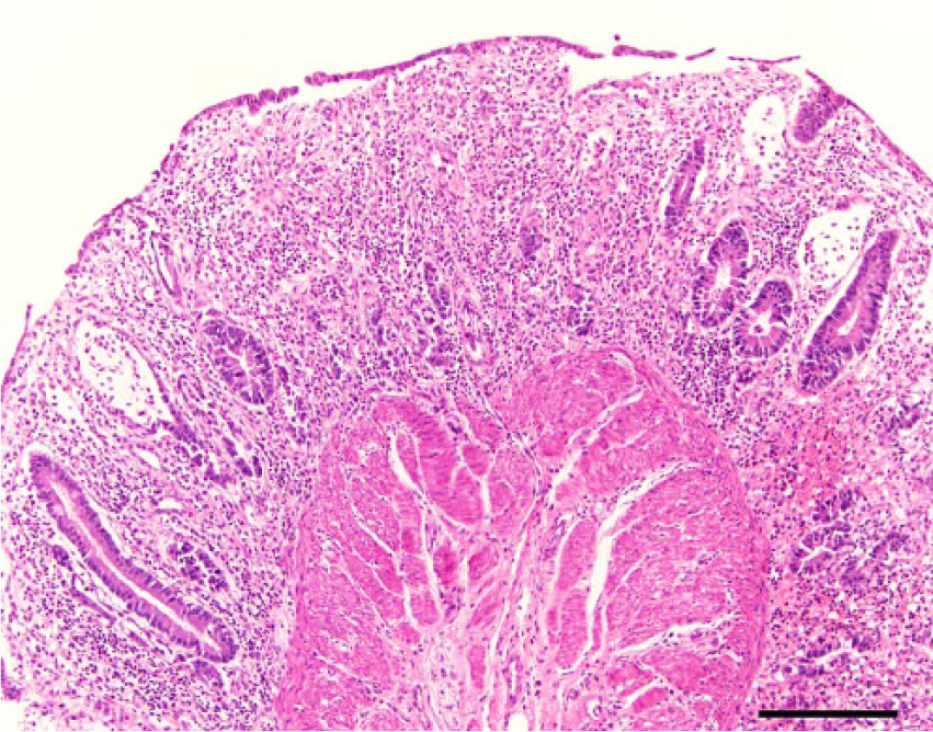
Colon; bovine. Lymphoplasmacytic colitis with regionally extensive crypt necrosis and loss, reepithelialization, and hemorrhage. Hematoxylin and eosin. Bar = 200 µm.
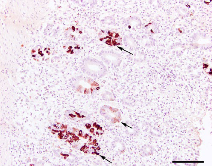
Colon; bovine. Immunohistochemical staining for Bovine coronavirus showing variably intense positive staining for epithelial intracytoplasmic viral antigen (arrows). Bovine coronavirus, horseradish peroxidase method, counterstained with hematoxylin. Bar = 100 µm.
Filtered intestinal swab samples were inoculated into freshly seeded human rectal tumor cells in 25-cm flasks containing 6 ml of cell culture medium, minimal essential medium containing antibiotics, and 7% BVDV-free bovine fetal serum. 5 Cultures were incubated for 6 days and observed daily for viral cytopathic effect (CPE). At the end of the incubation, the cultures were subjected to a freeze–thaw cycle, clarified by centrifugation, and stored frozen. All samples regardless of any viral CPE observed were tested for BoCV using a reverse transcription gel-based polymerase chain reaction (PCR) assay. Samples from the infected flasks were considered BoCV virus positive if the PCR results were positive, regardless of whether viral CPE was observed.
The BoCV respiratory viruses from the OSU studies along with reference strains and the MLV vaccine strain were examined for genomic diversity based on comparison of a region of the S gene. 4 In brief, RNA from the BoCV isolates from the OSU studies along with reference enteric and respiratory and MLV vaccine strains was prepared, S gene sequences amplified by PCR, and sequenced as described. Both of each amplicon were sequenced, and all sequencing reactions were run in duplicate. In addition to the Oklahoma samples in the current study, the following BoCV strains were included: BoCV from nasal swabs from New York dairy calves (OK AL 7490 NS, OK AL 7536 NS, OK SS 21564 NS, OK SS 2162 NS), and Australian BoCV (E146 and E147).
In the current study, there were 3 cases (4 animals) of enteric disease in postweaned beef calves occurring in different regions of the state of Oklahoma. Among the 3 cases were 2 different disease presentations from 1 beef herd in successive years (2012 and 2013). The clinical signs of bloody diarrhea gave rise to examinations for potential etiologic agents associated with this clinical presentation. The 4 animals were submitted for necropsy, which permitted additional testing to be performed. The findings of BoCV positives in the colon tissues from all calves, as determined by FAT and/or IHC along with pathology of colon disease, leads to the diagnosis of BoCV-induced naturally occurring fatal enteric disease in beef calves.
The BoCV isolated from these affected cattle were characterized based on sequence comparison of a genomic region coding for a variable portion of the spike gene. Comparison of this region has been used in prior studies to differentiate BoCV clades. Based on comparison of reference, vaccine, the strains from these outbreaks, and strains of other disease outbreaks, 4 clades of BoCV were identified. The 4 viruses isolated in the present study were classified as BoCV clade 2 (Fig. 3). In a previous study, it was found that all of the isolates of BoCV in Oklahoma belonged to this same clade. 4 The finding of BoCV clade 2 in these enteric diseases confirms the presence of this BoCV clade in Oklahoma cattle experiencing enteric disease in postweaned calves.
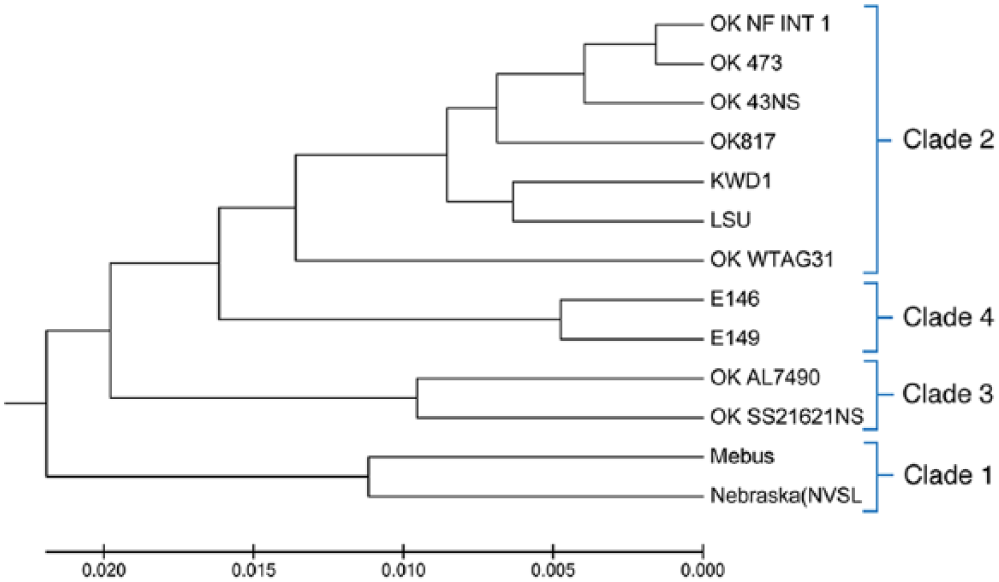
Dendrogram of Bovine coronavirus clades.
The recovery of BoCV from lesions of enteric disease occurring in postweaned calves adds to the list of clinical forms of BoCV in cattle. The other forms include neonatal enteric disease in very young calves, winter dysentery in adult dairy cows, and respiratory infections and or disease in cattle of varying ages. There have been prior reports of field cases in older calves with BoCV enteric infections and/or diseases and also experimental infections with BoCV with enteric disease.2,6,12 In an experimental BoCV challenge study, calves 3–63 days old were challenged by oral and intranasal route with a virulent intestinal origin strain of BoCV. Calves developed diarrhea and shed virus in the feces. 12 Approximately one-third of the calves evaluated regardless of inoculation route had petechiae on the luminal surface of the ileum and colon. A study of 7-month-old cattle entering a feedlot found respiratory and fecal shedding of BoCV during the first 21 days after arrival. 6 Sixty-two percent and 77% of cattle in the study had diarrhea and respiratory illness during the first 21 days. No descriptions of gross examination and histology were reported in the study. A winter dysentery–like disease in Iowa was reported with 3 feedlots consisting of 6–9-month-old calves with high morbidity and low mortality with detection of BoCV. 2 In the Iowa study, during necropsy, 3 calves from herd 1 and a calf from herd 3 had lesions with large blood clots and blood evident in the lumen of the spiral colon and rectum. Ulcers were not detected, but moderate numbers of petechial hemorrhages were evident on the mucosa of the colon of calves from one herd. Histologic lesions in the 3 calves were predominant in the large intestine. Approximately one-third of the crypts in the colon and rectum were dilated and lined with necrotic and metaplastic epithelium. Lamina propria was moderately expanded by neutrophils, macrophages, and increased numbers of lymphocytes and plasma cells. Bovine coronavirus antigen was detected by IHC in the intestines of all 3 calves. In the colon, BoCV antigen was associated with necrotic epithelium in the crypts. In that study, fecal samples and nasal swabs were collected from calves in these herds. Using ELISA, selected calves were positive in fecal samples in all 3 herds and also from 2 out of 3 calves in herd 2. Bovine viral diarrhea virus was isolated from tissues of 1 of 2 calves that died in herd 1. These experimental studies and reports from the diagnostic laboratory point out the inclusion of BoCV in older calves such as postweaned calves. Also, the need to test for other agents such as BVDV is emphasized. Clinicians and diagnosticians should include BoCV clade 2 in the differential diagnosis of enteric disease in postweaned beef calves. With the availability of reagents for FAT and IHC and PCR testing, the diagnostician is now better prepared to diagnose BoCV in clinical and necropsy cases.
The identification of BoCV clade 2 adds to the information on the ecology of BoCV in the Oklahoma beef cattle population. In a previous study, BoCV clade 2 was identified in postweaned calves using nasal swabs and lung washing fluids from calves with signs of BRD as well as from healthy calves. Control of BoCV in these postweaning clinical episodes relies on symptomatic treatment, using antimicrobials to minimize secondary bacterial disease. There are no licensed vaccines for BoCV other than those for controlling BoCV neonatal disease. The MLV vaccine strain in the United States is a member of BoCV clade 1 and is antigenically different than BoCV clade 2.
Future studies are varied in potential experimental design. First, additional information is needed on the pathogenicity of these BoCV isolates with challenge studies in susceptible calves. Second, BoCV strains from other regions of the United States and Canada recovered from similar postweaned beef calves with enteric disease should be tested for antigenic and genetic variability. Third, the efficacy of current U.S.-licensed BoCV vaccines should be tested in experimental models using the BoCV recovered from these studies or strains recovered by others. Finally, the BoCV isolates in the current study should be investigated as potential BoCV vaccine candidates as they might prove effective in protection of older cattle and neonates against BRD and/or enteric disease.
Footnotes
Declaration of conflicting interests
The authors(s) declared no potential conflicts of interest with respect to the research, authorship, and/or publication of this article.
Funding
The research was supported by the Oklahoma Agricultural Experiment Station and funds from the McCasland Foundation Endowed Chair for Food Animal Research.