
Abstract
A 7-year-old, intact female mixed-breed dog was presented for evaluation of hematuria. Physical examination revealed a suprapubic mass. Ultrasonographic examination showed a large lobular mass occupying the urinary bladder. At the owners' request, the dog was euthanatized and a postmortem examination was performed. Necropsy confirmed the presence of a lobular mass of about 5- to 6-cm diameter protruding into the lumen of the bladder. Histologically, the mass was composed of a large number of atypical lymphoid cells in the lamina propria and mucosal epithelium. Immunohistochemically, the neoplastic cells expressed CD3 but not CD79α or keratin and vimentin, supporting a diagnosis of T-cell lymphoma.
Primary malignant lymphoma of the urinary bladder is extremely rare in humans. 5 , 8 , 10 In veterinary medicine, only eight cases have been reported: two in dogs, two in cats, and four in cows. 9 In this paper we describe the histologic and immunohistochemical features of a primary malignant lymphoma of the urinary bladder with epitheliotropism in a dog, a finding not previously reported in this species.
A 7-year-old, intact female, mixed-breed dog was presented to the Veterinary Medicine Faculty, University of Naples, for examination after the owners complained that their pet had constant hematuria. The dog had no other clinical history. Physical examination revealed a suprapubic mass. Plain abdominal radiographs and ultrasonographic examination showed the presence of a large lobular mass of uniform density in the area of the urinary bladder. No other abnormalities could be seen. A tentative diagnosis of a tumor was made. In view of the poor prognosis, the owners requested euthanasia rather than an exploratory laparotomy.
At postmortem examination, abnormalities were confined to the urinary bladder. The bladder was grossly abnormal and the whole organ was removed. The gross pathologic examination confirmed the presence of a large, lobular, 5- to 6-cm-diameter, light tan mass protruding from the ventral mucosal surface, with no extension to the trigone evident. On the cut surface, the bladder mucosa was markedly thickened with prominent rugae and multiple foci of hemorrhage and ulcers. No evidence of metastasis or other gross abnormalities were found.
Representative areas of the tumor were fixed in 10% formalin for 48 hours and then processed for paraffin embedding. Four-micrometer sections were mounted on poly-
Histologically, the mucosa and submucosa of the urinary bladder were massively infiltrated by a large number of medium to large atypical lymphoid cells. The cells were usually round with scant pale eosinophilic or large optically clear cytoplasm. Nuclei were small, round to irregularly convoluted with finely aggregated and uniformly distributed chromatin, and contained rare nucleoli. Mitotic figures were frequent (two to four per high-power field). The neoplastic cells invaded the mucosal epithelium, with effacement of the stromal–epithelial junction, which was often eroded and replaced by the atypical lymphoid cell population expressing CD3 (Fig. 1). The intraepithelial lymphocytes were often surrounded by a halo of clear cytoplasm. A great number of histiocytic cells were also scattered throughout the neoplastic tissue imparting a “starry sky” appearance. The neoplastic cells were positive for the T-cell marker anti-human CD3 (Fig. 2) but not for vimentin, keratin, and CD79α, supporting a diagnosis of T-cell lymphoma. No evidence was found of bacterial infection.
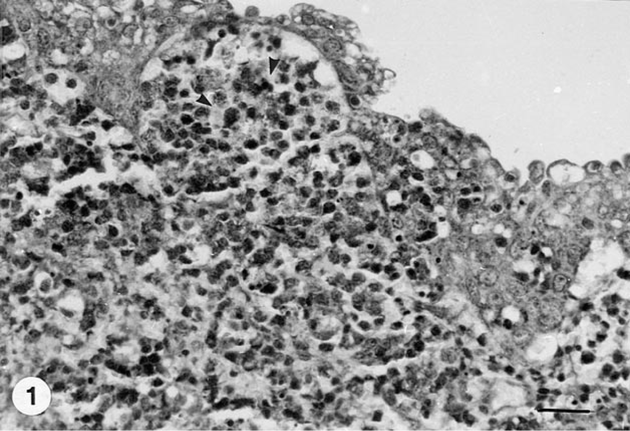
Urinary bladder, dog. Neoplastic lymphocytes, with a positive immunoreaction to CD3 antigen (arrowhead) infiltrate the mucosal epithelium with effacement of the epithelial–laminal propria junction. Streptavidin biotin peroxidase method, diaminobenzidine (DAB) chromogen, Mayer's hematoxylin counterstain. Bar = 30 µm.
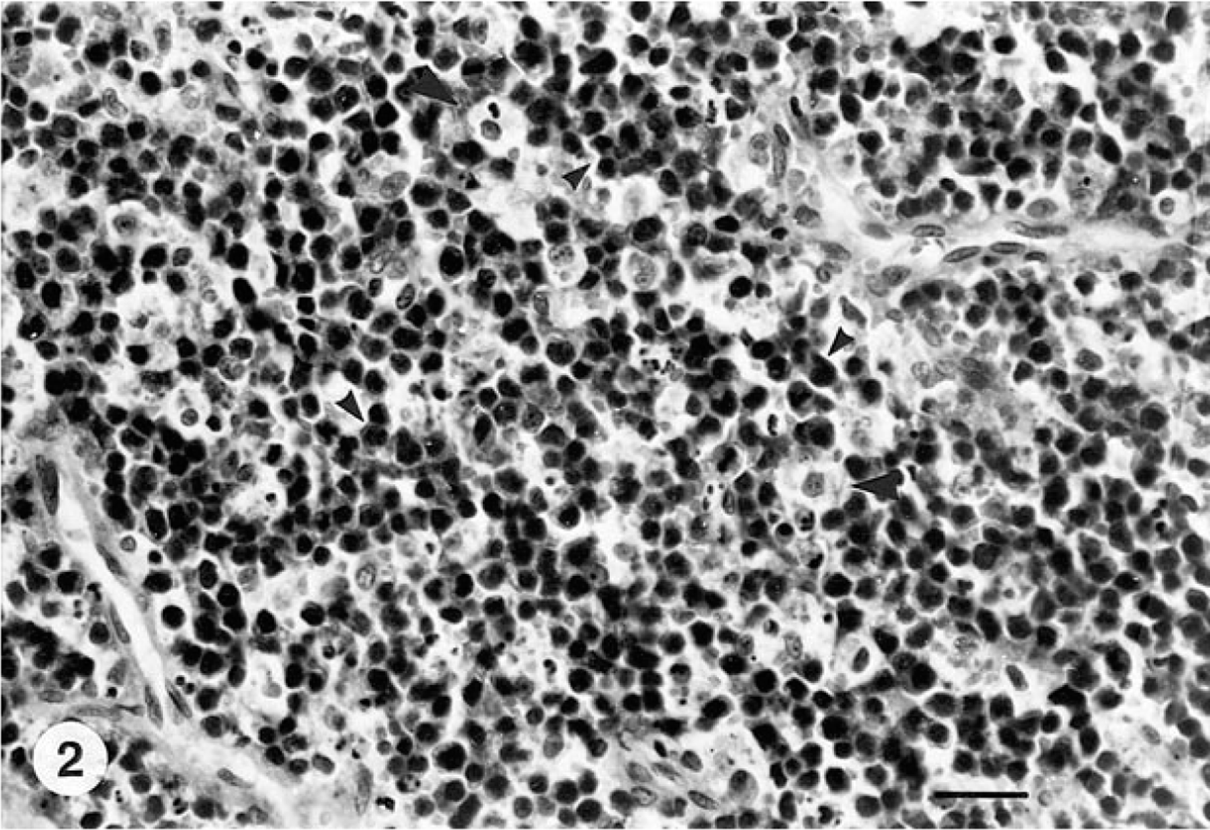
Urinary bladder, dog. Neoplastic lymphocytes, with a positive immunoreaction to CD3 antigen (small arrowheads). Note the presence of histiocytic cell CD3 negative (large arrowheads). Streptavidin–biotin peroxidase method, diaminobenzidine (DAB) chromogen, Mayer's hematoxylin counterstain. Bar = 25 µm.
Malignant lymphoma of the urinary bladder is known to occur in several domestic species as a part of a multicentric disease or as an extension from other sites. However, primary malignant lymphoma of the urinary bladder is known to be a rare pathologic event both in humans and animals. In humans, like most extranodal lymphomas, the neoplasms are usually considered to be of B-cell origin, and according to recent studies 1 the mucosa-associated lymphoid tissue lymphomas constitute a large proportion of these cases. However, to our knowledge, epitheliotropism in bladder lymphomas has never been described. The epitheliotropism refers to the affinity of the malignant lymphocyte to epithelial structures, mainly epidermis and cutaneous adnexae and usually occurs in cutaneous T-cell lymphomas (e.g., mycosis fungoides) in humans and animals 3 , 4 and in canine gastrointestinal T-cell lymphomas. 2 , 11 In canine cutaneous lymphoma, the epitheliotropism of the neoplastic elements has been suggested to result from the expression on their cell surface of specific adhesion molecules (e.g., integrins), which mediate a direct interation with keratinocytes. This surface pattern has also suggested that the neoplastic lymphoid T-cell could originate from a memory subpopulation. 6 , 7 In conclusion, the anatomical localization and the histologic and the immunohistochemical findings of present case, are consistent with the diagnosis of primary malignant T-cell lymphoma of the urinary bladder with epitheliotropism in a dog.
Footnotes
Acknowledgements
We wish to thank Mr. R. Ilsami for his technical assistance.