
Abstract
Cutaneous epitheliotropic T-cell lymphoma (CETL) is characterized by neoplastic T-cell infiltration of the epidermis, adnexal structures, and oral mucosa. The objective of this report was to describe the pathological findings of a canine case of terminal-stage CETL. A 10-year-old, mixed-breed, neutered male dog was presented with erosion of the oral mucosa and mucocutaneous junction. The dog was diagnosed with CETL with no evidence of metastasis. Despite chemotherapy, the dog was re-presented with oral pain, vomiting, and diarrhea, and died 17 months after the first visit to the hospital. A complete autopsy was performed. Histologic examination of the primary lesion and systemic organs was performed. Gross examination revealed an advanced-stage oral lesion. Distinct tumor formation was not observed in the primary sites and systemic organs. Histologically, the primary oral lesion was characterized by massive intraepithelial infiltration of a large number of neoplastic lymphocytes. The neoplastic cells in the metastatic sites also showed exclusive epitheliotropic proliferation in organs, including the trachea, tonsils, esophagus, stomach, small intestine, colon, anal mucosa, liver, pancreas, kidneys, urinary bladder, prostate gland, ear canals, and auricular and ventral skin. Immunohistochemically, the neoplastic cells were positive for CD3 and negative for CD20 as well as CD79α, supporting a diagnosis of CETL with systemic dissemination. In canine CETL with systemic signs, systemic metastasis should be considered even without evident mass formation. Neoplastic lymphocytes of CETL showed distinct epitheliotropism even in the systemic metastatic sites.
Cutaneous lymphomas originating from T or B lymphocytes are uncommon in humans and dogs, and are subdivided into epitheliotropic and non-epitheliotropic types.2,4,5,8,9 Cutaneous epitheliotropic T-cell lymphoma (CETL) is characterized by neoplastic T-cell infiltration of the epidermis, adnexal structures, and oral mucosa.4,5,9
In canine CETL, the spread of neoplastic cells to lymph nodes and various visceral organs may be observed during the tumor stage. 5 We recently encountered a case of canine CETL with characteristic systemic dissemination without mass formation in a dog. As seen in the cutaneous and oral lesions, the neoplastic cells also showed distinct epitheliotropism in almost all metastatic sites. Here we describe the pathological findings of a case of terminal-stage CETL in a dog.
A 10-year-old, mixed-breed, neutered male dog was examined at a veterinary hospital because of a 1-month history of oral pain. On physical examination, the dog showed swelling and erosion of the oral mucocutaneous junction (Fig. 1a); it was accompanied by erosion and reddening of the tongue base and soft palate (Fig. 1b), with mild swelling of the submandibular lymph nodes. Complete blood cell count (CBC), blood smear, serum chemistry panel, analysis of urine collected by cystocentesis, chest and abdominal X-rays, and abdominal ultrasound examination revealed no significant abnormalities. Fine-needle aspiration of the submandibular lymph nodes indicated reactive lymphoid hyperplasia. A punch biopsy of the skin and oral mucosa was performed, and the specimens were submitted to our laboratory for histologic examination. Histologically, the epidermis and oral mucosa were infiltrated by a large number of neoplastic lymphocytes with the formation of Pautrier microabscesses (Fig. 1c). Neoplastic lymphocytes displayed nuclei with moderately fine chromatin and 1 or 2 prominent nucleoli, along with a small amount of pale eosinophilic cytoplasm. Mitotic figures were frequent (2 per high power field). The neoplastic cells were positive for the T-cell marker cluster of differentiation (CD)3 (Fig. 1d) but not for the B-cell markers CD20 or CD79α. On the first day, the dog was diagnosed with CETL with no evidence of metastasis. Combination chemotherapy with cyclophosphamide, doxorubicin, vincristine,
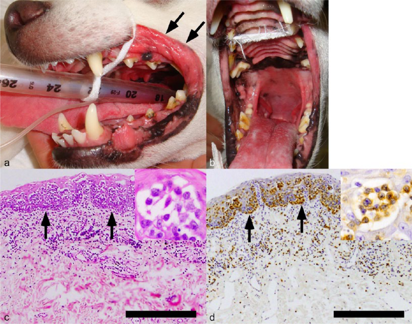
Presentation of cutaneous epitheliotropic T-cell lymphoma in the dog. (
A thorough autopsy was performed. Gross examination revealed that the oral mucocutaneous junction and the nasal, lingual, and velar mucosal surfaces were rough and pale in color (Fig. 2a, 2b). Erosion of the anal mucosa and the mucocutaneous junction was observed (Fig. 2c). Focal ulcers were observed in the left auricular and ventral skin (Fig. 2d). Marked atrophy of the skeletal muscle and subcutaneous fat, multiple small hemorrhages in the gastric mucosa, and marked splenomegaly were also observed. Distinct tumor formation was not observed in any organ. However, mild to moderate swelling of the systemic lymph nodes was observed. Organs including the heart, thoracic aorta, bone marrow, lymph nodes, spleen, trachea, lungs, oral mucosa, tonsils, esophagus, stomach, small intestine, colon, anal mucosa, liver, pancreas, kidneys, urinary bladder, testes, prostate gland, brain, ear canals, pituitary gland, adrenal grands, and auricular as well as ventral skin were collected, fixed in 10% neutral buffered formalin, embedded in paraffin, sectioned, and stained with hematoxylin and eosin (HE). Immunohistochemistry was performed using the immunoenzyme polymer method with the primary antibodies shown in Table 1. Peroxidase-conjugated anti-rabbit immunoglobulin (Ig)G a or peroxidase-conjugated anti-mouse IgG a was used as secondary antibody. Sections were counterstained with Mayer hematoxylin.
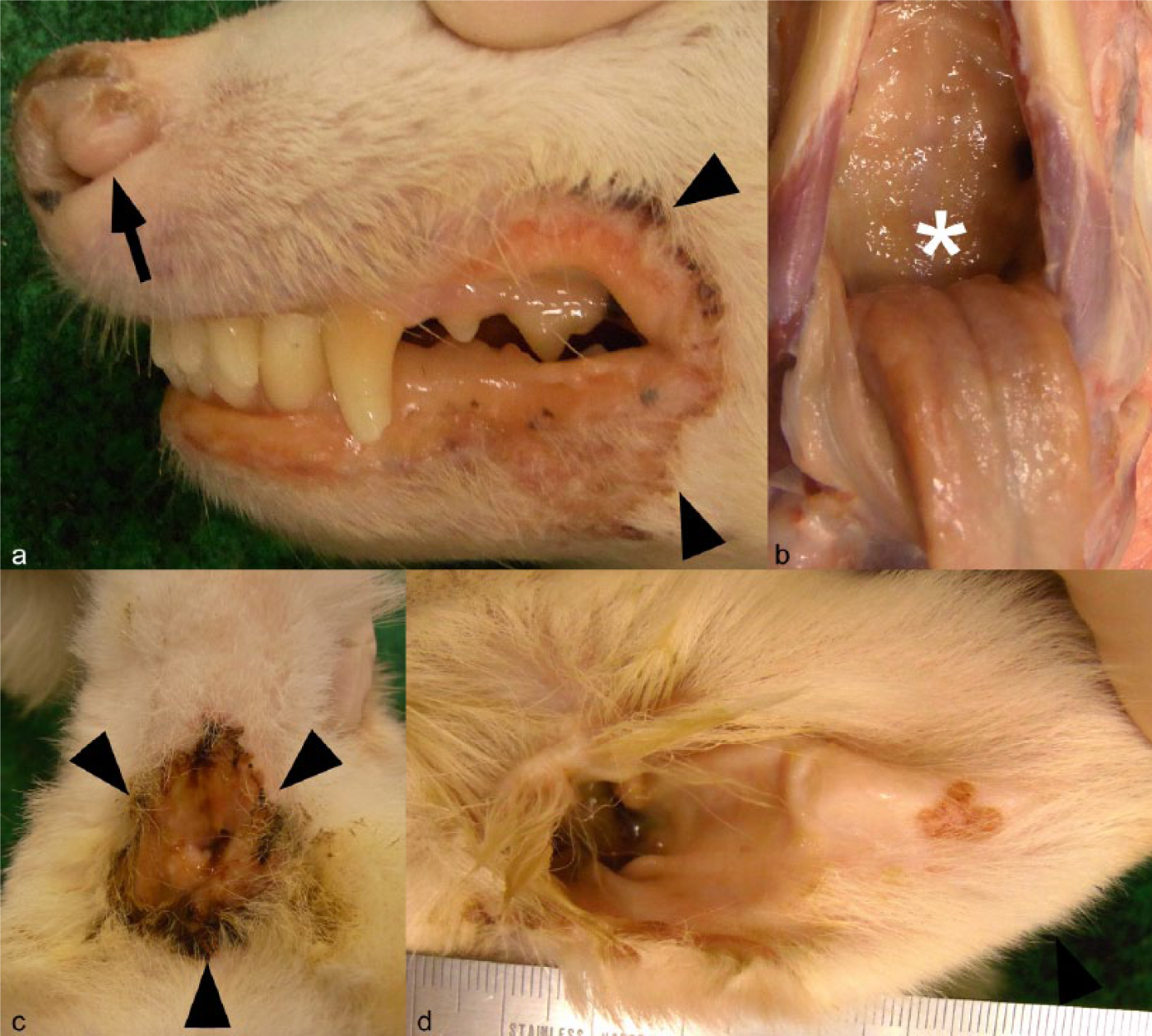
Autopsy images of the primary and metastatic sites of cutaneous epitheliotropic T-cell lymphoma in the dog. (
Primary antibodies and immunostaining protocol used in the current study.
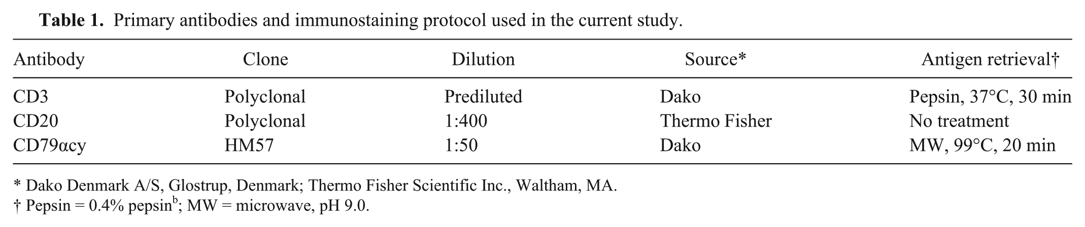
Dako Denmark A/S, Glostrup, Denmark; Thermo Fisher Scientific Inc., Waltham, MA.
Pepsin = 0.4% pepsin b ; MW = microwave, pH 9.0.
Histologic examination indicated that the neoplastic lymphocytes were positive for CD3 antigen and had infiltrated the oral mucosal epithelium, similar to the biopsy specimen. Distinct epitheliotropic proliferation of neoplastic lymphocytes was observed in the epithelium of the metastatic sites, including in the esophagus (Fig. 3a), kidneys (Fig. 3b), trachea, lungs, tonsils, stomach, small intestine, colon, anal mucosa, liver, pancreas, urinary bladder, prostate gland, ear canals, and auricular as well as ventral skin. In the liver, neoplastic lymphocytes were evident within sinusoids and bile duct epithelium (Fig. 3c). The respiratory epithelium was intensely thickened by a coalescing infiltration of neoplastic lymphocytes in the mucosa of the trachea and bronchi (Fig. 3d). In addition, the pulmonary interstitium was invaded by neoplastic lymphocytes. Diffuse infiltration of the bone marrow, lymph nodes, and spleen by neoplastic lymphocytes was detected, and follicular architectures were disrupted, particularly in the lymph nodes and spleen. Vascular and lymphatic invasion of neoplastic cells was frequently observed in primary and metastatic sites. The neoplastic lymphocytes in all metastatic sites were positive for CD3 antigen but not for CD20 or CD79α, similar to the oral mucosa specimen.
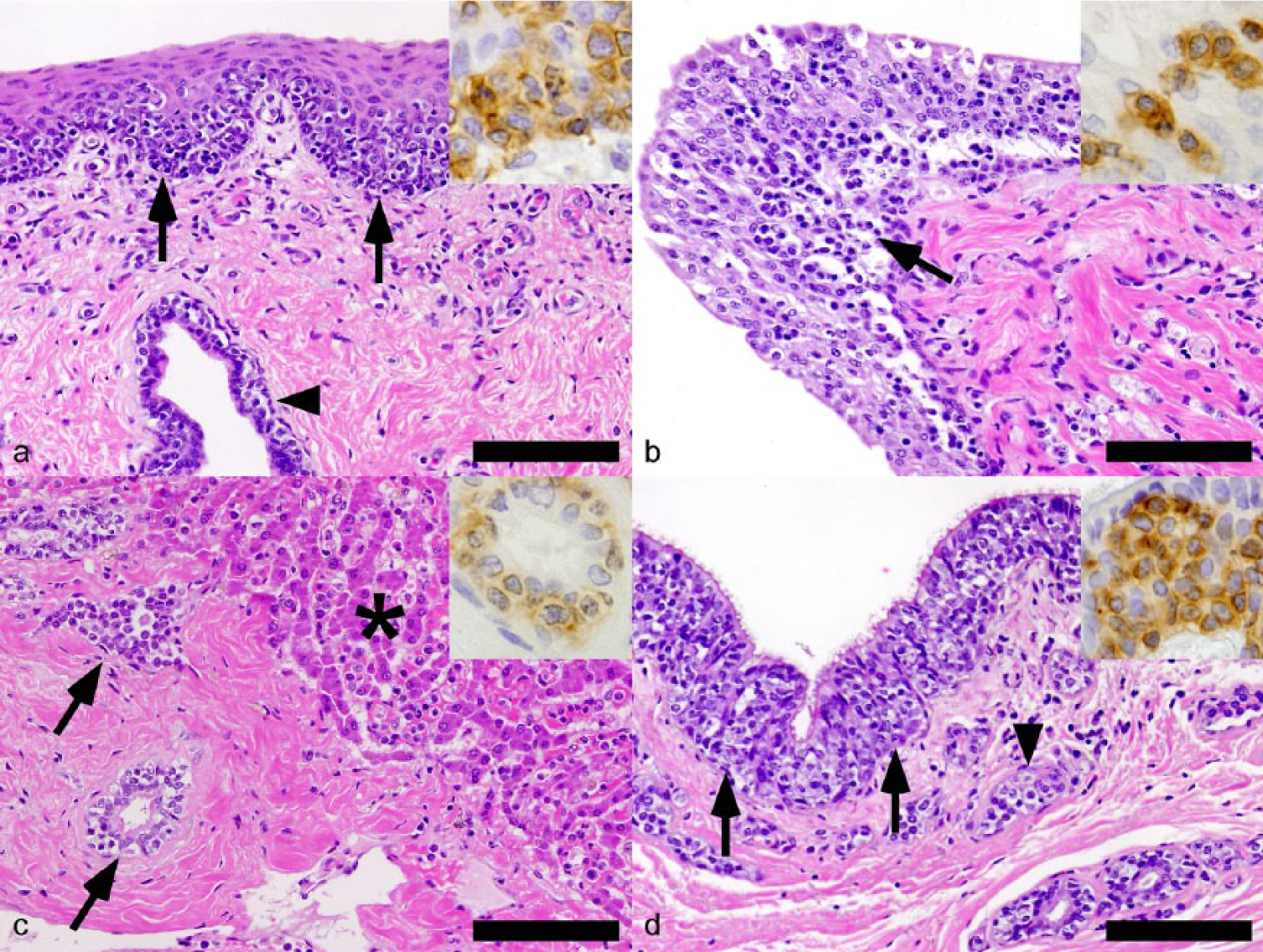
Histochemical analysis of the metastatic sites of cutaneous epitheliotropic T-cell lymphoma in the dog. (
Based on the histological and immunohistochemical findings, the dog was diagnosed with CETL. The results indicated that CETL occurred primarily in the oral mucosa and might have later metastasized to the visceral organs. However, the pattern of metastasis was characterized by intense infiltration and proliferation of neoplastic cells at the metastatic sites, without evident mass formation. In the present case, the metastatic lesions might have been associated with the observed clinical symptoms of vomiting, diarrhea, and asthenia. A previous study 1 reported a case of canine CETL that presented with a history of nervous symptoms. The case did not show evident mass formation in the nervous system but did show microscopic metastasis to the central nervous system. Therefore, in cases of canine CETL with systemic symptoms, existence of disseminated metastasis should be considered even without evident mass formation.
Furthermore, in the present case, microscopic examination of the tumor showed distinct epitheliotropic proliferation not only in the primary lesions but also in the systemic metastatic sites. Epitheliotropism refers to the affinity of neoplastic lymphocytes for epithelial structures, and it has mainly been observed in cutaneous T-cell lymphoma, gastrointestinal T-cell lymphoma, 3 and primary T-cell lymphoma of the urinary bladder in dogs. 7 To our knowledge, distinct epitheliotropism in metastatic sites has not been described in human or animal CETL. In human cutaneous T-cell lymphoma, several chemokine/chemokine receptor pairs, including CCL17/CCR4, have been implicated in cutaneous T-cell trafficking. 6 Thus, analyses focused on these factors might provide valuable information regarding cutaneous T-cell trafficking in canine CETL.
Footnotes
Acknowledgements
We thank Dr. Yoko Kakinuma and Dr. Tomotake Ikeda of Azabu University for their valuable assistance.
Authors’ contributions
T Mineshige contributed to conception and design of the study; contributed to acquisition, analysis, and interpretation of data; and drafted the manuscript. S Kawarai, K Segawa, S Neo, G Sugahara, J Kamiie, and M Hisasue contributed to conception of the study, and critically revised the manuscript. T Yauchi contributed to design of the study, and critically revised the manuscript. K Shirota contributed to conception and design of the study; contributed to acquisition, analysis, and interpretation of data; drafted the manuscript; and critically revised the manuscript. All authors gave final approval and agreed to be accountable for all aspects of the work in ensuring that questions relating to the accuracy or integrity of any part of the work are appropriately investigated and resolved.
a.
Histofine simple stain MAX-PO(R), Histofine simple stain MAX-PO(M); Nichirei, Tokyo, Japan.
b.
Pepsin, Sigma-Aldrich, St. Louis, MO.
Declaration of conflicting interests
The author(s) declared no potential conflicts of interest with respect to the research, authorship, and/or publication of this article.
Funding
The author(s) received no financial support for the research, authorship, and/or publication of this article.