
Abstract
An adult dog with ataxia and a lingual mass, previously diagnosed as protothecosis, was euthanized. At the postmortem examination, the lingual mass, regions of the lungs and hilar lymph nodes, liver, mesenteric and sublumbar lymph nodes, and spinal meninges had pronounced green discoloration. Histologically, pyogranulomatous inflammation and algal organisms were found in the tongue, spinal meninges, hilar and mesenteric lymph nodes, liver, and lung. The algae had cell walls positive for periodic acid-Schiff and cytoplasmic granules. Ultrastructurally, the algae had a well-defined cell wall, stacks of grana and thylakoid membrane, and dense bodies, typical of starch granules. The organisms were identified as Chlorella, a green alga, based on the results of histochemistical and electron microscopic examination. To the author's knowledge this is the first report of disseminated Chlorella infection and the first report in a companion animal.
A 9-year-old neutered female Golden Retriever dog developed progressive paraparesis and hind-limb ataxia of 1 week's duration. Six months previously, a mass on the tongue had been diagnosed as granulomatous glossitis with algal organisms, suspected to be Prototheca. The dog also had recent weight loss, dysuria, halitosis, and cough, with rapid deterioration during the week preceding this clinical episode.
A similar transient hind-limb ataxia that had developed 3.5 years previously was attributed to coccidioidomycosis based on a positive serum titer by agar gel immunodiffusion. Thereafter, fluconazole had been administered through the recent illness. Nevertheless, the dog remained seropositive for Coccidioides immitis, with a titer of 1 : 16 at 3 months before presentation. Serum biochemical abnormalities included hypoalbuminemia (2.4 g/dl; reference range, 2.5–4.0 g/dl) and hyperglobulinemia (4.6 g/dl; reference range, 2.1–4.5 g/dL).
On physical examination, the dog was found to be thin with a crouched hind limb posture, requiring assistance to walk. It exhibited discomfort in response to palpation over the lumbosacral spine. The tail was flaccid and hypesthetic; anal tone was reduced; and the urinary bladder was distended with urine, but easily expressed. An irregularly shaped, raised, green mass was in the dorsal aspect of the base of the tongue.
Using magnetic resonance imaging (MRI), an extradural mass surrounded and compressed the L4–L6 segments of the spinal cord. The mass was isointense to hyperintense on T2-weighted images, with heterogeneous enhancement of extradural tissue by gadolinium-enhanced postcontrast T1-weighted imaging. Similar enhancement occurred in the adjacent lumbar epaxial musculature and in the lamina and pedicle of the fourth and fifth lumbar vertebrae.
The dog was treated with fluconazole, cephalexin, and prednisone. Although its condition improved slightly over the next 4 days, the owner requested euthanasia 7 days after presentation. Grossly, an irregularly shaped, dark green, ulcerated mass elevated the dorsal surface of the base of the tongue (Fig. 1) and extended into underlying lingual tissue. The lumbar epaxial musculature was infiltrated by soft, poorly demarcated, greenish exudate that was contiguous with similar exudate in the vertebral canal. The exudate was loosely adhered to the dura mater along 4 vertebral segments. Cross sections of spinal cord demonstrated involvement of external and internal surfaces of the dura mater (Fig. 2). Sublumbar, mesenteric, and hilar lymph nodes were enlarged and green. Slight, patchy, green discoloration was also present on visceral pleura and in pulmonary parenchyma as well as on the hepatic capsule and in hepatic parenchyma.
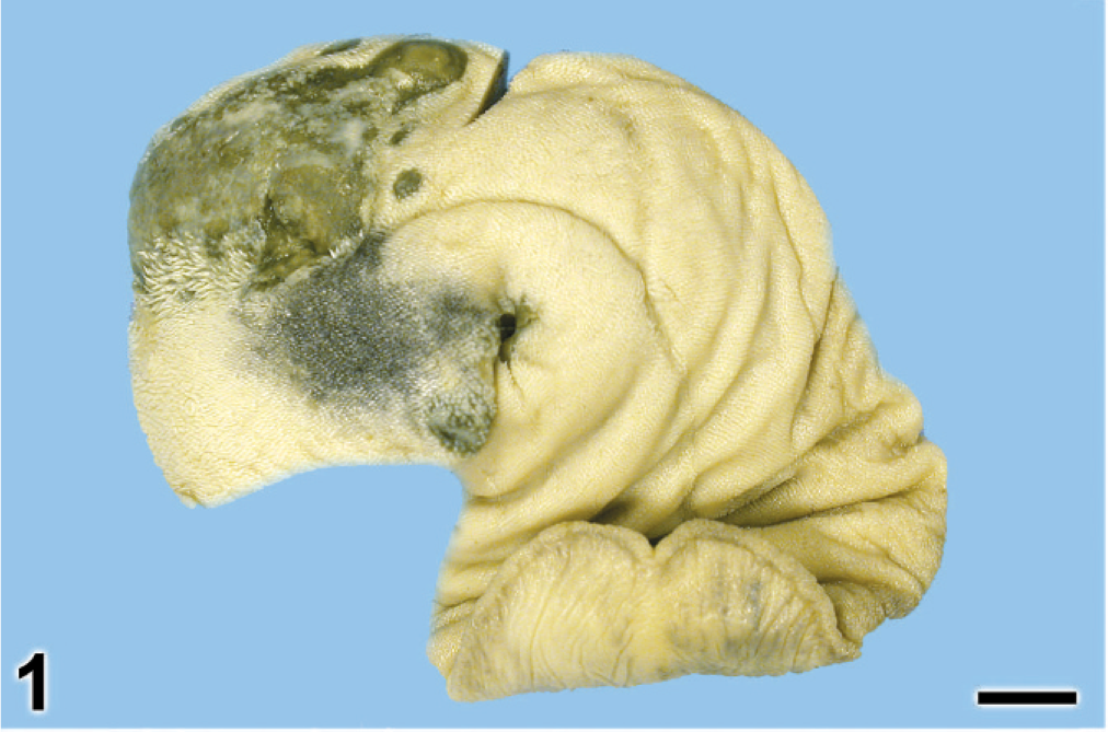
Tongue; dog. An irregular, focally ulcerated, slightly raised green mass is at the base of the tongue. Bar = 1 cm.
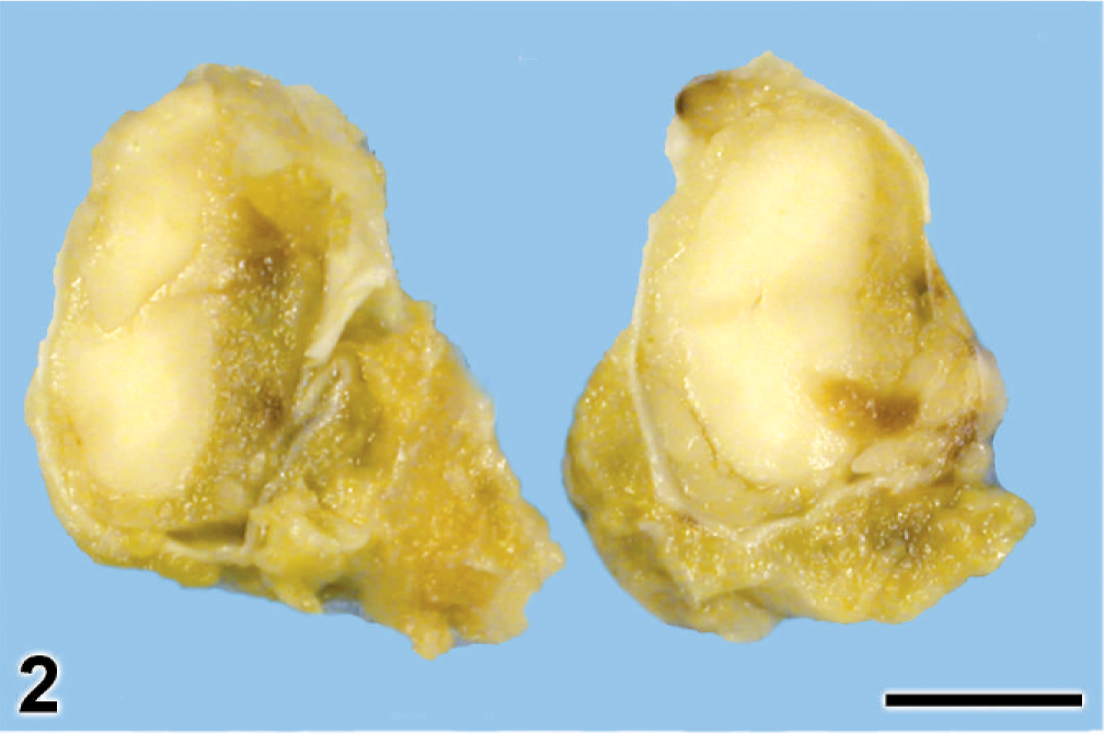
Lumbar spinal cord; dog. Exudate with green tint is present inside and outside the dura mater, extending into adjacent soft tissues. Prolonged formalin fixation has reduced the green color. Bar = 0.5 cm.
Samples of liver, lung, hilar and sublumbar lymph nodes, tongue, and lumbar spinal cord were fixed in 10% neutral buffered formalin, paraffin embedded, and stained with HE for light microscopic examination. Histochemistical examination of tongue and spinal cord sections included periodic acid-Schiff (PAS), Gomori methenamine-silver HE, Brown and Hopps tissue Gram stain, and Gridley fungal stain.
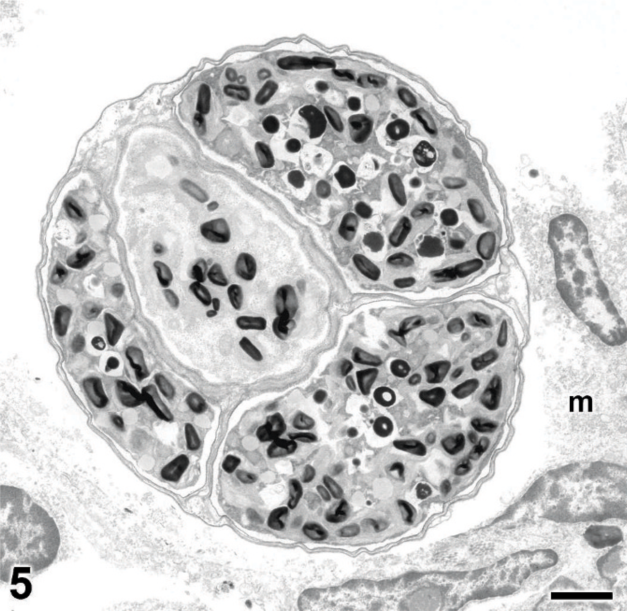
Histologically, sections of the affected spinal cord had intense pyogranulomatous inflammation with myriad organisms in the subarachnoid space and on the exterior surface of the dura mater. The inflammation extended only slightly into the perivascular spaces of the spinal cord and was composed mainly of neutrophils and macrophages with fewer lymphocytes and plasma cells. Individual organisms were most often free in the exudate, but some were in the cytoplasm of macrophages and multinucleated giant cells. The organisms were round and had a well-defined, narrow cell wall. Single large sporangia (Fig. 3), 7–25 µm in diameter, were mixed equally with compartmentalized organisms of equal or slightly larger diameter that contained a variable number of morula-like sporangiospores (Fig. 3). Eukaryotic nuclei were frequently observed in the organisms. Spinal nerve roots that ran through the inflamed segment of meninges contained swollen axons or empty axon sheaths, consistent with Wallerian degeneration. Organisms, particularly sporangiospores, had numerous strongly PAS-positive cytoplasmic granules that were PAS-negative after diastase treatment. The cell walls were also PAS-positive, but this feature was more easily observed in less granular sporangia (Fig. 4). Organisms stained poorly with Gram stain, but the cell walls were weakly positive with Gomori methenamine-silver HE (Fig. 3). Similar microscopic lesions were found in the mesenteric lymph node, liver, lung, and tongue. No evidence of coccidioidomycosis was found on histologic examination.
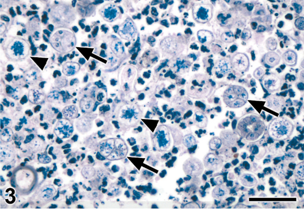
Meningeal exudate; dog. Algal organisms are in pyogranulomatous exudate. Numerous sporangia (arrowheads) and morula-like endosporulating organisms (arrows) have well-defined cell walls. Gomori methenamine-silver HE stain. Bar = 30 µm.
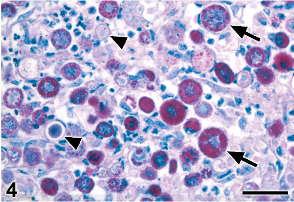
Meningeal exudate; dog. Sporangiospores (arrows) and, to a lesser extent, sporangia (arrowheads) contain many PAS-positive granules. PAS reaction. Bar = 30 µm.
Meningeal samples were fixed in formalin, divided into 1-mm cubes, transferred to 2% glutaraldehyde-2% paraformaldehyde in 0.1 M cacodylate buffer, postfixed in 1% osmium tetroxide and embedded in Epon-Spurr medium. Thin sections were stained with uranyl acetate and lead citrate and then examined with a JEOL 1400 electron microscope.
Ultrastructurally, the organisms were identified as algae. Individual sporangiospores had well-defined cell walls that were closely associated with a cell membrane (Figs. 5,6). The outer wall of the sporangium had a similar structure but was more wrinkled, probably an artifact of fixation. The cytoplasm of individual organisms had numerous polyhedral electron-dense starch granules with adjacent stacks of membranes (Fig. 6), consistent with the ordered grana and less ordered thylakoid membranes of chloroplasts. The ultrastructural features were those of a photosynthetic alga, with characteristics of Chlorella. Confirmatory isolation was not attempted.
Meningeal exudate; dog. A sporulated alga, surrounded by host macrophages (m), contains electron-dense, angular starch granules. Lead citrate and uranyl acetate. Bar = 2 µm.
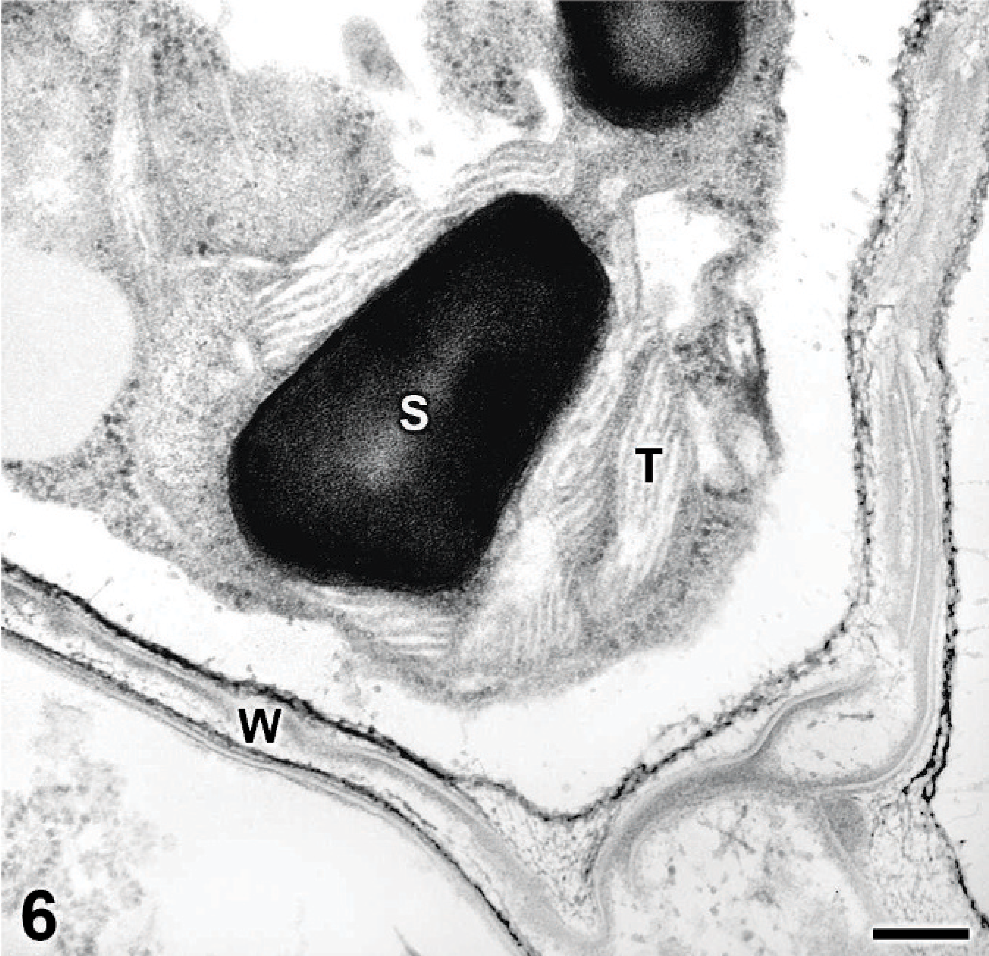
Algal sporangiospore, meningeal exudate; dog. The cell wall (W) is present peripherally. Stacks of grana and thylakoid membrane (T) occur between starch granules (S) within the chloroplast. Lead citrate and uranyl acetate. Bar = 0.2 µm.
Green algal infection of animals was first reported as the cause of green hepatitis and lymphadenitis in a slaughtered lamb. 3 Since the initial report, Chlorella sp. infections have been reported rarely in various herbivorous species, fish, and a human. 3, 7– 9, 16, 18, 19, 22 Many animal cases were incidentally detected during meat inspection at abattoirs. 18, 22 Collectively, lymph nodes, liver, lung, skin, and intestines were involved. 3, 8, 9, 12, 16, 18, 19, 22 To the author's knowledge, chlorellosis has not been reported in dogs before and never as a cause of dysfunction of the central nervous system.
Chlorella is a chlorophyll-containing green alga closely related to Prototheca, which is thought to be its achlorophyllous mutant. 1, 6, 15 Chlorella is unique among algae in that a single large chloroplast dominates its cytoplasm. 21 Unlike Prototheca, Chlorella spp. contain numerous starch granules that are strongly PAS-positive.∗ The granules become PAS-negative after diastase digestion, 11 and under polarized light they are birefringent in unstained or HE-stained sections. 1 Prototheca lacks chloroplasts or starch granules, although smaller related organelles, protoplasts, are present. Therefore, only the cell wall of Prototheca is PAS-positive. 1 The life cycles of Chlorella and Prototheca are similar. 1, 15 Both produce hyaline cells (sporangia) that mature to produce 2–20 sporangiospores (endospores) asexually. After endosporulation, the outer sporangial wall ruptures, releasing the sporangiospores to repeat the cycle.
Light microscopy demonstrates similarities between Chlorella and Prototheca in shape, size, wall, and endosporulating reproductive mode. Because chlorophyll dissolves during fixation and embedding, the green color is lost with light microscopy. 1 Wet mounts or Giemsa-stained smears are needed to observe the bright green pigment granules of Chlorella. 22 The finding of chloroplasts with electron microscopy supports a diagnosis of algal infection. The dog in this report had lesions in the tongue, lymph nodes, lung, liver, spinal meninges, and epaxial muscle. This disseminated infection is more extensive than that previously reported for chlorellosis in any species. Lymph nodes, lung, and liver have been target organs of infection in sheep and cattle. 3, 9, 16, 18, 22 The primary portal of entry in cases of disseminated chlorellosis is often thought to be the gastrointestinal tract. 12, 16, 22 A single portion of jejunum obtained from the dog of this report was not affected; it is possible that additional sections may have revealed algae. Secondary hematogenous dissemination and lymphatic drainage were thought to account for the localized visceral lesions in previous cases. 12, 16, 22
Algae had been detected in the tongue of this dog 6 months before the onset of neurologic signs. This protracted course is not unusual. 13 Dogs with disseminated protothecosis often have a chronic history of intermittent bloody diarrhea 1–10 months before ocular or neurologic signs develop. 5, 6, 13, 20 It is possible that the lingual lesions were the primary portal of entry, leading to secondary hematogenous and lymphatic dissemination. Alternatively, another primary lesion on the surface of the tongue could have allowed secondary invasion of Chlorella. An analogous situation occurred in the single human Chlorella infection, in which a surgical wound was exposed to contaminated river water. 8
It is speculated that defective cell-mediated immunity is responsible for some cases of disseminated protothecosis in dogs. 2, 4, 14, 17, 20 Immunosuppressive medications and neoplasia are often cited as predisposing factors. Evidence of heavy environmental exposure is also frequently present. 9, 12, 16 Both Chlorella and Prototheca are ubiquitous, and Prototheca spp. have been isolated from diverse environmental sources, including tree sap, fresh and marine water, potato skins, fingernails, sludges, and animal feces. 9, 12, 13, 15, 20 Chlorella is considered an opportunistic pathogen, often requiring immunosuppression, wound inoculation, or heavy exposure to organisms for infection to occur. 3, 12 Cases of mammalian chlorellosis are much less frequent than cases of protothecosis.
Sheep with chlorellosis had prior access to stagnant water that was covered by mats of bright green algae. 9 However, the dog of this report was kept mainly indoors and had no known contact with stagnant water. Undetected primary immunodeficiency or immunosuppression secondary to chronic Coccidioides immitis infection are potential contributing factors for the infection in this dog. Immunosuppression can predispose human beings to coccidioidomycosis, 4 but no evidence of active coccidioidomycosis was found at the postmortem examination in this dog. The failure of dogs with disseminated protothecosis to respond to treatment 2 may reflect a delay in commencement of therapy and/or underlying immunodeficiency.
This case demonstrates the potential of Chlorella organisms to infect dogs and cause widely disseminated disease. Although rare, disseminated chlorellosis should be considered, particularly when lesions have a green discoloration. Chlorella should be differentiated from Prototheca by cytochemical or ultrastructural means.
Footnotes
Acknowledgements
We gratefully thank Ms. Cheryl Jensen and Mr. Howard Wilson for assistance with electron microscopy and preparation of illustrations, respectively. The patience of technicians in the VMDL histopathology laboratory in performing histochemistry is also appreciated.