
Abstract
The 2019 novel coronavirus, known as COVID-19, has greatly affected the way sonographers care for their patients. Sonography can be a useful imaging tool for surveillance and diagnosis of various conditions associated with COVID-19 patients or patients under investigation (PUIs). Currently, there are limited resources and protocols for preventing the transmission of COVID-19 from ultrasound equipment. Our institution has created a detailed protocol for scanning COVID-19 patients or PUIs to address this important issue.
The novel coronavirus (known as COVID-19) is a disease that is caused by the severe acute respiratory syndrome coronavirus 2 (SARS-CoV-2). It was first recognized in China in December 2019 after a spike in acute respiratory symptoms, specifically pneumonia.1,2 Since then, the virus has rapidly spread, and the World Health Organization declared a global pandemic in March 2020. 3 COVID-19 is a highly infectious virus, which is easily transmitted through human contact. 1 Furthermore, COVID-19 has fundamentally changed the way health care workers provide care for patients. 4 Health care organizations have quickly compiled policies and procedures to minimize exposure for patients and their workers from COVID positive (COVID-19+) and patients under investigation (PUIs). 5 Diagnostic medical sonography is a safe and effective tool that has been used in this patient population to monitor and assess COVID-19 complications. 6 However, conventional methods for conducting sonography can increase the risk of transmitting the virus from patient to sonographer and potentially to an unaffected patient. Therefore, a systematic protocol designed to limit transmission and maximize the advantages of this imaging modality should be required.6,7 This includes appropriate protection for sonographer, ultrasound equipment, and minimizing patient contact by shortening the examination time. The authors share their experience in creating a detailed disinfectant protocol that their workplace uses for COVID-19+ patients and PUIs.
Sonography is an imaging modality that has unique benefits due to its portability, safety, and cost-effectiveness. These advantages make sonography an effective portable diagnostic tool to evaluate COVID-19+ patients but can lead to an unintended transmission.6–10 In 2019, Westerway et al. 11 found a lack of knowledge among clinicians concerning proper disinfection and hygiene practices with ultrasound equipment. This raises concerns, especially when performing bedside ultrasonography for COVID-19+ patients. The global COVID-19 pandemic lays bare the problem of limited detailed protocols and disinfection processes in sonography departments.
The authors implemented a process for sonographers to create imaging protocols under the supervision of the division director and manager. Hence, two experienced (American Registry for Diagnostic Medical Sonography [ARDMS] credentialed) sonographers created this COVID-19 disinfectant and equipment management protocol. Appendix A details the protocol that provides a thorough explanation of the process of disinfecting the ultrasound equipment when scanning a COVID-19+ patient or PUI. This protocol describes the preparation of the ultrasound equipment before leaving the department, as well as the steps for disinfecting the equipment inside and outside the patient room. It also details the cleaning process for the equipment and transducer upon returning to the department, after the examination.
In addition to this COVID-19 disinfectant and equipment management protocol, the institution has created specific protocols for COVID-19+ patients or PUIs. These protocols were shortened to limit exposure to the sonographer and target images that answer the clinical question. Sonographers should acquire specific clinical information needed for the appropriate treatment. One example of specific imaging is visualizing thrombus in the common femoral vein and continuing into the external iliac vein. This triggers treatment for deep vein thrombus (DVT) in the common femoral and femoral veins, which may differ from the iliac veins. Additional imaging of the iliac veins with magnetic resonance imaging (MRI) or computed tomography (CT) can be avoided. In addition, for some COVID-19 protocols, the department has shortened the examination to one side (the symptomatic side) and replaced the number of static images with video clips to reduce staff’s exposure time. Appendices B, C, and D highlight the COVID-19 protocols for renal, upper extremity, and lower extremity, respectively.
The authors’ experience has underscored the need for sonographers to fully prepare the ultrasound equipment and transducers, in the department, before performing a bedside examination on a COVID-19+ patient or PUI. Proposed guidelines have suggested having dedicated ultrasound equipment(s) for COVID-19+ patients or PUIs. 7 In addition, it would be important to remove any unnecessary items from the equipment except for those transducers that are necessary for the examination. 7 Ultrasound equipment(s) should be disinfected with low-level disinfectant (LLD) wipes (the brand of wipes would be specified under each institution’s guidelines). 7 A single large equipment cover that protects the entire ultrasound machine needs to be used. The cover minimizes contaminating small crevices (e.g., small spaces between the keys on the keyboard) and cooling fans that are designed to prevent overheating (e.g., in the machine and perhaps in monitors). Transducer covers should also be used to effectively implement this protocol. At the authors’ institution, sterile transducer covers are used, similar to the biopsy covers. These covers extend the cord length to minimize contamination. After the covers are placed on the transducer(s), they should sit on the outside of the machine cover in the designated equipment holder. In addition, sonographers should prepare for the examination by using single-use disposal gel packets, if available. If single gel packets are not available, use only one gel bottle, and discard it after the examination. 7 The single gel packets or gel bottle should be placed on the outside of the machine cover in its designated equipment holder. Figure 1 illustrates an example of proper equipment preparation, in the department, prior to scanning a COVID-19+ patient.
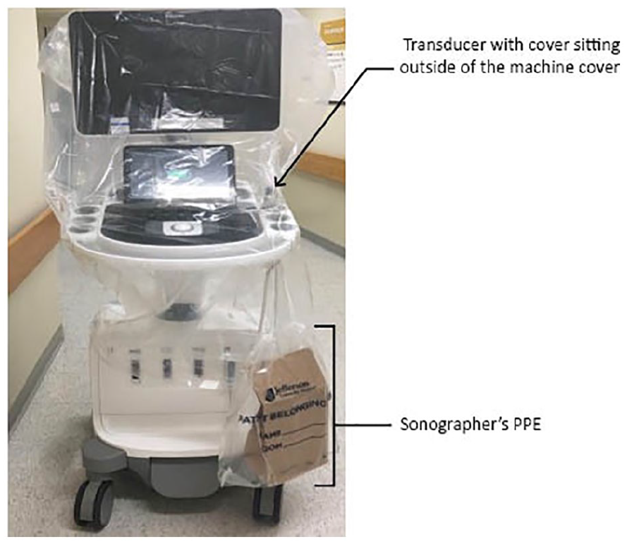
Demonstration of an ultrasound machine setup for coronavirus disease 2019 (COVID-19) patients or patients under investigation (PUIs). This setup is performed within the department before going to the designated COVID-19+ patient unit. The plastic bag connected to the ultrasound machine contains the sonographer’s personal protective equipment, which is put on just before entering the patient’s room.
When sonographers arrive to perform a sonogram on a COVID-19+ patient or PUI, they must protect themselves with proper personal protective equipment (PPE). The type or brand of PPE will depend on the specific institution’s recommendations and guidelines, as well as availability. Before entering the patient’s room, the sonographer should put on a N95 mask, powered air-purifying respirator (PAPR), or face shield; gown; and two sets of gloves. The outer pair of gloves is used to scan the patient. Keep one hand dedicated to manipulating the transducer and the other for the equipment. At the authors’ institution, it is mandatory to wear a face shield with a N95 mask or PAPR with a N95 mask (in case of PAPR failure) when scanning COVID-19+ patients or PUIs.
After completing the sonogram, the machine should be moved away from the patient, so that there is at least a 6-foot distancing. Achieving this physical distancing may be harder in some patient rooms, and this is based on room size and presence of other equipment. The outer pair of gloves, equipment cover, transducer covers, and gel should be removed and discarded at the completion of the sonogram. Before the sonographer exits the patient’s room, the inner pair of gloves is used to disinfect the ultrasound equipment and transducers. When the sonographer is leaving the patient’s room, they should remove their gown and gloves and wash their hands for 20 seconds. When the sonographer is outside of the patient’s room (with the ultrasound equipment), they should put on a clean pair of gloves, remove their remaining PPE, and wash their hands again. While still outside of the patient’s room, the sonographer should disinfect the ultrasound equipment and all parts of the transducer with LLD wipes. Finally, they should remove the gloves and wash their hands for another 20 seconds. In addition, some institutions report incorporating a “clean sonographer” and a “dirty sonographer” system to assist in the disinfectant process. The “dirty sonographer” would take off the equipment and transducer covers while in the patient’s room, clean the equipment, and then push the machine outside the room. The “clean sonographer” would wait outside the patient’s room for the machine and disinfect equipment in the hallway.
Upon returning to the department after a COVID-19 examination, the machine(s) should be disinfected again with LLD wipes. Transducers are wiped down with LLD wipes and placed in a Trophon machine (Nanosonics, Inc., Indianapolis, IN) for an additional layer of disinfection. When the dedicated ultrasound machine(s) and transducer (s) are not being used, they should be stored in a designated examination room with appropriate signage. By leaving the dedicated ultrasound machine(s) in one examination room, an additional layer of isolation is provided, and the sonographers know where the COVID-19 ultrasound machine(s) are at all times.
The intent of this article is to inform the broader sonographer community about this COVID-19 disinfectant and equipment management protocol as it is useful and easy to understand. This example protocol should not replace any existing COVID-19 standards. Instead, it should act as an adjunct to an already approved hospital protocol. The current situation is in flux, and providing care during this pandemic is evolving as more information is provided. Sonographers are on the frontline providing care for COVID-19+ patients and PUIs and cannot distance themselves, given the nature of the work. 12 It is important to continue providing safe and quality care to patients, while keeping sonographers safe. This article and the associated protocols are designed to aid in that process.
Footnotes
Appendix A
Appendix B
Appendix C
Appendix D
Declaration of Conflicting Interests
The author(s) declared no potential conflicts of interest with respect to the research, authorship, and/or publication of this article.
Funding
The author(s) received no financial support for the research, authorship, and/or publication of this article.