
Abstract
Molecular ultrasound imaging provides the ability to detect physiologic processes non-invasively by targeting a wide variety of biological markers in vivo. The current study investigates the novel application of molecular ultrasound imaging for the detection of neural inflammation. Using a murine model with acutely injured spinal cords (n=31), subjects were divided into four groups, each being administered ultrasound contrast microbubbles bearing antibodies against various known inflammatory molecules (P-selectin, vascular cell adhesion protein 1 [VCAM-1], intercellular adhesion molecule 1 [ICAM-1], and isotype control) during molecular ultrasound imaging. Upon administration of the targeted contrast agent, ultrasound imaging of the injured spinal cord was performed at 40MHz for seven minutes, followed by a bursting pulse. We observed significantly enhanced signals from contrast targeted to P-selectin and VCAM-1, using a variety of outcome measures. These findings provide preclinical evidence that molecular ultrasound imaging could be a useful tool in the detection of neural inflammation.
Keywords
Introduction
Ultrasound contrast agents are composed of micron-sized bubbles, which change the contrast in sonograms by enhancing the amplitude of the reflected ultrasonic signal. Contrast enhanced ultrasound (CEUS) imaging is commonly used in situations where knowledge of blood perfusion is important, as ultrasound contrast agents are restricted to the vascular compartment. Knowledge of the presence and amount of tissue perfusion is valuable in a wide variety of clinical conditions and can be provided conveniently, safely, at a low cost, and in real- time using CEUS imaging.1,2 Currently, the properties of ultrasound contrast are well established and have been characterized by their acoustic behavior.3,4 This has facilitated their clinical use in multiple areas of medicine, most commonly in radiology and cardiology.5-7
Current research is examining the utility of ultrasound contrast imaging to detect and examine biochemical processes at the molecular level. 8 Through a nanoengineering process, targeting ligands can be attached to the microbubble surface. 9 This allows contrast agents to be targeted to specific human antigens in vivo, creating an opportunity to extend CEUS capabilities from the tissue to the molecular level. 10 Targeting ligands act not only to attract and attach ultrasound contrast microbubbles to specific biochemical markers, but to also hold the contrast in close association, regardless of blood flow.11-13 This enables the presence of disease-indicative molecular signatures to be revealed, which can then be imaged and quantified, allowing for an early assessment of disease processes.14,15 However, before this imaging technique can be feasible and easily performed in the clinical setting, multiple factors must be examined and studied to ensure appropriate dose, targeted marker, and imaging parameters, among others, are being employed.
Early investigations of targeted contrast enhanced ultrasound (TCEUS) were completed for the detection of thrombus formation, in which successful binding of targeted ultrasound contrast to the thrombus was achieved, both in vitro and in vivo. 16 These findings, although successful, represented the detection of a disease process in its late stage. Therefore, TCEUS research shifted to the investigation of early stage disease markers, as this was thought to be a more worthwhile application of targeted ultrasound contrast technology. 17
Because of the bubble’s micrometer radius (~ 1 – 5 m) and restriction to the vascular compartment, ultrasound contrast agents are ideal for targeting disease-indicative biomarkers that over-express on the endothelial surface. 18 Thus, the focus of TCEUS research has included the detection and evaluation of pathological mechanisms such as angiogenesis, atherosclerosis, thrombus, and inflammation.1,19
Inflammation is a common pathophysiologic process that occurs in a vast majority of diseases. 20 The inflammatory process is mediated by the initial capturing and adhesion of rolling leukocytes by cell adhesion molecules (CAMs) on the endothelial surface. 21 Vascular cell adhesion protein 1 (VCAM-1), intercellular adhesion molecule 1 (ICAM-1), and P-selectin are CAMs that perform these roles, and are known to be present in the early stages of inflammation; all of which have been successfully targeted and detected on TCEUS, in a variety of tissues.4,15,17,19,22,23 For this reason, they have all been considered to be early marker of inflammation.18,24-26
In our pilot work, we explored the utility of TCEUS imaging to detect neural tissue inflammation of the spinal cord. Using a mouse model that had undergone a traumatic spinal cord injury we report the feasibility of detecting the presence of inflammatory biomarkers while imaging the neural tissue of the acutely injured spinal cord. Furthermore, qualitative and quantitative evaluation of the TCEUS sonograms provided insight into which inflammatory biomarkers were able to be imaged with the most sensitivity. The reason for selecting the acutely injured spinal cord for investigation, was in part due to the gap in the literature examining this imaging technology in neural tissue, particularly neural inflammation. Additionally, the model of induced injury used in this study, similarly represents the pathophysiological effects present in humans following a moderate-severe spinal cord injury. Thus, this application could become a frequently used imaging technique, due to the high prevalence of traumatic back injuries. Furthermore, this could provide the opportunity for physicians to quickly confirm a diagnosis and begin treatment to reduce further damage to the spinal cord. The goal of the current research is to more rigorously investigate the use of the aforementioned biomarkers (P-selectin, ICAM-1, and VCAM-1) within neural tissue of the acutely inflamed spinal cord by gauging their response upon targeted contrast administration. Based on the high success rate of TCEUS in detecting inflammatory markers in other tissue types, it is hypothesized that ultrasound contrast microbubbles targeted to P-selectin, VCAM-1, and ICAM-1 will tether in vivo, and be detected by enhanced signal intensities compared to control injections. This will provide preclinical proof of principle evidence regarding the utility of TCEUS imaging for the detection of neural inflammation.
Materials and Methods
Mouse Model
This research protocol was approved by our university’s Institutional Animal Care and Use Committee (IACUC). Thirty-one adult female C57BL/6 mice underwent a contusive spinal cord injury, followed by a TCEUS sonogram seven days later. This animal population is commonly used in preclinical molecular ultrasound studies, and was therefore selected so that appropriate comparisons could be possible. 27 The average subject weight was 17.1 grams (g) (15.0-18.8g), and the average subject age was 65.9 days (62-70 days). All surgeries were performed under aseptic conditions. Briefly, under deep anesthesia with ketamine and xylazine (intraperitoneal injection 90 mg/kg and 10 mg/kg, respectively), mice received a dorsal laminectomy of the 9th thoracic vertebra (T9). An Infinite Horizon Impactor device administered a moderate-severe contusion injury using an experimentally controlled force of 75 kdynes (mean actual force 76.8 ± 1.8 kdynes). After securing the lateral processes of the 8th thoracic vertebra (T8) and 10th thoracic vertebra (T10), the stainless steel impactor tip displaced the cord 598.2 ± 54.8 micrometers. 28 The contusion severity was derived from previous studies with C57BL/6 mice.29,30 Pain management was provided to the subjects according to the protocol stipulated by the university’s IACUC as well as closely assessed by our veterinarian technologist. This consisted of administrations of Baytril (enrofloxacin) and gentamicin, neither of which contains an anti-inflammatory component. The structure of the spinal cord lesion was characterized as a central lesion with a small peripheral rim of spared white matter within the T9 segment. 29 The exposed microcirculation at the injury epicenter caused the expression of CAM inflammatory biomarkers after the induced moderate-severe contusion.31,32
The subjects were divided into 4 groups. Each group was administered a bolus injection of an ultrasound contrast targeted against a different CAM (P-selectin (n=8), ICAM-1 (n=7), VCAM-1 (n=8), and isotype control (n=8)). (Figure 1) Monoclonal rat anti-mouse antibodies were used for each of the targeted inflammatory biomarker groups. The isotype control injection consisted of a bolus of ultrasound contrast microbubbles conjugated to rat IgG antibodies, which possess no specificity for any of the targeted antigens, or any other markers of inflammation in the species used. This served as a negative control.
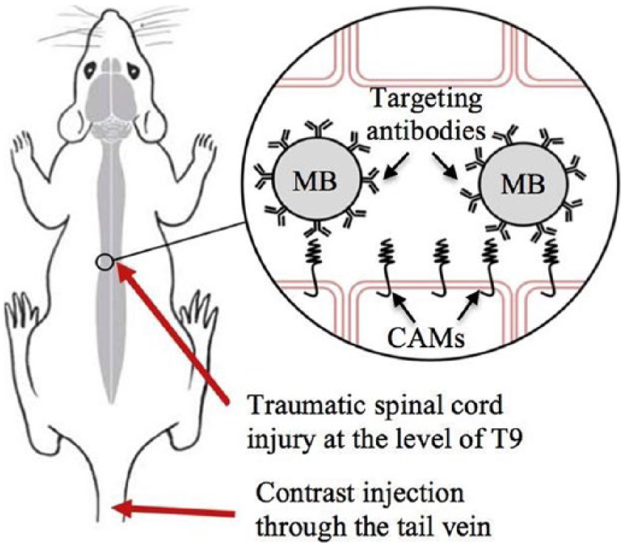
Methodological diagram of the hypothesized binding of biotinylated ultrasound contrast microbubbles (MBs) to the targeted cell adhesion molecules (CAMs) within the vasculature of the inflamed spinal cord in a mouse model.
A TCEUS sonogram was performed seven days after the traumatic spinal cord injury. This allowed for an appropriate amount of time for suture healing on the skin surface, however was still early enough to capture CAM expression. 33 During TCEUS imaging, subjects were anesthetized with 2.5% isofluorane mixed with one liter per minute carbogen, and were maintained with 0.5%-1.5% isofluorane. Once anesthetized, a tail vein catheter was placed to provide a route for targeted ultrasound contrast administration. The animal was then positioned prone, to allow for a posterior scanning approach. The hair of the animal was also removed to optimize sonographic imaging. A small animal monitoring system integrated into the ultrasound machine monitored physiologic parameters such as cardiac rhythm, respiration, and temperature during the entire TCEUS. Heart rate was maintained at the range of 300-450 beats per minute by adjusting the level of anesthesia. Core temperature was maintained at 37 C using a heated warming pad.
Ultrasound Contrast
A 30 microliter (µL) bolus injection of Targestar SA (Targeson, San Diego, California) microbubble ultrasound contrast solution, nanoengineered to target the selected CAMs, was used for TCEUS imaging. Targeting ligands were bioconjugated and attached to the surface of the microbubbles via a biotin-streptavidin-biotin interaction. The nanoengineering process began by dispersing the microbubble base solution until a uniformly opaque color is achieved. This opaque color is due to the suspended microbubble formation, which is now ready to be attached to the targeting antibody (anti P-selectin, anti ICAM-1, anti VCAM-1, or isotype control). Two hundred microliters of the biotinylated targeting antibody solution were then injected into 1.0 mL of the microbubble base. This was then re-dispersed by shaking of the vial. A washing process was used to remove any excess and non-attached antibodies. Buffer solution was then added to the vial to achieve a total volume of 5.0 mL. Next, the solution was centrifuged at 400xG for four minutes, causing the microbubbles to form a cake at the top of the solution. Excess solution was drained without disturbing the cake to attain a final volume of 1.0 mL. The final step in the nanoengineering process was to re-disperse the solution of now targeted microbubbles by shaking the vial. The isotype control injections were prepared and administered in the exact same manner as the antibody injections to allow for a proper comparison to be made.
TCEUS
A Vevo 2100 ultrasound unit (VisualSonics, Toronto, ON, Canada) was used to perform TCEUS imaging. A linear transducer operating at 40 MHz and 2% power was employed to collect sagittal images at the level of the T9 spinal cord. Quality control data was collected using a tissue mimicking phantom to ensure consistent axial and lateral resolution parameters throughout the course of the research study.
Sagittal images were acquired using a posterior approach on the animal. A posterior approach allowed for the least possible distance between the transducer face and the spinal cord, allowing the use of the highest possible operating frequency. Once an elongated image of the spinal cord was obtained, the transducer was locked into a transducer mounting system that secured the transducer in a stationary position. This allowed for consistent imaging throughout the entirety of the TCEUS sonogram. Sonographic imaging began ten seconds prior to contrast administration to acquire non-enhanced images. These images serve as the reference for non-enhanced pixel intensity, and provide a baseline for contrast enhanced images to be compared. The contrast bolus was then administered, followed by seven minutes of imaging. At the seven-minute time point, the burst mechanism was activated, to theoretically rupture all microbubbles within the beam profile. Imaging continued for two more minutes in order to monitor the effect of the bursting pulse on the contrast distribution. This was done to confirm ultrasound contrast microbubble binding to the targeted inflammatory biochemical marker. 34 All images were recorded in the form of video clips (AVI), which allowed for the option of selecting individual frames during the analysis process. All TCEUS sonograms were performed by a credentialed sonographer, with experience in human and animal musculoskeletal imaging.
Data Analysis
Retrospective TCEUS analysis was performed using the Vevo analysis computer software. A region of interest (ROI) was drawn around the spinal cord at the level of T9 using electronic calipers. The ROI was then applied to the entire seven minute AVI of the TCEUS sonogram. This provided a quantified value of average pixel intensity within the T9 spinal cord for each frame of the TCEUS. Enhanced pixel intensity is representative of microbubble binding and CAM expression. Pixel intensities were plotted over time to generate contrast time intensity curves (TICs). These were constructed to provide a semi-quantitative display of signal intensity data, representing the temporal activity of the targeted contrast agent throughout the entire TCEUS sonogram. 35 (Figure 2)
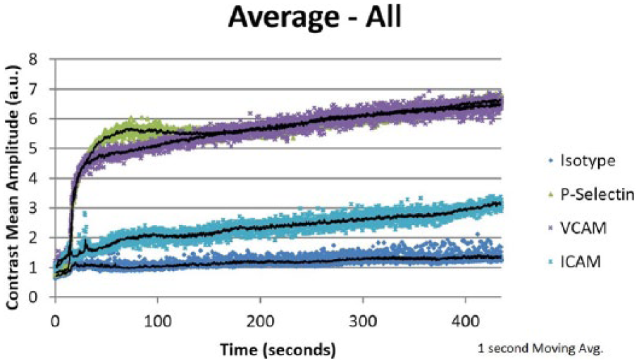
Time intensity curves representing the temporal behavior of all the molecular targets from each experimental condition, derived from the average signal intensity values from the subjects within each targeted antibody group.
From the generated TICs, outcome measures of area under the curve (AUC), maximum pixel intensity (MPI), time to peak intensity (TTP), rise time (RT), positive gradient (PG), and signal due to bound contrast (SDBC) were calculated.36-41 This allowed inferential statistics to be used for the comparison of outcome measures between antibody groups. Outcome measures are defined as follows: AUC (area under the TIC), MPI (highest pixel value in the TIC), TTP (time from zero to maximum pixel intensity, aka mean transit time), RT (time from 10% to 90% of maximum pixel intensity), PG (MPI/TTP), and SDBC (average pixel intensity obtained 100 frames before the burst minus the average pixel intensity from the 100 frames after the burst mechanism). 40
Prior to performing inferential statistical procedures, a Shapiro-Wilk test of normality was used to determine the nature of the data for each of the outcome measures within each antibody group. Failure of an outcome measure’s data to demonstrate normality (<0.05) resulted in the use of non-parametric testing for antibody group comparison for that outcome measure. Non-parametric comparison testing consisted of a Kruskal-Wallis analysis of variance. Alternatively, outcome measure data that was found to be normally distributed was then analyzed with parametric statistical methods, specifically a one-way analysis of variance (ANOVA), with post hoc Tukey multiple comparison testing. Parametric testing was able to be used due to the balanced design of the experiment. 42 Finally, paired one-way Student t-tests were used to determine if a significant drop in pixel intensity obtained from the 100 frames prior to, and the 100 frames following the burst mechanism, for each subject. All statistical analyses were conducted in SPSS (version 21), with an a priori α ≤ 0.05.
Results
Normality testing revealed non-normality within the outcome measures of TTP, RT and PG. Therefore, non-parametric testing was used to obtain inferential statistical information on these measures. Normality testing revealed normally distributed data in the outcome measures of AUC, SDBC, and MPI, as such, parametric testing was used to obtain inferential statistical information on these measures.
ANOVA testing identified statistically significant differences in AUC values among the antibody groups (F=13.61, p<0.001). A Tukey post hoc test revealed greater AUC values of statistical significance in the P-selectin group (2453.07±331.50) when compared to the ICAM-1 ((880.08±211.46), p=0.005, and the isotype control groups (511.57±103.50), p<0.001. Similarly, a Tukey post hoc test revealed greater AUC values of statistical significance in the VCAM-1 group (2401.64±351.16) when compared to the ICAM-1 (880.08±211.46), p=0.007, and the isotype control groups (511.57±103.50), p<0.001. (Figure 3)
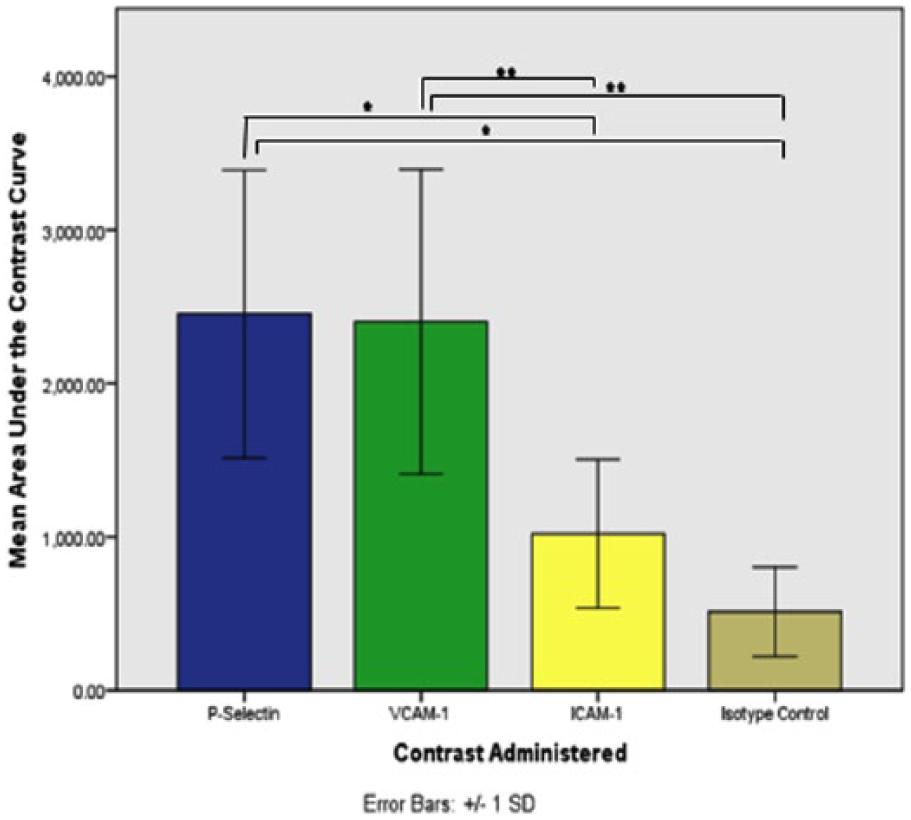
Bar graph comparing mean area under the curve (AUC) among the targeted contrast antibody groups. These data demonstrate P-selectin and VCAM-1 targeted microbubbles to have significantly greater AUC values than those of ICAM-1 and the isotype control. *Statistically significant difference at P < .05. a.u., arbitrary units; ICAM-1, intercellular adhesion molecule 1; SD, standard deviation; VCAM-1, vascular cell adhesion molecule 1.
ANOVA testing identified statistically significant differences in SDBC values among the antibody groups (F=9.32, p<0.001). A Tukey post hoc test revealed greater SDBC values of statistical significance in the P-selectin group (2.87±0.71) when compared to the ICAM-1 (0.06±0.05), p=0.006, and the isotype control groups (-0.04±0.02), p=0.003. Similarly, a Tukey post hoc test revealed individual greater SDBC values of statistical significance in the VCAM-1 group (2.77±0.74) when compared to the ICAM-1 (0.06±0.05), p=0.008, and the isotype control groups (-0.04±0.02), p=0.004. (Figure 4)
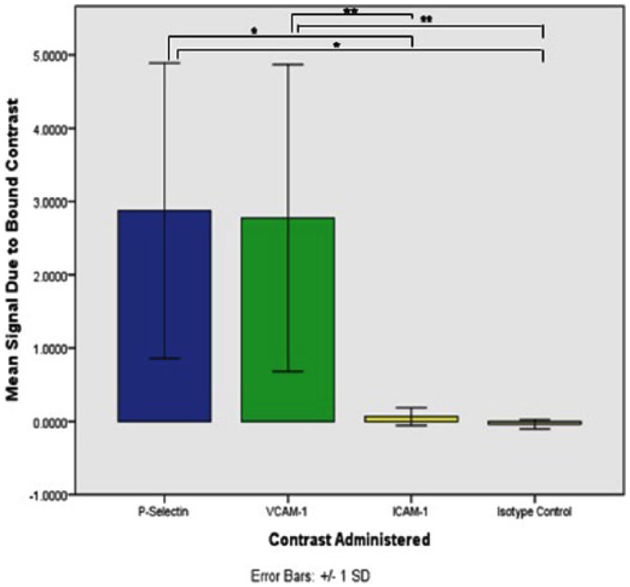
Bar graph comparing mean signal amplitude due to bound contrast among the targeted contrast antibody groups. These data demonstrate P-selectin and VCAM-1 targeted microbubbles to have significantly greater SDBC values than those of ICAM-1 and the isotype control. *Statistically significant difference at P < .05. a.u., arbitrary units; ICAM-1, intercellular adhesion molecule 1; SD, standard deviation; VCAM-1, vascular cell adhesion molecule 1.
ANOVA testing identified statistically significant differences in MPI values among antibody groups (F=4.47, p=0.011). A Tukey post hoc test revealed greater MPI values of statistical significance in the P-selectin group (7.77±0.92) when compared to the isotype control group (3.47±1.05), p=0.030. Similarly, a Tukey post hoc test revealed greater MPI values of statistical significance in the VCAM-1 group (7.84±1.00) when compared to the isotype control group (3.47±1.05), p=0.027. (Figure 5)
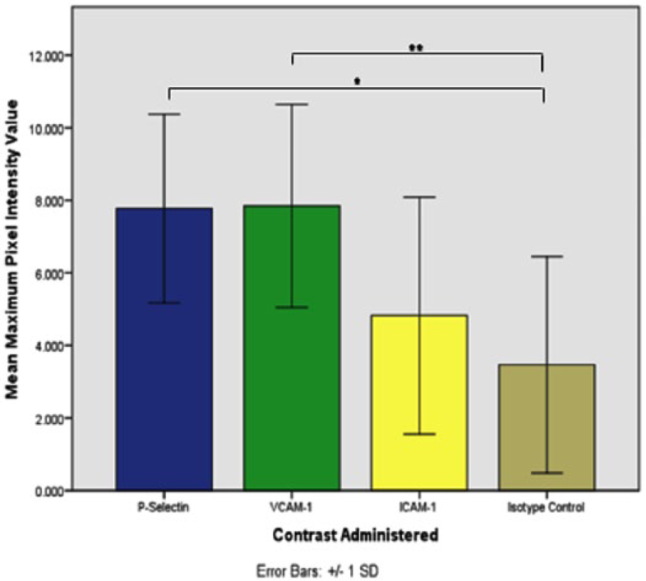
Bar graph comparing the average maximum signal amplitude among the targeted contrast antibody groups. These data demonstrate P-selectin and VCAM-1 targeted microbubbles to have significantly greater maximum pixel intensity (MPI) values than those of the isotype control. *Statistically significant difference at P < .05. a.u., arbitrary units; ICAM-1, intercellular adhesion molecule 1; SD, standard deviation; VCAM-1, vascular cell adhesion molecule 1.
Kruskal-Wallis testing revealed no significant differences among the antibody groups for the measures of TTP (p=0.11), RT (p=0.71), and PG (p=0.73). A descriptive data summary of the values within each antibody group for each outcome measure can be found in Table 1.
Descriptive Data Table of Targeted Antibody Group Means and Standard Deviations for Each of the Targeted Contrast-Enhanced Ultrasound Outcome Measures.
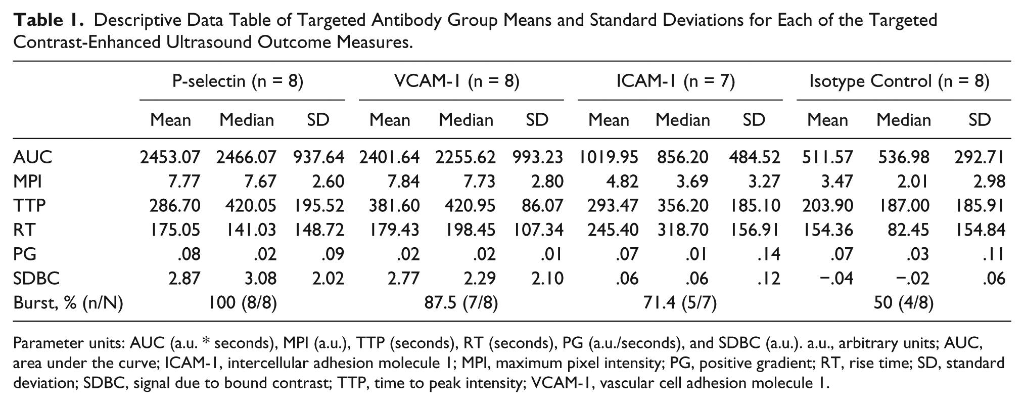
Parameter units: AUC (a.u. * seconds), MPI (a.u.), TTP (seconds), RT (seconds), PG (a.u./seconds), and SDBC (a.u.). a.u., arbitrary units; AUC, area under the curve; ICAM-1, intercellular adhesion molecule 1; MPI, maximum pixel intensity; PG, positive gradient; RT, rise time; SD, standard deviation; SDBC, signal due to bound contrast; TTP, time to peak intensity; VCAM-1, vascular cell adhesion molecule 1.
Finally, burst mechanism testing revealed a statistically significant drop in pixel intensity after the burst was applied in 100% (8/8) of the subjects in the P-selectin group, 87.5% (7/8) in the VCAM-1 group, 71.4% (5/7) in the ICAM-1 group, and 50% (4/8) in the isotype control group.
Discussion
Molecular sonographic imaging, or TCEUS, has been used to target a myriad of biomarkers in a variety of physiological processes. Once targeted ultrasound contrast is introduced into circulation, microbubbles will bind and remain tethered to the targeted molecule, via the attached ligand located on the microbubble surface. As acoustic energy is applied to the targeted area, compression and rarefaction of the tethered microbubbles begins, causing them to produce an enhanced harmonic signal. 43 This is due to the large acoustic impedance of the gas-tissue interface of the microbubble and its surroundings. 44 The enhanced signal can then be subtracted from the surrounding non-enhanced ultrasound signals, to allow for the detection of the targeted molecules. (Figure 6) The enhanced signal can also be displayed in the form of a TIC, which allows for the evaluation of its temporal behavior, as well as for a semi-quantitative evaluation of the TCEUS to be made.
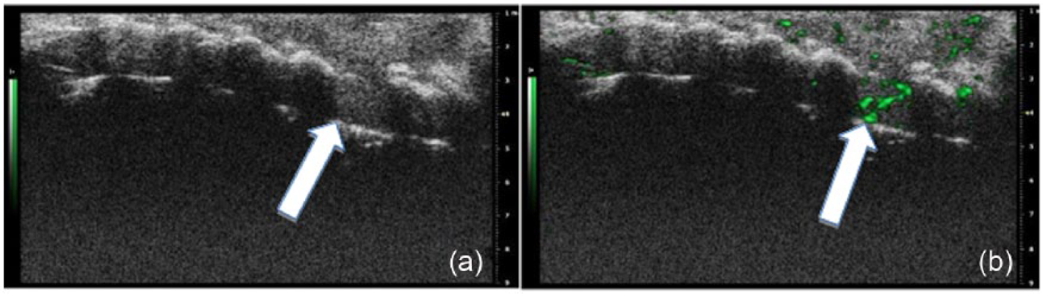
(a) Sagittal unenhanced noncontrast gray-scale image of a mouse spinal cord demonstrating the T9 laminectomy and T9 portion of the spinal cord (indicated by the white arrow). (b) Sagittal contrast enhanced image of a mouse spinal cord, demonstrating contrast enhancement at the level of T9, representative of microbubble presence, for P-selectin targeted microbubbles seven days after spinal cord injury.
Inflammation is a common pathological condition that has been investigated with TCEUS, as inflammatory biomarker expression is located on the vascular endothelium, and ultrasound contrast agents are restricted to the vascular compartment. In the present research, we have reproduced the application of TCEUS imaging to detect inflammation in neural tissue. TCEUS scans of an acutely inflamed spinal cord were performed in a murine population. The findings reported on our four groups of subjects, with three paired to known inflammatory biomarkers (P-selectin, ICAM-1, and VCAM-1), represent preclinical proof of principle evidence that informs the utility of targeting three different inflammatory markers in inflamed neural tissue.
Our results suggest that ultrasound contrast microbubbles targeted against P-selectin and VCAM-1 tether to molecular markers of inflammation and produce an enhanced signal for their detection. These findings are consistent with prior studies that reported the ability to successfully detect these two biomarkers with TCEUS in the presence of inflamed cardiac tissue. Kaufmann et al. demonstrated ten times more binding of microbubbles to targeted to P-selectin, than controls, while examining myocardial ischemia and reperfusion. 45 A similar study conducted by Villaneuva el at., also examining P-selectin targeting in myocardial ischemia and reperfusion, produced similar findings. 46 Likewise, Hernot et al. investigated the targeting and evaluation of VCAM-1 with TCEUS, using a murine model with inflamed tumor vasculature, and reported findings of enhanced signal intensities in the experimental subjects compared to controls. 47
Other findings in the present study include significantly greater MPI values in the P-selectin and VCAM-1 groups when compared with the control group. However, significant differences were not found between the P-selectin and VCAM-1 groups with the ICAM-1 group, in contrast with the AUC and SDBC outcome measures. A possible explanation could be that MPI is not as appropriate as the other measures since it is a measure of only one point (maximum pixel intensity), and therefore does not provide a comprehensive representation of targeted contrast temporal behavior throughout the entire TCEUS sonogram.
Based on the existing literature, ultrasound contrast microbubbles targeted against P-selectin have demonstrated great success. This has made P-selectin a biomarker of interest while evaluating inflammation with molecular ultrasound imaging. 48 This success may be attributed to attributed to P-selectin maintaining a high-on and high-off rate, its presence in the early stages of the inflammatory cascade, and/or its uniqueness of being expressed on the surfaces of platelets and the endothelium.21,49,50 To this end, P-selectin has been effectively targeted and evaluated with ultrasound contrast in the detection of inflammation for a variety of tissues, including kidney, bowel, venous endothelium and myocardium.11,45,46,51,52
However, in an unpublished study conducted by Rychak et al., the feasibility of a variety of inflammatory biomarkers were evaluated in inflamed bowel tissue. That study reported findings similar to the current research being presented, in which ultrasound contrast targeted against VCAM-1 produced greater pixel amplitudes than contrast targeted against P-selectin (J. Rychak [Targeson] unpublished data). Therefore, a hypothesis could be made that microbubbles targeted against inflammatory biomarkers behave similarly in bowel, myocardial, and neural tissues. However, additional research is required in this area to make any further conclusions.
The presented findings also suggest that ICAM-1 may not be an appropriate marker for targeted ultrasound contrast imaging in the evaluation of neural inflammation, as significant differences between this group and the isotype control group were only seen in the AUC outcome measure in the current study. Additionally, multiple outcome measures revealed P- selectin and VCAM-1 to be statistically significantly greater than ICAM-1. This is supported in the literature as in vitro studies have reported that ultrasound contrast targeted against selectins contained significantly more bound microbubbles than that of ICAM-1. 53 Furthermore, other in vitro have also found the microbubble saturation of targeted P-selectin and VCAM-1 to be similar, and much more than that of ICAM-1.54-56 Although in vivo studies, including Weller et al., have reported that microbubbles targeted to ICAM-1 provided statistically significant enhancement in imaging acute cardiac transplant rejection, in comparison to controls. 57 One possible explanation for the disagreement in findings regarding the ability to detect ICAM-1 targeted microbubbles, could be due to the differences in CAM expression between the two tissue types. The inflammatory response varies among tissues, and the role of ICAM-1 following a contusive spinal cord injury is not well defined, therefore it is possible that higher levels of ICAM-1 are expressed in the tissue of the myocardium than in neural tissue of the spinal cord.58,59 This would indicate that our results not suggest ICAM-1 targeted microbubbles’ poor ability to bind in vivo, but rather would suggest the lack of ICAM-1 molecules being expressed, resulting in limited available microbubble binding sites. Additional research and replication studies are needed in this area to provide more information regarding the properties of ICAM-1 targeted microbubbles in various tissue types.
In addition to investigating the ability of targeted ultrasound contrast to detect inflammatory biomarkers in inflamed neural tissue, this study also incorporated multiple analysis methods that are novel to TCEUS. This was done to provide parameters representing different characteristics of targeted ultrasound contrast agent temporal behavior during TCEUS imaging. Outcome measures of AUC, MPI, SDBC, and the burst evaluation, are commonly used in TCEUS analysis. However, the derived parameters of TTP, RT, and PG are commonly used methods of analysis in magnetic resonance (MRI) contrast imaging.37-39 These were translated to TCEUS analysis to begin building the body of evidence regarding their utility. However, findings of this preclinical study suggest that these measures may not be appropriate for TCEUS imaging, as no statistically significant differences were found during comparison testing between antibody groups. Although it is important to note that this was the first attempt to translate these parameters from MRI contrast to TCEUS imaging, and therefore larger, more in depth studies are needed to more accurately define their role.
Limitations
This study adds to the body of preclinical evidence, as it reports results in the novel application for the use of TCEUS in the detection of neural inflammation. However, this study is limited to providing evidence of CAM targeting in the extreme end of the neuro-inflammatory disease process.31,32 Therefore, the utility of this molecular ultrasound imaging application in the presence of only mild neural inflammation remains unknown. Furthermore, when comparing targeted antibody conditions, it is important to underscore that the expression of the inflammatory biomarkers is assumed to be similar. This advocates that microbubbles targeted to each of the CAMs have an equal chance to encounter and tether to its selective molecular marker. The diminished expression of a CAM may falsely suggest the inability of microbubble adherence to that selective biochemical marker, although may provide insight into the effectiveness of targeting that CAM in the particular tissue environment being investigated. Lastly, this study was performed using an animal model, and therefore should be considered as low-level evidence.
Future Directions
The next step in the investigation of TCEUS for the detection of neural inflammation must build upon the preclinical evidence being reported from this study. The current study should be replicated in a larger population to provide more information, which may support or refute these findings, and to ensure that eventual clinical translation is done so based on appropriate preclinical evidence. Furthermore, this study investigates only three inflammatory biomarkers. Other members of the CAM family and other known molecular markers of inflammation that have proven to be successfully targeted for the detection of inflammation in other tissues, such MAdCAM-1, E-, and L-selectin must also be investigated to determine the best targeting molecule.60-62
Conclusions
In summary, this study demonstrates the proof of principle that microbubbles modified with ligands targeted known inflammatory markers (P-selectin, VCAM-1, and ICAM-1) will selectively adhere and produce an enhanced and sustained ultrasonic signal within acutely inflamed neural tissue of the spinal cord. Although all three inflammatory markers demonstrated greater pixel intensity measures than control injections, only P-selectin and VCAM-1 were significantly greater while comparing all three of the commonly and largely accepted measures (AUC, MPI, SDBC) used to analyze TCEUS imaging. Differences were not seen while using MRI contrast measures (TTP, RT, PG) that were translated to molecular ultrasound imaging. This preclinical research represents the first step in the evaluation of early inflammatory markers for the detection of neural inflammation. The findings of this study create a potential opportunity for the advancement of molecular ultrasound imaging into applications involving the early detection of disease processes within neural tissue and warrant further investigation of its utility.
Footnotes
Acknowledgements
Our research team would like to thank Dr. D. Michele Basso and her research team, for their support and advice during the analysis of the imaging data.
Declaration of Conflicting Interests
The authors declared no potential conflicts of interest with respect to the research, authorship, and/or publication of this article.
Funding
The authors received financial support from the ASRT Foundation and Targeson LLC for their research.